Download

1 / 16
160 likes | 400 Views
Aggregation phenomena relevant to Alzheimer ’ s Disease: Statistical physics approach. Sijung Yun. Collaborators: Brigita Urbanc, Luis Cruz, Shouyong Peng, Jose Borreguero, Alfonso Lam . Advisor: H.E. Stanley. Outline. Alzheimer ’ s disease & A β - protein

E N D
Aggregation phenomena relevant to Alzheimer’s Disease: Statistical physics approach Sijung Yun Collaborators: Brigita Urbanc, Luis Cruz, Shouyong Peng, Jose Borreguero, Alfonso Lam Advisor: H.E. Stanley
Outline • Alzheimer’s disease & Aβ- protein • Simulation of Aβ-proteins folding and aggregation
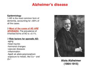
Alzheimer’s disease is related to Amyloid β-proteins(Aβ) • Over 50% for the people over 85 years old • Increasing forgetfulness, etc. • Clinically, a dementia characterized by fibril made of amyloid β-proteins(Aβ) and tangles made of τ-protein in brain • Amyloid β-proteins(Aβ) come in two forms: • Aβ40: 1DAEFR6HDSGY11EVHHQ16KLVFF21AEDVG26SNKGA31IIGLM36VGGVV • Aβ42: 1DAEFR6HDSGY11EVHHQ16KLVFF21AEDVG26SNKGA31IIGLM36VGGVV41I42A • Oligomers of Aβ-40 and Aβ-42 are neurotoxic • Oligomers of Aβ-42 are more neurotoxic than that of Aβ-40
What did we do? Why? • We used “Discrete Molecular Dynamics” for the study of oligomerization of Aβ-40 and Aβ-42 in atomic detail • Why? • Experiment cannot show how Aβ-40 protein and Aβ-42 protein oligomerize in atomic detail • Conventional molecular dynamics cannot study oligomerization (too much CPU time)
Conventional Molecular Dynamics Discrete Molecular Dynamics Discrete Molecular Dynamics(DMD)
P.E. Hydrophobic attraction P.E. Hydrophilic repulsion 3.07Å 7.5 Å Distance(Å) Distance(Å) 3.07Å 7.5 Å DMD with hydrophobic interaction • Hydrophobicity is the driving force of the protein folding and aggregation • Hydrophobicity appears as “the effective attraction” between hydrophobic particles • Hydrophilicity appears as “the effective repulsion” between hydrophilic particles
Running DMD Simulation Prepare 8 sets of 32 proteins for each Aβ40 and Aβ42 Get 8 trajectories for each Aβ40 and Aβ42 Finally, Statistical analysis
Simulation results (1)Oligomer Size distribution Ref) Bital et al. (2003) Proc. Natl. Acad. Sci. USA 100(1) 330-5
How to read a contact map (35,18) (39,36) 1DAEFR6HDSGY11EVHHQ16KLVFF21AEDVG26SNKGA31IIGLM36VGGVV41I42A
Simulation results(2)How “monomers” fold as time goes on 1DAEFR6HDSGY11EVHHQ16KLVFF21AEDVG26SNKGA31IIGLM36VGGVV41I42A Monomers fold from “C terminal region”(Around 40 or 42) to “N terminal region”(around 1)
Simulation results (3)“Pentamers” of Aβ-40 and Aβ-42 Aβ-40 Aβ-42 “N terminal”(Around 1) of Aβ42 is more stretched than that of Aβ40 Ref) Urbanc B, Cruz L,Yun S, Buldyrev SV, Bitan G, Teplow DB, Stanley HE, (2004) Proc. Natl. Acad. Sci. USA 101 17345-17350
Ongoing work: Adding Coulombic interaction • Some amino acids are charged. • Negatively charged amino acids: D-, E- • Positively charged amino acids: R+, K+ 1D-AE-FR+6HD-SGY11E-VHHQ16K+LVFF21AE-D-VG26SNK+GA31IIGLM36VGGVV41I42A
Analysis ongoing:When electrostatic interaction is added Sharper better defined structure
Conclusions • “Monomers” fold from C terminal to N terminal • N terminals of Aβ-42 oligomers are more stretched out than those of Aβ-40 oligomers • Simulation shows there is a significant difference between aggregation of Aβ-40 and Aβ-42 • Simulation gives insights why Aβ-40 and Aβ-42 aggregate differently; • Reason; The hydrophobicity of 41th and 42th amino acids causes the structural difference in folding
Primary structure of Aβ(1-42) 1D-AE-FR+6HD-SGY 11E-VHHQ 16K+LVFF 21AE-D-VG 26SNK+GA 31IIGLM 36VGGVV {41I42A} • (Hydrophobic) I:Ile V:Val L:Leu F:Phe C:Cys M:Met A: Ala • (Neutral in hydrophobicity) S:Ser T:Thr W:Trp P:Pro Y:Tyr G:Gly • (Hydrophilic) R+:ArgK+:LysD-:AspE-:Glu N:Asn Q:Gln H:His
Simulation additional resultsPrediction of secondary structure of monomers (no electrostatic interaction) Initial 103 Steps 104 Steps 105 Steps Black: Aβ40 Red: Aβ42