Endoscopic Vessel Harvesting System Market Will Be Worth $3.4 Billion By 2025: Grand View Research, Inc.
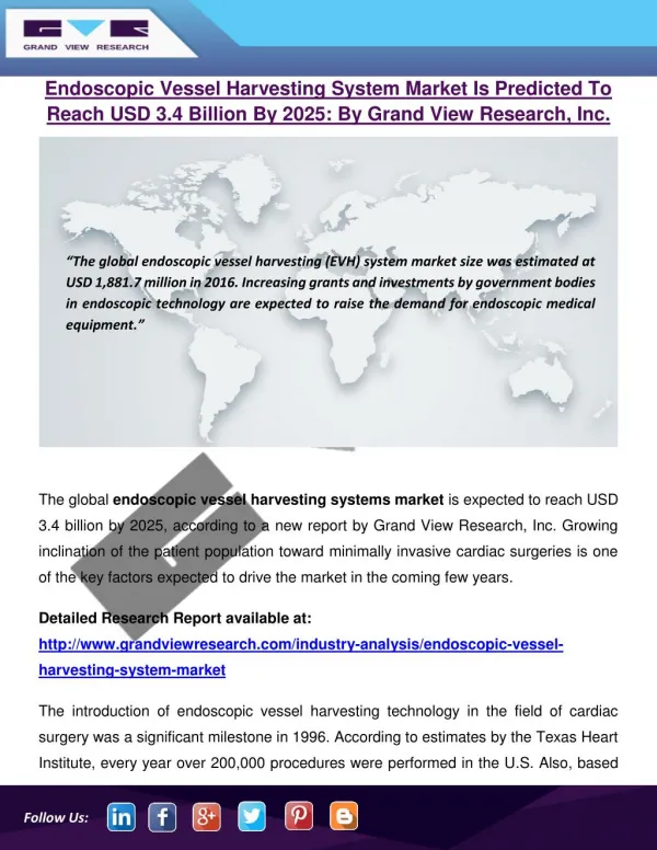
The global endoscopic vessel harvesting systems market is expected to reach USD 3.4 billion by 2025, according to a new report by Grand View Research, Inc. Growing inclination of the patient population toward minimally invasive cardiac surgeries is one of the key factors expected to drive the market in the coming few years. The introduction of endoscopic vessel harvesting technology in the field of cardiac surgery was a significant milestone in 1996. According to estimates by the Texas Heart Institute, every year over 200,000 procedures were performed in the U.S. Also, based on the statistics published by the department of surgery (Washington University School of Medicine in St. Louis), they perform Coronary Artery Bypass Grafting (CABG) surgeries on approximately 0.5 million people very year. The growing demand for these procedures and technologically advanced surgical procedures are among few factors anticipated to boost the market over the forecast period. The presence of favorable reimbursement policies in the U.S. pertaining to CABG surgeries is anticipated to increase the number these surgeries. On July 2016, the Department of Health & Human Services announced that it will begin implementing bundled payment models for high-quality, coordinated cardiac and hip fracture care. The market is highly concentrated with a limited number of products and market players operating in this space. Many studies stated that the open vessels harvesting technique is as effective as minimally invasive vessels harvesting technique. However, the growing awareness regarding benefits of Endoscopic Vessels Harvesting (EVH), such as minimally invasive nature, less complications, and quick recuperation are some of the factors that are anticipated to propel its growth during the forecast period.
126 views • 8 slides