Download

1 / 8
0 likes | 2 Views
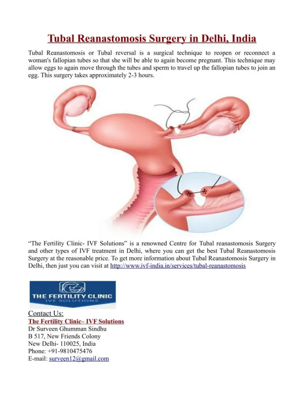
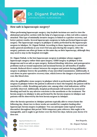
Tubal diseases are one of the frequent causes of infertility. The<br>most common predisposing factor is pelvic inflammatory<br>disease (PID). Distal tubal obstruction has been managed<br>previously by open surgery using microsurgical technique.<br>The pregnancy rate after reconstructive surgery is 20u201330%<br>2 years postoperatively. Laparoscopy for tubal infertility has<br>been a significant factor in reducingu2014costs, hospitalization,<br>and recuperation. Recently, in women with severe tubal<br>damage, in vitro fertilization (IVF) offers a better chance for<br>term pregnancy (72.3%) compared to reconstructive surgery<br>(27.

E N D
Laparoscopic T ubal Surgery Prof. Dr. R. K. Mishra INTRODUCTION Tubal diseases are one of the frequent causes of infertility. The most common predisposing factor is pelvic inflammatory disease (PID). Distal tubal obstruction has been managed previously by open surgery using microsurgical technique. The pregnancy rate after reconstructive surgery is 20–30% 2 years postoperatively. Laparoscopy for tubal infertility has been a significant factor in reducing—costs, hospitalization, and recuperation. Recently, in women with severe tubal damage, in vitrofertilization (IVF) offers a better chance for term pregnancy (72.3%) compared to reconstructive surgery (27.3%). Fimbrioplasty and lysis of peritubal and periovarian adhesions have been associated with good pregnancy rates. In these patients, IVF is appropriate when pregnancy is not achieved postoperatively after a few years. From anterior to posterior, following important tubular structures are found crossing the brim of true pelvis; the round ligament of the uterus, the infundibulopelvic ligament, which contains the gonadal vessels and the ureter. The ovaries and fallopian tube is found between the round ligament and the infundibulopelvic ligament. The ovarian ligaments run from the ovaries to the lateral border of the uterus. Ovary is attached to the pelvic side wall with infundibulopelvic ligament, which carries ovarian artery. One of the common mistakes that a surgeon can land into is injury of the ureter during dissection of the infundibulopelvic ligament. If the uterus is deviated to the contralateral side with the help of uterine manipulator, infundibulopelvic ligament is spread out and a pelvic side wall triangle is created. The base of this triangle is the round ligament, the medial side is the infundibulopelvic ligament, and the lateral side is the external iliac artery. The apex of this triangle is the point at which the infundibulopelvic ligament crosses the external iliac artery. The ureters enter the pelvis in close proximity to the female pelvic organs and are at risk for injury during laparoscopic surgery of these organs. As the ureter courses medially over the bifurcation of the iliac vessels, they pass obliquely under the ovarian vessels and then run in close proximity to the uterine artery. LAPAROSCOPIC TUBAL ANATOMY The fallopian tubes arise from the superior portion of the uterus just above the attachment points of the round ligament. Laparoscopically, the round ligaments overhang the fallopian tube because of uterine manipulation and can be easily mistaken for them. The fallopian tubes toward its lateral end encircle the ovaries partially with their fimbriated ends (Fig. 1). Patient Position Patient should be in steep Trendelenburg’s and lithotomy position. One assistant should remain between the legs of patient to do uterine manipulation whenever required. Port Position Port position should be in accordance with baseball diamond concept. If the left side of tube has to be operated, one port should be in right iliac fossa and another below left hypochondrium (Fig. 2). Fig. 1: Tubal anatomy.
404 SECTION3: Laparoscopic Gynecological Procedures Tubo-ovarian abscess is a severe sequelae and occurs in as many as 34% of patients hospitalized with PID. Symptomatic or subclinical infections can progress rapidly into a TOA. These abscesses can rupture, resulting in severe peritonitis. At present, surgical intervention is used to treat a tubo- ovarian mass only when medical management is ineffective. The laparoscopic procedure for managing pelvic abscesses has been described by several authors. Once TOA is diagnosed laparoscopically, two 5 mm trocars are placed in both the flanks according to baseball diamond concept through which a suction-irrigator probe and grasping forceps are inserted. The pelvis, upper abdomen, and pelvic gutters should be examined for free or loculated purulent material and the course of both ureters should be identified. Purulent fluid is aspirated from the pelvis, and cultures are taken from the aspirated fluid and the inflammatory exudates. If necessary, the suction-irrigator is used to mobilize the omentum, small bowel, rectosigmoid junction. Tubo-ovarian adhesions should be dissected until the abscess cavity is localized. After the abscess cavity is drained, the suction-irrigator is used to separate the bowel and omentum completely from the reproductive organs. Chromotubation is not indicated in case of PID because edema in the interstitial tissue of the tube occludes the lumen. At the end of the procedure, whole peritoneal cavity is irrigated with normal saline until the effluent is clear. Between 300 and 400 mL of irrigation fluid are left in the pelvis to distance these organs during the early healing phase. Adhesiolysis is technically difficult and associated with a high risk of complications. Hydrodissection decreases the potential for intestinal or ureteral injury; the laser and electrosurgery should be used sparingly. Fig. 2: Port position tubal surgery left side. OPERATIVE PROCEDURE Management of Acute Pelvic Inflammatory Disease Pelvic inflammatory disease usually results from sexually transmitted diseases caused by Chlamydia or Gonococcus infection, an intrauterine device (IUD), postpartum endometritis, or hysteroscopy at the time of endometrial infection. Pelvic inflammatory disease has four primary sequelae: 1. Infertility 2. Ectopic pregnancy 3. Chronic pelvic pain 4. Recurrent upper UTI. One of the worst outcomes of PID is adhesions of reproductive organ leading to infertility and pain. The degree of tubal damage and pelvic adhesions often depends on the severity of the infection, the number of PID episodes, and etiology. Severe peritonitis is associated with a 17% risk of infertility compared to 3% for mild infection. With each successive episode of PID, the risk of infertility doubles. Despite its typically mild presentation, chlamydial PID results in a threefold increase in infertility compared to gonococcal PID. The risk of ectopic pregnancy is 6–10 times higher in women who have had PID. In addition, chronic pelvic pain has been shown to occur in 15–18% of patients after PID, usually because of adhesions. Up to 20–25% of patients will have at least one recurrent infection because damaged fallopian tubes are more susceptible to infection. Laparoscopy is being used increasingly in patients suspected of having PID to make a precise diagnosis and thereby avoid the potential sequelae. Prompt surgical confirmation of the diagnosis is possible with laparoscopy. A tubo-ovarian abscess (TOA) can be drained, reducing the risk of serious morbidity associated with rupture. The clinical diagnosis of PID is difficult because of the wide variation in symptoms and signs. Many women with PID report subtle, vague, or mild symptoms that are not specific, such as dyspareunia, postcoital spotting, or abnormal uterine bleeding. In these situations, a bimanual examination may demonstrate cervical mobility or adnexal tenderness. Laparoscopy for Adnexal Torsion Adnexal torsion is a rare gynecologic emergency of women who are mostly in reproductive ages. So there is an increasing trend toward conservative approach for preservation of fertility in young women. Literature has all come to a point of agreement that, as minimal surgery as possible and sparing of adnexa for these women of reproductive age should be done, since torted adnexa has a benign histopathology in most cases. Operative laparoscopic procedures are being performed increasingly in gynecology in recent years. It delivers some main advantages over laparotomy. Smaller surgical scars which have better healing process than a single big scar, reduced postoperative pain and morbidity; and shorter hospital stays, recovery periods with a lower cost are the major advantages of laparoscopy for a number of gynecologic conditions (ectopic pregnancy, benign ovarian cysts, tuba peritoneal infertility, etc). The results of laparoscopic treatment are comparable with those of
405 CHAPTER30: Laparoscopic Tubal Surgery laparotomy. For these reasons operative laparoscopy has become the surgical treatment of choice for the conditions listed above. Like all surgeries, operative laparoscopy does bring with it a risk of complications which need to be assessed. Since mostly the lesions are benign in nature, simply detorsion of the torted adnexa and if necessary the cyst excision is the preferred procedure. But what is very important, is the time period between the diagnosis and treatment of the pathology. Since the torsion of adnexa causes a relative ischemia of the ovarian tissue, it may result in failure and loss of ovarian function. In older studies, it was advised to remove the necrotic adnexa since they thought that it might lead to pulmonary emboli. But in recent literature, it is not advised to remove the adnexa even if the adnexa looks necrotic because even severely necrotic looking adnexa may save its function after surgery. Some literature suggest that only simple detorsion procedure may cause retorsion and the rate of retorsion is higher patients with normal looking adnexa and is lower in the patients who have pathological adnexa and some other procedure should also applied along with detorsion. Ovariopexy may be applied additionally, especially if there is a long ovarian pedicle, but more studies are needed to evaluate its value. In the literature, it is shown in retrospective studies that in the patient selection for laparoscopic surgeries, size of the adnexal cystic pathology is an important criteria that is the mean size of the cyst, which is smaller in laparoscopic surgeries compared to laparotomy. Risk factors for conversion to laparotomy are studied in some articles and it is found that the most important risk factor for conversion to laparotomy is previous pelvic surgery and especially hysterectomy. In cases where laparoscopic access cannot be performed, minilaparotomy is an alternative method and the results are comparable to laparoscopic surgery. Adnexal torsion in pregnant patients may occur due to drugs used for ovarian hyperstimulation at infertility therapy that increases the size of ovary or due to persistence of corpus luteum or other pathologies of the adnexa. Laparoscopic detorsion and cyst excision procedures were safely applied in pregnant patients even in third trimester of pregnancy. The maternal and fetal outcomes after procedure were satisfactory and comparable to laparotomy. Open laparoscopy technique is advised in literature as more safe procedure even in advanced pregnancy. In case of premenarchal and adolescence period, although very rare, adnexal torsion may occur and sometimes an additional congenital malformation may accompany. With the advent of new and smaller instrumentation, laparoscopic surgery has extended to include the neonate as well as the pediatric patient. Laparoscopic detorsion and the sparing of the adnexa is the type of treatment encouraged in the literature in case of benign neoplasm, although the patient’s numbers are very limited. Some authors suggest contralateral oophoropexy in case of normal appearing adnexa. In postmenopausal women due to increase in rate of malignant transformations, preoperative investigations for predicting malignant and benign lesions is very important. Literature supports that in case of good analyses of the patient preoperatively and the criteria for the lesion to be benign are fulfilling, laparoscopic surgery is safe and if the intraoperative histopathological diagnosis is also benign, bilateral salpingo-oophorectomy is the treatment of choice. But in advanced centers with a skilled surgeon at malignant procedures, laparoscopic surgery and laparoscopic staging may be performed in case there is suspicion of malignancy. Laparoscopic Tubal Reconstruction and Anastomosis Prior to laparoscopy, most tubal recanalization operations were performed by an operating microscope or with magnifying loupes (Figs. 3 and 4). These reduced tissue trauma and increased the detection of abnormalities. Magnification, which enabled the use of microsurgical instruments and fine, nonreactive sutures, was an Fig. 3: Tubal recanalization surgery.
406 SECTION3: Laparoscopic Gynecological Procedures A B Figs. 4A and B: Tubal recanalization. (Figs. 5A to E). Gynecologists who want to perform recanalization surgery should have good practice of intracorporeal suturing. However, it is possible to prepare the tubes through the laparoscope and either bring the ends through a minilaparotomy incision or bring the entire uterus out to perform the anastomosis under the operating microscope, using 8-0 polydioxanone suture. Successful tubal anastomosis depends on precise apposition of tissues to ensure and restore anatomic integrity (Fig. 6). Fine suture material can minimize tissue reaction and excessive scar formation. Several obstacles have limited the performance of tubal anastomosis at laparoscopy. One of the limiting factors of lesser success rate of laparoscopic anastomosis is inappropriate intracorporeal suturing skill. In Europe, the availability of fibrin glue has increased the options for joining tissues without suture. Recently performed randomized prospective study has compared microsurgical tubal anastomosis with anastomosis using fibrin glue. Postoperative adhesions and pregnancy rates did not differ between the two groups. If surgeon does not have sufficient laparoscopic intracorporeal suturing skill, the ends of the tube are exteriorized through a minilaparotomy incision and the lumen is approximated with 8-0 or finer Vicryl. Exteriorization is aided by using traction on the uterine manipulator to properly position the uterus. Patients are discharged the same day or the following morning. The anastomosis is considered as complete after four 6/0 sutures have been placed. Methylene blue dye injected into the uterine cavity emerges from the end of the tube with no leakage at the joint. With continuous progress in development of laparo- scopic microinstruments, with refinement of video cameras, and with further improvement of endoscopic surgical suturing skills, it is now possible to perform tubal anastomosis entirely by laparoscopy. However, the success of the anastomosis should never be sacrificed for the sake of performing the procedure by laparoscopy and increasing skill on human patients. improvement over macrosurgical techniques. The combi- nation of the laparoscope and the video monitor make it possible to perform tubal microsurgery using laparoscopic instruments due to the magnification. The serosa of the fallopian tube is delicate and easily traumatized, especially when graspers are used to apply traction. Although laparoscopic Babcock’s clamps allow atraumatic manipulation of the tube, it is still possible to tear the mesosalpinx and lacerate vessels. It is preferable to use a manipulating probe, a closed grasper, or the suction-irrigator to position the tube and apply traction. If really necessary, the tubal serosa should be held behind the fimbria on the antimesenteric aspect using atraumatic grasping forceps. The fimbria is very vascular and bleeds with little provocation. The bleeding is difficult to localize precisely and frequent attempts to achieve hemostasis may damage tubes. An injection of 3–5 mL of dilute Pitressin in the mesosalpinx can be used to decrease bleeding. The removal of any large clots is vital to prevent adhesion formation. Outcome of tuboplasty depends on the extent of adnexal disease and the degree of postoperative adhesion formation. The adhesions can be filmy, dense, and vascular and involve the tubes and ovaries. Tubal abnormality and other pelvic disease (i.e., endometriosis, fibroids) also affect the outcome. Judicious use of suture can improve the operative outcome. A monofilament suture is recommended (i.e., 4-0 PDS or similar type) (Fig. 3). Desire to perform minimally invasive surgery has resulted in the continued better performance of anastomosis by laparoscopy. However, the reproductive outcome after tubal anastomosis by laparoscopy has been slightly poor than open procedure. Patients who want reversal of sterilization should show the documentation of the sterilization procedure previously. If previous sterilization is performed near fimbria, the reversal is seldom successful and in these patients, IVF is recommended. If the mechanical occlusion was used for previous sterilization, the tube is not much destroyed and reversal is more successful. The ability to perform laparoscopic tubal reversal is limited by the fine suture and needles required for anastomosis
407 CHAPTER30: Laparoscopic Tubal Surgery A B D C E Figs. 5A to E: 6-0 Vicryl is used for tubal recanalization surgery. Laparoscopic Management of Distal Tubal Occlusion A hydrosalpinx is caused by distal tubal occlusion and is characterized by a dilated tube filled with clear fluid. It is usually a consequence of infectious salpingitis and is associated with intrinsic tubal disease. Distal tubal obstruction also can be caused by ruptured appendix, adhesions from previous pelvic surgery, or endometriosis, all of which result in extrinsic disease and do not significantly affect the delicate tubal mucosa. The pregnancy outcome following tuboplasty is related to many variables that reflect the severity of pre-existing disease. Only a small percentage of patients achieve intrauterine pregnancy. Fig. 6: Chromotubation after recanalization.
408 SECTION3: Laparoscopic Gynecological Procedures In contrast, pregnancy rates approached zero when there were numerous dense adhesions. No clear pattern was associated with the risk for ectopic pregnancy. Several scoring systems have been proposed to predict the probability of conception. To free the fimbria, a closed 3-mm forceps is inserted into the fallopian tube through the phimotic opening. The jaws of the forceps are opened within the tube; the open forceps are withdrawn. This procedure is repeated until satisfactory. it creates space and makes the anatomy of the mucosa fold more visible. The distal end of the tube can be occluded with an atraumatic grasper if distention of the tube is inadequate. The scope is slowly and gently advanced under direct vision into the tubal infundibulum where the major and minor folds can be seen. In a normal tube, the folds are well formed, parallel to each other, and freely move in the distending fluid. The tubal lumen is followed into the ampulla by advancing the scope and carefully negotiating the bends. In the ampulla there are 4–6 major folds, each about 4 mm in height, with accessory folds arising from them. Between the major folds there are several minor folds approximately 1 mm in height. When the junction of ampulla and isthmus is reached, the major folds give way to three or four rounded folds. With experience, it is usually possible to follow the lumen as far as the isthmic-ampullary junction. Salpingoscopy revealed various lesions such as synechiae and denuded areas that were unsuspected from HSG appearance, for a false-negative rate of 45%. If the tubal mucosa seemed to have an abnormality on HSG, a normal mucosa was discovered at salpingoscopy 21% of the time. These data suggest that salpingoscopy more accurately indicates the condition of the tubal mucosa than HSG and that assessment of tubal status by salpingoscopy allows a better assessment of treatment options. Currently, more infertility surgery is performed by laparoscopy than by laparotomy. Laparoscopic salpingoscopy permits detailed examina- tion of the ampullary portion of the tubal mucosa and is particularly useful to: ■ Detect unsuspected tubal lesions not previously identified on HSG. ■ Evaluate the extent of mucosal damage in a woman who has PID. ■ Evaluate the status of tubal mucosa in patients who have known tubal disease with or without a hydrosalpinx. ■ Decide on management of the contralateral tube in a woman with an ectopic pregnancy. ■ Examine tubes before the granulocyte immuno- fluorescence test. Complications from this operation are rare; however, it is possible to damage the fimbriae with the forceps, causing minor bleeding or adhesion formation. The most serious complication, perforation of the tubal mucosa, may occur when the scope is advanced blindly or with unnecessary force. Occasionally, bleeding will occur at the level of the fimbria, but usually ceases spontaneously. Neosalpingostomy Once the laparoscope is inserted, two more suprapubic trocars are placed; the suction-irrigator and grasping forceps can be introduced. The distal portion of the tube is manipulated into position with the grasper or the uterine fundus. Fluid distention of the tube using chromotubation allows identification of the avascular central point, which is generally the thinnest portion of the tube. A cruciate incision should be given using the scissor. Neosalpingostomy also can be performed by opening the distended end of the tube and then grasping the endosalpinx with an atraumatic grasping forceps, pulling it out and back over the tube like a sleeve. The defocused laser may be used to further evert the edges. The edges of the tube are sutured to the tubal serosa using 6-0 PDS. Tubal patency is confirmed by injecting diluted indigo carmine through the cannula of the uterine manipulator. The presence of fimbrial adhesions may be assessed on close-up view of the fimbria as the dye is injected. The condition of the tubal mucosa can be evaluated by salpingoscopy. Salpingoscopy Until recently, it was not possible to examine the tubal mucosa endoscopically. As previously noted, the degree of tubal mucosa damage is probably the major factor in establishing a prognosis for tubal reconstructive surgery. It has been assumed that tubal patency on hysterosalpingogram (HSG) indicated tubal normality. Thus, the selection of patients who could benefit from tubal reconstructive surgery was based on preoperative HSG and laparoscopic appearance of the tubes. Although the mucosal folds can be outlined by HSG, the correlation between radiological studies and endoscopy in assessing the tubal mucosa is poor. For salpingoscopy first laparoscopy is performed by introducing the laparoscope through the umbilicus and inserting two other accessory ports in both the iliac fossa. The tube is manipulated gently with atraumatic forceps applied to the antimesenteric serosal surface close to the fimbria. Once the fimbrial end is in the line, a 3-mm telescope is inserted through the ipsilateral accessory port and gently inserted in the tubal lumen. Normal saline is infused through the Cohen cannula, which has been attached to the cervix. The saline infusion is an essential part of the procedure because Salpingectomy There are occasions when a fallopian tube is damaged to such an extent that its removal is indicated (Figs. 7A and B). Circumstances that frequently require salpingectomy
409 CHAPTER30: Laparoscopic Tubal Surgery A B Figs. 7A and B: Laparoscopic salpingectomy. B A D C F E Figs. 8A to F: Salpingectomy by harmonic scalpel. include pathologic conditions such as ruptured ectopic pregnancy, more than two ectopic pregnancies in the same tube, severe tubal damage, particularly if the contralateral tube is normal, severe pelvic adhesions, pain caused by recurrent hydrosalpinx, and large hydrosalpinx or torsion with nonviability of the tube. Salpingectomy is a relatively easy procedure, requiring those instruments commonly used to perform tubal electrocoagulation for sterilization. The minimum instruments necessary are bipolar electrocoagulator, grasping forceps, scissors, and laparoscope. Once the patient is anesthetized, the laparoscope and two other
410 SECTION3: Laparoscopic Gynecological Procedures ports are placed, through which the graspers and bipolar electrocoagulator are inserted. Adhesions that limit mobility of the fallopian tube are lysed and it is grasped at the isthmic portion. The most proximal portion of the isthmus is coagulated and cut using bipolar. If scissors are used, the bipolar electrocoagulator must be removed and replaced with the scissors through the same secondary trocar, or a third accessory trocar is placed. The harmonic scalpel or LigaSure generally is faster and more precise than the scissors with monopolar (Figs. 8AtoF). Cutting is performed in layers so there is less chance to cut beyond the coagulated area. Once the isthmus of the tube is transected, the mesosalpinx is alternatively coagulated and cut at intervals of 1–2 cm in the direction of the tubo-ovarian ligament. Alternatives to bipolar electrocoagulation of the mesosalpinx are the automated stapling device, and Endoloop suture. The stapling device is introduced through a 12-mm trocar incision. After lysing significant adhesions and mobilizing the tube, it is pulled up and put under traction. The stapler is used from the proximal to the distal end to staple and cut the tube. One to two applications are sufficient for the entire tube. Before using the Endoloop ligature, both the proximal portion of the tube and its distal attachment to the ovary are coagulated and cut. The Endoloop is passed around the tube, and the mesosalpinx is ligated with one Endoloop and removed. The mesosalpinx is cut above the ligature. Once detached, the fallopian tube is removed from the pelvis through one of the 10-mm cannula or the operating channel of the laparoscope. The removal of a larger tube, ruptured tubal pregnancy or hydrosalpinx, may require an endobag. 10. DeCherney AH, Kase N. The conservative surgical manage ment of unruptured ectopic pregnancy. Obstet Gynecol. 1979;54:451-5. 11. DeCherney AH, Romero R, Naftolin F. Surgical management of unruptured ectopic pregnancy. Fertil Steril. 1981;35:21-4. 12. Fernandez H, Yves Vincent SC, Pauthier S, Audibert F, Frydman R. Randomized trial of conservative laparoscopic treatment and methotrexate administration in ectopic pregnancy and subsequent fertility. Hum Reprod. 1998;13:3239-43. 13. Fujishita A, Ishimaru T, Hideaki M, Samejima T, Matsuwaki T, Chavez RO, et al. Local injection of methotrexate dissolved in saline versus methotrexate suspensions for conservative treatment of ectopic pregnancy. Hum Reprod. 1995;10:101-4. 14. Ghosh S, Mann C, Khan K, Gupta JK. Laparoscopic management of ectopic pregnancy. Semin Laparosc Surg. 1999;6:68-72. 15. Gross M, Blumstein SL, Chow LC. Isolated fallopian tube torsion: a rare twist on a common theme. Am J Radiol. 2005;185:1590-2. 16. Hajenius PJ, Mol F, Mol BW, Bossuyt PM, Ankum WM, van der Veen F. Interventions for tubal ectopic pregnancy. Cochrane Database Syst Rev. 2007;2007:CD000324. 17. Hansen OH. Isolated torsion of the fallopian tube. Acta Obstet Gynecol Scand. 1970;49:3-6 18. Kawauchi H, Iino J, Ishii T, Nakai M, Kenmochi M. Laparoscopic salpingotomy for tubal pregnancy. Jpn J Gynecol Obstet Endos. 1994;10:140. 19. Krissi H, Shalev J, Bar-Hava I, Langer R, Herman A, Kaplan B. Fallopian tube torsion: laparoscopic evaluation and treatment of a rare gynecological entity. J Am Board Fam Pract. 2001;14:274-7. 20. Lang PF, Tamusssion K, HoÈnigl W, Ralph G. Treatment of unruptured tubal pregnancy by laparoscopic instillation of hyperosmolar glucose solution. Am J Obstet Gynecol. 1992;154:1216-21. 21. Lavy G, Diamond MP, DeCherney AH. Ectopic pregnancy: its relationship to tubal reconstructive surgery. Fertil Steril. 1987;47:543-56. 22. Lindblom B, Halin M, Lundorff P, Thorbun L. Treatment of tubal pregnancy by laparoscope-guided injection of prostaglandin F2a. Fertil Steril. 1990;54:404-8. 23. Lundorff P, Thorburn J, Lindblom B. Second-look laparoscopy after ectopic pregnancy. Fertil Steril. 1990;53:604-9. 24. Mecke H, Semm K, Lehmann-Willenblock E. Results of operative pelvicoscopy in 202 cases of ectopic pregnancy. Int J Fertil. 1989; 34:93-100. 25. Nelson LM, Margara RA, Winston RM. Primary and secondary closure to ampullary salpingotomy compared in the rabbit. Fertil Steril. 1986;45:292-5. 26. Pouly JL, Mahnes H, Mage G, Canis M, Bruhat MA. Conservative laparoscopic treatment of 321 ectopic pregnancies. Fertil Steril. 1986;46:1093-7. 27. Raziel A, Mordechai E, Friedler S, Schachter M, Pansky M, Ron-El R. Isolated recurrent torsion of the fallopian tube: case report. Hum Reprod. 1999;14:3000-1. 28. Reich H, Freifeld ML, McGlynn F, Reich E. Laparoscopic treatment of tubal pregnancy. Obstet Gynecol. 1987;69:275-9. 29. Semm K. Advances in pelviscopic surgery. Curr Probl Obstet Gynecol. 1982;5:20-5. 30. Tulandi T, Guralnick M. Treatment of ectopic pregnancy by salpingostomy with or without tubal suturing and salpingectomy. Fertil Steril. 1991;55:53-5. 31. Tulandi T, Saleh A. Surgical management of ectopic pregnancy. Clin Obstet Gynecol. 1999;42:31-8. 32. Vermesh M, Presser SC. Reproductive outcome after linear salpingostomy for ectopic gestation: a prospective 3-year follow up. Fertil Steril. 1992;57:682-4. 33. Vermesh M. Conservative management of ectopic gestation. Fertil Steril. 1989;51:559-67. 34. Yao M, Tulandi T. Current status of surgical and nonsurgical management of ectopic pregnancy. Fertil Steril. 1997;67:421-33. BIBLIOGRAPHY 1. American Fertility Society. The American Fertility Society classi- fication of adnexal adhesions, distal tubal occlusion, secondary to tubal ligation, tubal pregnancies, Müllerian anomalies, and intrauterine adhesions. Fertil Steril. 1988;49:944-9. 2. Baumgartel PB, Fleischer AC, Cullinan JA, Bluth RF. Color Doppler sonography of tubal torsion. Ultrasound Obstet Gynecol. 1996;7:367-70. 3. Bernardus RE, Van der Slikke JW, Roex AJ, Dijkhuizen GH, Stolk JG. Torsion of the fallopian tube: some considerations on its etiology. Obstet Gynecol. 1984;64:675-8. 4. Blair CR. Torsion of the fallopian tube. Surg Gynecol Obstet. 1962;114:727-30. 5. Bland-Sutton T. Salpingitis and some of its effects. Lancet. 1890;2:1146. 6. Bruhat MA, Manhes H, Mage G, Pouly JL. Treatment of ectopic pregnancy by means of laparoscopy. Fertil Steril. 1980;33:411-4. 7. Clasen K, Camus M, Tounaye H, Devroy P. Ectopic pregnancy: let’s cut! Strict laparoscopic approach to 194 consecutive cases and review of literature on alteratives. Hum Reprod. 1997;12:596-601. 8. Cropp CC, Cowell PD, Rock JA. Failure of tubal closure following laser salpingostomy for ampullary tubal ectopic pregnancy. Fertil Steril. 1987;48:887-8. 9. DeCherney AH, Diamond MP. Laparoscopic salpingostomy for ectopic pregnancy. Obstet Gynecol. 1987;70:948-50.