Download

1 / 20
0 likes | 1 Views
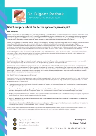
Inguinal hernia results from a hole or defect in the muscles,<br>through which the peritoneum protrudes, forming the<br>sac (Figs. 1 to 5). Inguinal herniorrhaphy is one of the<br>most common operations that general surgeons perform.<br>Minimally invasive surgical approaches are increasingly<br>popular because they offer the potential for less<br>postoperative pain and a quick return to normal activities.<br>Laparoscopic herniorrhaphy is being done at a time when<br>laparoscopic cholecystectomy has shown definite benefits<br>over the open technique.

E N D
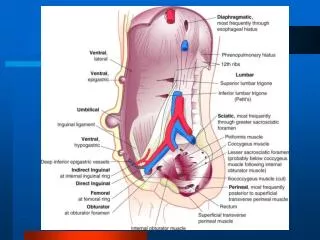
Laparoscopic Repair of Inguinal H ernia Prof. Dr. R. K. Mishra INTRODUCTION Inguinal hernia results from a hole or defect in the muscles, through which the peritoneum protrudes, forming the sac (Figs. 1 to 5). Inguinal herniorrhaphy is one of the most common operations that general surgeons perform. Minimally invasive surgical approaches are increasingly popular because they offer the potential for less postoperative pain and a quick return to normal activities. Laparoscopic herniorrhaphy is being done at a time when laparoscopic cholecystectomy has shown definite benefits over the open technique. Laparoscopic repair of inguinal and femoral hernia is no exception, with laparoscopic approach. The laparoscopic approach to inguinal hernia repair is theoretically possible in nearly all inguinal hernias. However, the precise role of laparoscopy in inguinal hernia repair remains somewhat controversial given the increased costs and greater technical demands. Ger in 1982 attempted minimal access groin hernia repair by closing the opening of an indirect inguinal hernial sac using Michel clips. Bogojavlensky in 1989 modified the technique by intracorporeal suture of the deep ring after plugging a polypropylene mesh (PPM) into the sac. Toy and Smoot in 1991 described a technique of intraperitoneal onlay mesh (IPOM) placement, where an intra-abdominal piece of polypropylene or expanded polytetrafluoroethylene (e-PTFE) was stapled over the myopectineal orifice without dissection of the peritoneum. The present day techniques of laparoscopic hernia repair evolved from Stoppa concept of preperitoneal reinforcement of fascia transversalis over the myopectineal orifice with its multiple openings by a prosthetic mesh. In the early 1990s, Arregui and Doin described the transabdominal preperitoneal (TAPP) repair, where the abdominal cavity is first entered, peritoneum over the posterior wall of the inguinal canal is incised to enter into the avascular preperitoneal plane which is adequately dissected to place a large (15 × 10 cm) mesh over the hernial orifices. After fixation of the mesh, the peritoneum is carefully sutured or stapled. TAPP approach has the advantage of identifying missed additional direct or femoral hernia during the first operation itself. Around the same time, Phillips and McKernan described the totally extraperitoneal (TEP) technique of endoscopic hernioplasty where the peritoneal cavity is not breached and the entire dissection is performed bluntly in the extraperitoneal space with a balloon device or the tip of the laparoscope itself. An advanced knowledge of the posterior anatomy of the inguinal region is imperative. Once the dissection is complete, a 15 × 10 cm mesh is stapled in place over the myopectineal orifice. It appears to be the most common endoscopic repair today. In both these repairs, the mesh in direct contact with the fascia of the transversalis muscle in the preperitoneal space, allows tissue in growths leading to the fixation of the mesh as opposed to being in contact to the peritoneum as in IPOM repair where it is prone to migrate. TOTALLY EXTRAPERITONEAL REPAIR Totally extraperitoneal repair is performed in the preperitoneal space and was developed to avoid the risks associated with entering the peritoneal cavity (Fig. 2A). The surgeon develops a space between the peritoneum and the anterior abdominal wall so that the peritoneum is never violated. In experienced hands and smaller direct hernia, this approach has the advantage of eliminating the risk of intra-abdominal adhesion formation. Fig. 1: Bilateral direct hernia.
220 SECTION2: Laparoscopic General Surgical Procedures A B Figs. 2A and B: (A) Totally extraperitoneal (TEP) versus transabdominal preperitoneal (TAPP) hernia repair; (B) Diagrammatic representation of ligaments. A B Figs. 3A and B: Important landmarks in laparoscopic hernia repair: (1) Medial umbilical ligament; (2) Inferior epigastric vessels; (3) Spermatic vessels; (4) Vas deferens; (5) External iliac vessels in “triangle of doom”; (6) Indirect defect. Fig. 4: Left side indirect hernia. Fig. 5: Triangle of doom. TRANSABDOMINAL PREPERITONEAL REPAIR (Fig. 2A). Because TAPP is performed transabdominally, it has a larger working space than TEP, with ready access to both groins, and can be attempted in patients with prior lower abdominal surgery. However, TAPP rarely results in injuries to adjacent intra-abdominal organs, adhesions resulting in intestinal obstruction, or bowel herniation. Transabdominal preperitoneal repair involves the placement of mesh in a preperitoneal position, but peritoneal incision is given after entering in abdominal cavity, which is covered by peritoneum to keep the mesh away from the bowel
221 CHAPTER16: Laparoscopic Repair of Inguinal Hernia For patients in whom the TEP technique is not appropriate or fails due to inability to develop the preperitoneal space, conversion to a TAPP approach can be performed. On occasion, conversion to an open surgical approach may be necessary. Larger hernias, especially huge scrotal hernias, are probably best repaired open. In female patients with indirect inguinal hernia, a TAPP approach may be easier. Indirect inguinal hernia sacs are frequently much more intimately attached to the round ligament in women than are indirect sacs to the cord structures in males. posteriorly and the posterior rectus sheath and pubic bone anteriorly. This is the space first entered in extraperitoneal repair of hernia. Three dangerous areas where stapling and electro surgery should be avoided are described below. Triangle of Doom (Fig. 5) The triangle of doom is defined by vas deferens medially, spermatic vessels laterally, and external iliac vessels inferiorly. This triangle contains external iliac artery and vessels, the deep circumflex iliac vein, the genital branch of genitofemoral nerve, and, hidden by fascia, the femoral nerve. Staple should not be applied in this triangle otherwise chances of mortality are there if these great vessels are injured. LAPAROSCOPIC ANATOMY A clear understanding of the anatomy of the groin and its anatomic approaches is important for successful laparoscopic hernia repair. In the lower abdomen, there are five peritoneal folds or ligaments which are seen through the laparoscope in umbilicus. These ligaments are generally overlooked at the time of open surgery. Triangle of Pain Triangle of pain is defined as spermatic vessel medially, the iliopubic tract laterally, and inferiorly the inferior edge of skin incision. This triangle contains lateral femoral cutaneous nerve and anterior femoral cutaneous nerve of thigh. The staple in this area should be less because nerve entrapment can cause neuralgia. One Median Umbilical Ligament In the midline, there is median umbilical ligament which extends from the mid of urinary bladder up to the umbilicus. Median umbilical ligament is obliterated urachus (Fig. 2B). Circle of Death This is also called as corona mortis and refers to vascular ring form by the anastomosis of an aberrant obturator artery with the normal obturator artery arising from a branch of the internal iliac artery. At the time of laparoscopic hernia if this vessel is torn, both ends of vessel can bleed profusely, because both arise from a major artery. The surgeon should remember these anatomic landmarks and the point of mesh fixation should be selected superiorly, laterally, and medially. Two Medial Umbilical Ligament One on Either Side The paired medial umbilical ligament is obliterated umbilical artery except where the superior vesical arteries are found in the pelvic portion. The medial umbilical ligaments are the most prominent fold of the peritoneum. Sometimes, it hangs down and obscures the vision of lateral pelvic wall. These ligaments are important landmarks for the lateral extent of the urinary bladder (Figs. 3A and B). Two Lateral Umbilical Ligaments Lateral to the medial umbilical ligament, the less prominent paired lateral umbilical fold contains the inferior epigastric vessels. The inferior epigastric artery is lateral border of Hesselbach’s triangle and hence is a useful landmark for differentiating between direct and indirect hernia. Any defect lateral to the lateral umbilical ligament is in direct hernia and medial to it is direct inguinal hernia (Fig. 4). The femoral hernia is below and slightly medial to the lateral inguinal fossa, separated from it by the medial end of the iliopubic tract internally and the inguinal ligament externally. Important landmarks for extraperitoneal hernia dissection include the musculoaponeurotic layers of the abdominal wall, the bladder, Cooper’s ligament, and the iliopubic tract. The inferior epigastric artery and vein, the gonadal vessels, and vas deferens should also be recognized. The space of Retzius lies between the vesicoumbilical fascia INDICATIONS OF LAPAROSCOPIC REPAIR OF HERNIA The indications for performing a laparoscopic hernia repair are essentially the same as repairing the hernia conventionally. There are, however, certain situations where laparoscopic hernia repair may offer definite benefit over conventional surgery to the patients. These include: ■ Bilateral inguinal hernias ■ Recurrent inguinal hernias In recurrent hernia, surgery failure rate is as high as 25–30%, if again repaired by open surgery. The distorted anatomy after repeated surgery makes it more prone to recurrence and other complications such as ischemic orchitis. In recurrent hernia, the laparoscopic approach offers repair through the inner healthy tissues with clear anatomical planes and thus, a lower failure rate. In laparoscopic bilateral repair with three ports technique, there is simultaneous
222 SECTION2: Laparoscopic General Surgical Procedures ■ access to both sides without any additional trocar placement. Even in patients with clinically unilateral defect after entering inside the abdominal cavity, there is 20–50% incidence of a contralateral asymptomatic hernia being found which can be repaired, simultaneously, without any additional morbidity of the patient. Recurrent inguinal hernias are very difficult to operate open, and more liable to complications. The size of mesh used in open methods is limited by natural fusion of muscles. All meshes and plugs shrink with time, and this works against all open methods. Any method of repair must achieve two fundamental goals—removal of the sac from the defect and durable closure of the defect. In addition, the ideal method should achieve these with the least invasion, pain, or disturbance of normal anatomy. Laparoscopic repair in expert hands is now quite safe and effective, and is an excellent alternative for patients with inguinal hernia. It is confusing that laparoscopic repair is more complex and is not widely available. The public needs to be educated as to its advantages. All surgeons agree that for bilateral or recurrent inguinal hernias, laparoscopic repair is unquestionably the method of choice. The argument against its use for unilateral or primary inguinal hernias is unfounded if it is the best for bilateral or recurrent hernias. ■ ■ CONTRAINDICATIONS OF LAPAROSCOPIC REPAIR OF HERNIA ■ ■ ■ ■ ■ ■ ■ ■ ■ ■ ■ Nonreducible, incarcerated inguinal hernia Prior laparoscopic herniorrhaphy Massive scrotal hernia Prior pelvic lymph node resection Prior groin irradiation Inability to tolerate general anesthesia (GA) Prior pelvic surgery in the preperitoneal space Incarcerated inguinal hernia Large scrotal hernia Ascites Active infection TYPES OF LAPAROSCOPIC HERNIA REPAIR Many techniques were used to repair hernia such as: ■ Simple closure of the internal rings ■ Plug and patch repair ■ IPOM repair ■ TAPP mesh repair ■ TEP repair The technique of TAPP repair was first described by Arregui in 1991. In the TAPP repair, the peritoneal cavity is entered, the peritoneum is dissected from the myopectineal orifice, mesh prosthesis is secured, and the peritoneal defect is closed. This technique has been criticized for exposing intra-abdominal organs to potential complications, including small bowel injury and obstruction. The TEP repair maintains peritoneal integrity, theoretically eliminating these risks while allowing direct visualization of the groin anatomy, which is critical for a successful repair. The TEP hernioplasty follows the basic principles of the open preperitoneal giant mesh repair, as first described by Stoppa in 1975 for the repair of bilateral hernias. Both approaches (TAP or TEP) are acceptable, and one approach may be preferred over the other under specific clinical circumstances. TAPP was the original approach, and TEP evolved to minimize some of the problems associated with TAPP, but TEP repair is technically more challenging because of the limited working space, which may explain higher conversion rates. TAPP approach is more popular and commonly used procedure all over the world for inguinal hernia repair. Although surgeons should learn both techniques, they should use the technique with which they are most familiar. ADVANTAGES OF LAPAROSCOPIC APPROACH ■ Tension-free repair that reinforces the entire myopectineal orifice Less tissue dissection and disruption of tissue planes Three ports are adequate for all type of hernias. Less pain postoperatively Low intraoperatively and postoperative complications Early return to work ■ ■ ■ ■ ■ DISADVANTAGES OF OPEN METHOD ■ ■ ■ Requires 4–6 inches of incision at the groin Generally very painful, because of muscle spasm Considerable postoperative swelling of tissues in groin, around the wound Requires cutting through the skin, fat, and good muscles in order to gain access for repair, which in itself causes damage Frequent complications of wound hematomas, wound infection, scrotal hematomas, and neuroma Usually takes 6–8 weeks for recovery. Sometimes long-term disability may follow, e.g., neuralgia, neuroma, and testicular ischemia. Whether a flat mesh or a plug is used from the front, they do not hold themselves in place; what holds them in place are stitches, so the strength of the repair still depends on the stitches, not so much on the mesh or plug. Bilateral inguinal hernias require two incisions, doubling the pain; or two operations. ■ ■ ■ ■ ■ ■
223 CHAPTER16: Laparoscopic Repair of Inguinal Hernia Patient Selection The GA and the pneumoperitoneum required as part of the laparoscopic procedure do increase the risk in certain groups of patients. Most surgeons would not recommend laparoscopic hernia repair in those with pre-existing disease conditions. Patients with cardiac diseases and chronic obstructive pulmonary disease (COPD) should not be considered good candidates for laparoscopy. The laparoscopic hernia repair may also be more difficult in patients who have had previous lower abdominal surgery. The elderly may also be at increased risk for complications with GA combined with pneumoperitoneum. If the patient is young or the hernia is small, it does not matter how the hernia is repaired. Many surgeons agree that for bilateral or recurrent inguinal hernias, laparoscopic repair is unquestionably the method of choice. Laparoscopic surgery is not recommended for big irreducible and incarcerated hernia. Hernia repair like many other laparoscopic procedures should not be performed under local anesthesia. Small direct hernia can be performed under spinal anesthesia if TEP is planned, but best anesthesia for laparoscopic hernia repair is GA. Fig. 6: Position of surgeon in right-sided hernia. fossa and left port in left hypochondrium so that both the instruments should make a manipulation angle of 60°. In right-sided hernia surgery, right port should move up toward hypochondrium and left port will come down to make the triangle. TRANSABDOMINAL PREPERITONEAL REPAIR OF INGUINAL HERNIA As with most laparoscopic procedures, the peritoneal cavity is entered during TAPP hernia repair. The major advantage of the TAPP approach to groin hernias is that all three hernia defects (direct, indirect, and femoral) are well visualized and in close proximity to each other, allowing easy repair of any type of groin hernia. PROCEDURE OF TRANSABDOMINAL PREPERITONEAL REPAIR After access, diagnostic laparoscopy is performed to rule out any adhesion or other intra-abdominal lesion. All the important anatomical landmarks of hernia surgery are identified with the help of telescope and one atraumatic grasper. The important landmarks of laparoscopic hernia repair are the pubic bone and inferior epigastric vessels. The defect should be seen carefully and if any content is present inside the sac, it should be reduced gently (Figs. 9A to D). A sliding hernia of colon should be carefully reduced because chances of perforation of large bowel are more than other viscus so the assistant should reduce the hernia by pressing it from outside. Any adhesion between bowel and omentum should be divided carefully using harmonic scalpel or bipolar and scissors. The next step of TAPP repair of hernia is the creation of pre- peritoneal space. Many surgeons like to do hydrodissection to create this preperitoneal space just by injecting normal saline into preperitoneal space. Some surgeons think that it is easy to create preperitoneal space with sharp dissection as well. The peritoneum is cut at a distance of minimum 4 cm lateral to the outer margin of deep ring at 2 o’clock position if the hernia is right side and 10 o’clock position for the left side of hernia. Medial dissection of peritoneal incision should continue up to medial umbilical ligament (Figs. 9E and F). Going medial to medial umbilical ligament is risky because there is risk of injury of the urinary bladder. The flap Position of Surgical Team and Patient Surgeon stands toward the opposite side of the hernia, near the shoulder. Camera assistant should stand either right to the patient or on the opposite side of the patient (Fig. 6). The patient is usually placed in 15–20° of Trendelenburg position to improve exposure of the hernia defect, which is particularly important with TAPP hernia repair to move the small bowel or omentum away from the area of dissection. Port Position The position of port in a laparoscopic repair of transabdominal hernia repair should be again according to baseball diamond concept (Figs. 7A to C). Please refer Chapter 6: Abdominal Access Techniques. The telescopic port should be in umbilicus. A 30º telescope is a better choice for laparoscopic hernia surgery. A 10 mm umbilical port is used. Two other ports, usually 10 mm for dominant hand and 5 mm for nondominant hand, are placed lateral to the umbilicus (Figs. 8A to C). In a left- sided hernia, the right lateral port should be in right iliac
224 SECTION2: Laparoscopic General Surgical Procedures A B C Figs. 7A to C: (A) Port position of right-sided hernia; (B) Port position of bilateral hernia; (C) Port position of left-sided hernia. A B C Figs. 8A to C: (A) Port position of right-sided hernia; (B) Port position of surgical team; (C) Port position right hernia (1—Camera; 2 and 3—Instruments). of peritoneum is separated from above downward as soon as it reaches the site of internal ring, the hernia sac will be encountered. Dissection should be started with opening the peritoneum lateral to the medial umbilical fold in order to identify Cooper’s ligament. Stoppa’s parietalization technique should be used for dissection of the spermatic cord from the peritoneum by separating the elements of the spermatic cord from the peritoneum and peritoneal sac (Figs. 10A to E). In case of indirect defect, the hernial sac has to be either gently dissected free or inverted or else if it is completely adhered with the transversalis fascia and cord structures, it can be transected. Surgeons should use both blunt and sharp dissection and the sac is dissected off the anterior abdominal wall. After being reduced partially, it is ligated using an endoloop and then transected with scissors. In case of bilateral hernias, the procedure is repeated on the other side. The vas and spermatic vessels also need to be separated from the sac. The next step is separation of the sac from cord structures and dissection for creation of proper lateral space for the placement of mesh. Lateral limit of dissection is the anterosuperior iliac spine, while inferior limit laterally is the psoas muscle. Dissection should be avoided in the “triangle of doom” which is bounded medially by the vas deferens and laterally by the gonadal vessels. A large hernial sac creates multiple planes and it is easy for the beginners to get disoriented with sac, vas, and vessel. The best way to avoid this confusion is that surgeon should keep himself as close as possible to the outer surface of peritoneum. If the spermatic vessels are injured accidentally, it can be clipped. Even if the testicular vessel is injured, the testes will get the blood supply from collateral vessels developed through cremasteric vessels. In direct hernias, the creation of preperitoneal space is comparatively easy as there is no chance of injury of spermatic vessels and vas. The bulge in the transversalis fascia may be repaired by suturing or stapling. The tacker application and application of electro surgery should be done very carefully in the triangle of doom, triangle of pain, and trapezoid of disaster. In case of massive complete indirect scrotal hernias, no attempt should be made to reduce the sac completely as it may increase the risk of testicular nerve injury and hematoma formation.
225 CHAPTER16: Laparoscopic Repair of Inguinal Hernia A B C D E F Figs. 9A to F: (A to D) Reduction of the content of hernia sac; (E and F) Incision over peritoneum. Placement of the Mesh Mesh is a necessary element of laparoscopic inguinal hernia repair to provide a tension-free hernia repair, which is the recommended method. Preformed mesh that conforms to the preperitoneal space is available and is preferred by some surgeons over a flat piece of mesh that needs to be trimmed to accommodate the patient’s anatomy. Criteria for laparoscopic mesh are as follows: ■ Nonabsorbable ■ Adequate size ■ Adequate memory Polypropylene woven mesh (e.g., Marlex, Prolene, and SurgiPro) has been used in laparoscopic inguinal hernia repair and is preferred over other prosthetic materials. e-PTFE is another material that is also used extensively for incisional hernias, but it has not been used for the laparoscopic inguinal and femoral hernia repair, except for the IPOM technique. PPM is commercially available in light, medium, or heavy weight. Light weight mesh is associated with a lower incidence of chronic groin pain, groin stiffness, and foreign body sensations without any increased risk for hernia recurrence. A Prolene mesh of appropriate size, usually 15 × 15 cm should be taken and one corner of mesh should be tailored (Fig. 11). Mesh is placed inside the abdominal cavity through 10 mm port. Mesh should be rolled and loaded backward in this port. If surgery is being performed by 10 mm port only the port should be removed and rolled mesh should be introduced though the port wound directly (Fig. 12). After introduction of mesh, it is unrolled when it reaches in the peritoneal cavity. The mesh is fixed medially over the Cooper’s ligament and pubic bone using a tacker or anchor (Figs. 13A and B). The tailored corner of the mesh should be positioned inferomedially. No lateral slit should be made in the mesh and it should not be fixed lateral to cord structures to prevent injury to lateral cutaneous nerve of thigh. The mesh in this position covers the direct, indirect, and femoral defects. It is essential that mesh should extend below the pubic tubercle so that it covers the femoral orifice. Mesh should also extend medially to cover all the possible orifices of hernia. Laterally mesh should project at least 2–3 cm beyond the margin of deep ring. If mesh is not of appropriate size, the chances of recurrence increase. Sometimes, the surgeon may be disoriented and mesh is placed with its long axis vertical
226 SECTION2: Laparoscopic General Surgical Procedures B A D C E Figs. 10A to E: Creation of preperitoneal space. instead of transverse. If the mesh is cut at one of the corners, chances of this disorientation are minimum. Implant for Fixing Mesh Many preloaded devices are available for fixing mesh in hernia surgery. Mesh is fixed medially over the Cooper’s ligament and pubic bone using an implant. Currently, three popular brands of implants to fix the mesh are available. These are Tacker, Protack, and Anchor. The comparative chart of these implants is shown in Table 1. After adjusting the mesh properly, it should be fixed by stapling first its middle part three fingers above the superior limit of the internal ring. With mesh duly stapled, pneumoperitoneum is reduced to 9 mm Hg. It is important to avoid pricking of the inferior epigastric artery or the testicular vessels. Intracorporeal suturing can also be used for fixation of mesh if surgeon has sufficient suturing skill. After fixing the mesh properly, the peritoneum flap is replaced over the mesh. It is important that mesh should be completely covered by the peritoneum. Ideally, peritoneum should be opposed by overlap fashion and peritoneum Fig. 11: Cutting the corner of mesh. defect is closed either by staples or by continuous suturing and Aberdeen termination (Figs. 14A and B). Repair of Bilateral Inguinal Hernia In laparoscopic surgery, postoperative recovery of bilateral hernia is same as that of unilateral hernia. The technique
227 CHAPTER16: Laparoscopic Repair of Inguinal Hernia of bilateral laparoscopic repair of hernia is same as that of unilateral hernia. Patients with bilateral hernia are good candidates of laparoscopy. The two sides may be repaired using two meshes, but single long mesh also can be used and is pushed across from one side behind the bladder, and across the inguinal orifice on the opposite side. The size of the mesh for bilateral hernia should be 30 × 15 cm (Fig. 15). Surgeon should avoid twisting of the mesh. After placing the mesh in bilateral hernia surgery, it should look just like a bow tie. be taken that the vas or vessels should not be caught in the ligature (Figs. 16A and B). Ending of the Operation At the end of surgery, the abdomen should be examined for any possible bowel injury or hemorrhage. The entire instrument should be removed followed by all the ports. Each port should be removed under direct observation through telescope. Ports larger than 10 mm should be sutured. Telescope should be removed at last after releasing all the gas keeping in mind that last port should not be pulled without putting telescope or any blunt instrument in, to prevent entrapment of bowel or omentum and formation of omental or intestinal adhesion. Wound should be closed with suture, especially 10 mm wound. Repair of Recurrent Inguinal Hernia Recurrent laparoscopic hernia after open surgery is better to be repaired laparoscopically, because external anatomy is disrupted and open repair has more chances of recurrence. Laparoscopy is method of choice for recurrent hernia. The defect is usually direct and more than one in recurrent hernia. The result of laparoscopic repair is excellent even in case of multiple hernias. TOTALLY EXTRAPERITONEAL HERNIA REPAIR The technique of TEP repair of inguinal hernia was described even before the TAPP technique; however, technical difficulties of working in closed space and anatomy with very limited working space hindered its popular acceptance. The effectiveness of this type of repair has been well established by the open operation of Stoppa. Laparoscopic Hernia in Children Laparoscopy has been tried in small children. Only closure of ring and herniotomy is possible in pediatric age group. The sac is simply inverted and tied internally. Care should ADVANTAGES OF TOTALLY EXTRAPERITONEAL REPAIR ■ Pneumoperitoneum is not required. ■ Less chance of dangerous vessel injury or bowel injury ■ The view of groin is better for dissection around the neck of the sac. ■ Continuity of peritoneum is not breached so it need not be closed. DISADVANTAGES OF PREPERITONEAL REPAIR ■ The identification of correct plane of dissection is difficult. Fig. 12: Introduction of mesh in preperitoneal space. A B Figs. 13A and B: (A) Hernia secure trap Ethicon stapler; (B) Hernia tacker from Covidien.
228 SECTION2: Laparoscopic General Surgical Procedures TABLE 1: Comparison of ESS Endoanchor, Tyco Protack, and Tyco Tacker. Feature ESS Endoanchor Tyco Protack Tyco Tacker Number of implants 20 30 20 Geometry of implant Anchor Helical fastener Helical fastener Implant material Nitinol Titanium Titanium Implant length 5.9 mm 3.8 mm 3.6 mm Implant width 6.7 mm 4 mm 3.4 mm Port size required 5 mm 5 mm 5 mm Shaft length 360 mm 356 mm 356 mm Trigger fire orientation Release to deploy Depress to deploy Depress to deploy A B Figs. 14A and B: (A) Closure of peritoneum by suturing; (B) Closure of peritoneum by tacker. ■ There is always a chance of breach of peritoneum continuity and this will reduce the view. Four ports generally are necessary for bilateral hernia surgery. Whereas, in TAPP only three ports are sufficient. ■ Preparation of the Patient Preparation of the patient in totally preperitoneal hernia repair is same as of the transabdominal hernia repair. Knowledge of the anatomy of the abdominal wall muscle and recognition of the transition zone that occur at the arcuate line of Douglas is very important for totally preperitoneal hernia repair. Fig. 15: Introduction of mesh for bilateral hernia. Approach to Preperitoneal Space In TEP repair of hernia, the main concern is to make an extraperitoneal space. The extraperitoneal space is made possible by the fact that the peritoneum in suprapubic region can easily be separated from anterior abdominal wall, thereby creating enough space for dissection. A 2 cm longitudinal skin incision is made just below the umbilicus 1 cm lateral to the midline on the side of hernia (Figs. 17A and B). The incision is deepened down to reach up to the anterior rectus sheath. All the subcutaneous fat is cleared and the rectus is opened under direct vision. Two-stay ■ The landmarks of hernia dissection can only be identified when they are encountered. Reduction of content of sac is difficult to ensure. Sliding hernia is difficult to recognize from outside of the sac. If the sac gets accidentally cut, it is difficult to close it again. In recurrent hernia, extensive adhesion makes the dissection difficult because peritoneum may be adherent to the under surface of the scar. ■ ■ ■ ■
229 CHAPTER16: Laparoscopic Repair of Inguinal Hernia A B Figs. 16A and B: Closure of defect with intracorporeal suturing in pediatric age. A B Figs. 17A and B: Access technique of totally extraperitoneal hernia repair. Sweeping Movement of Telescope Once the telescope is placed properly, a 10 mm port is inserted under direct view approximately halfway between the symphysis pubis and the umbilicus (Figs. 19A to D). Another 5 mm port should be placed two fingers below and medial to the right anterior iliac spine. If the secondary port site is not seen clearly through the telescope, one can infiltrate the port site with local anesthetic and look for the tip of the needle internally (Fig. 20). This will insure the exact placement of port and allow the tip of trocar to be seen by telescope at the time of insertion. suture on each leaf of rectus sheath is placed and the rectus muscle is retracted by two retractors downward toward symphysis pubis in an oblique fashion; we should never cross the posterior fascia of the rectus muscle while dissecting. By finger or swab toward the hernia, dissection should performed carefully, and preperitoneal space will be found below the arcuate line of Douglas. Insertion of Port A balloon dissector should be introduced with telescope and balloon is inflated for further dissection of the preperitoneal space. An 11 mm port is introduced without its sharp tip with a laparoscope of 30°. A small preperitoneal pocket is created by manipulating laparoscope in sweeping manner. If balloon dissector is not available, the glove finger can be tied around the suction irrigation instrument and can be used to create some preperitoneal space (Figs. 18A and B). Dissection of Preperitoneal Space and Cord Structures in TEP Repair In TEP repair of hernia, Stoppa parietalization technique is used for dissection of the spermatic cord from the peritoneum by separating the elements of the spermatic cord from the peritoneum and peritoneal sac should be done (Fig. 21).
230 SECTION2: Laparoscopic General Surgical Procedures A B Figs. 18A and B: Making balloon dissection with finger of gloves. B A C D Figs. 19A to D: Balloon dissection. The dissection is started by tracing the inferior epigastric vessels toward the deep ring. The upper border of the hernia sac is readily recognized because indirect hernia is lateral to the inferior epigastric vessels and direct hernia is medial to it. As the inguinal region is approached, the dissection is continued all around the sac to encircle the neck. The surgeon should try to remain close to peritoneum and dissection continues medially to separate vas from the sac. Under the neck of the sac, care should be taken to avoid injury of iliac vessels. In case of direct inguinal hernia, the dissection is carried out from above downward and progressed medially to the
231 CHAPTER16: Laparoscopic Repair of Inguinal Hernia Fig. 20: Introduction of secondary port. Fig. 21: Dissection of preperitoneal space. Fig. 22: Introduction of mesh. Fig. 23: Placement of mesh. inferior epigastric vessels. The direct sac is freed from the transversalis fascia. Dissection should be continued until the peritoneum has reached the iliac vessels inferiorly. Care should be taken that any hole in peritoneum is not made, otherwise it will be difficult to maintain good working space because the gas will escape into the abdominal cavity increasing the intra-abdominal pressure. If the hole is made accidentally, it should be identified and enlarged as this will equalize the pressure on both sides of peritoneum and allows the peritoneum to drop back down due to gravity. A venting 5 mm port or Veress needle can be placed in the right upper quadrant at Palmer’s point to decompress the abdominal cavity. The technique of insertion of mesh in TEP repair of hernia is same as that of transabdominal preperitoneal. Mesh of appropriate size usually 15 × 15 cm is used and rolled and loaded backward in one of the port. Mesh should be fixed by stapling first in its middle part, three fingers above the superior limit of the internal ring (Figs. 22 and 23). In TEP repair, some surgeons do not use staple, because peritoneum is not breached and once the gas from the preperitoneal space is removed, it will hold the mesh in its proper position. In 1–2% of cases of TEP, conversion to open or TAPP may be necessary due to large peritoneal tear making the vision difficult or in the cases where content is not reduced completely. Ending of the Operation At the end of the surgery, the abdomen should be examined for any possible bowel injury or hemorrhage. The entire instrument should be removed and then all the ports. Generally, vicryl is used for rectus and stapler for skin. Adhesive sterile dressing should be applied over the wound. LAPAROSCOPIC REPAIR OF FEMORAL HERNIA Laparoscopic repair of femoral hernia is same as that of laparoscopic direct or indirect hernia. It can be performed by both TAPP and TEP methods. In case of laparoscopic femoral hernia repair, the sac should be carefully excised because rigid femoral ring makes it difficult to mobilize the sac. The dissection should be done very carefully because there is increased risk of injury of abnormal obturator artery on the lateral side of the sac. The femoral hernia defect is
232 SECTION2: Laparoscopic General Surgical Procedures Fig. 24: Postoperative scrotal hematoma. Fig. 25: Perforation bowel during hernia surgery. ■ ■ ■ between the iliopubic tract and pubic ramus and can be easily identified. Repair of the femoral canal should be done by approximating iliopubic tract to the Cooper’s ligament by Prolene stitches. Massive hernias Pregnancy Unfit for GA Inguinal Hernia Repair in Pediatric Patients COMPLICATIONS OF LAPAROSCOPIC HERNIA REPAIR Like any other laparoscopic procedures, several complications have been recorded during the learning curve. The major problems include: ■ Recurrence ■ Neurovascular injury ■ Urinary tract injury ■ Injury to vas ■ Testicular complications ■ Problems due to mesh The mechanism of recurrence can be related to lack of understanding of the difficult laparoscopic anatomy, wrong hernia repair technique, or the wrong prosthesis. These include incomplete dissection without proper pocket formation, missed sac, migration of mesh due to small sized mesh which may be prone to get displaced once fixed, inadequate fixation with rolling up of the mesh, and hematoma formation leading to infection. The complication of laparoscopic hernia repair can be summarized as follows: ■ Immediate: Visceral injury, vascular injury, and injury to vas and spermatic vessels (Fig. 24) ■ Late: Bowel adhesions to mesh, intestinal obstruction, fistulization, orchitis, testicular atrophy, nerve entrap- ment, and incisional hernia recurrence (Fig. 25). Small children gain little benefit from laparoscopic hernia repair as inguinal skin crease incision used in the herniotomy is one of best incisions as far as cosmesis is concerned. It is hardly visible after a few months. Also, it is covered by underwear. Compared to this, three stab incisions, however small, are in the visible area. Inguinal Hernia Repair in Obese Patients Operations in patients with BMI above 27 may be difficult for less experienced surgeons, particularly when trying to encircle an indirect sac. Patients with BMI of above 30 should be encouraged to lose weight or should even be turned down for the laparoscopic approach. They are incidentally more likely to develop recurrence after even an open hernia repair. It is also easy for the laparoscopic surgeon to become disoriented when the patient is very obese. Inguinal Hernia Repair in Recurrence Generally, the short-term recurrence rate of laparoscopic inguinal hernia repair is reported to be <5%. In both the open and laparoscopic repair procedures, the aim is to cover the whole inguinofemoral area by a preperitoneal prosthetic mesh, so that recurrences should not occur. When they do occur, recurrences must be regarded as technical failures. Recurrences after laparoscopic repair most often result from using too small a mesh, or not using staples to fix the mesh. Most recurrences after laparoscopic hernia repair occurred medially, and the technique was needed modifications. The mesh is now placed at least until the midline, and occasionally hernia staples are used when an adequate overlap (2 cm) cannot be achieved medially. The TEP technique is now used more often, allowing for better visual control in the medial part of the operating field. Relative Contraindications for Laparoscopic Approach ■ ■ ■ ■ Obesity with body mass index (BMI) > 30 Significant chest disease Patient on anticoagulants Adhesions
233 CHAPTER16: Laparoscopic Repair of Inguinal Hernia Operating Time Operating times of surgical techniques varies between surgeons and also vary considerably between centers. It reduces with experience and comparison between laparoscopic and open surgery is subject to bias due to pre- existing familiarity with open techniques. It is less important to the patient than a successful operation. The time taken to perform the surgery can have cost implications. The operative time to perform unilateral primary inguinal repair has frequently been reported as longer for laparoscopic compared to open repair, however, the mean difference in 36 of 37 randomized trials is 14.81 minutes. These differences disappear in bilateral and recurrent hernia repairs. sheath. Also, the balloon trocar is inserted gently, parallel to the abdominal wall, to avoid puncturing the peritoneum. The balloon must be inflated slowly with saline to ensure smooth and even distention and prevent its rupture. Precautions During Port Placement The trocars should be short and threaded in proportion to the less workspace and to ensure a snug fit, respectively. The skin incisions should be just adequate to grip the trocar and prevent its slipping. The patient should empty their bladder before surgery as the suprapubic trocar could injure a filled bladder. The pressure in the preperitoneal space must be such as to offer sufficient resistance during trocar insertion to avoid puncturing the peritoneum. Postoperative Pain and Amount of Narcotics Used The open tension-free mesh repair is found to cause less postoperative pain than open nonmesh repairs; however, most randomized trials assessing postoperative pain between open tension-free repairs and laparoscopic repairs report less pain in the laparoscopic groups. In many cases, this also results in less analgesia being consumed by the patient. Correct Identification of the Anatomical Landmarks The next most important and crucial step in any hernia surgery is the correct identification of anatomical landmarks. This is difficult for beginners as the anatomy is different from that seen in open surgery. The first most important step is to identify the pubic bone. Once this is seen, the rest of the landmarks are traced keeping this as a reference point. One is advised to keep away from the triangle of doom, which contains the iliac vessels, and to avoid placing tacks in the triangle of pain laterally. Complication Rates Complications in endoscopic inguinal hernia surgery are more dangerous and more frequent than those of open surgery, especially in inexperienced hands and hence are best avoided. It is possible to avoid most of these complications if one follows a set of well-defined steps and principles of endoscopic inguinal hernia surgery. Complications of laparoscopic repair of inguinal hernia can be divided into: ■ Intraoperative ■ Postoperative Bladder Injuries Bladder injury most commonly occurs during port placement, dissecting a large direct sac or in a sliding hernia. It is mandatory to empty the bladder prior to an inguinal hernia repair to avoid a trocar injury. It is advisable that beginners catheterize the bladder during the initial part of their learning curve. The diagnosis is evident when one sees urine in the extraperitoneal space. Repair is done with vicryl in two layers and a urinary catheter inserted for 7–10 days. INTRAOPERATIVE COMPLICATIONS AND PRECAUTION During Creation of Preperitoneal Space This is the most important step for beginners. ■ A wide linea alba may result in breaching the peritoneum; in such a situation, it is best to close the rectus and incise the sheath more laterally. ■ Improper placement of balloon trocar causing dissection of muscle fibers ■ Entry into peritoneum causing pneumoperitoneum ■ Rupture of balloon in preperitoneal space ■ The Hassan trocar must snugly fit into the incision to avoid CO2 leak. To avoid these, one must ensure that the balloon is made properly and the correct space is entered by retracting the rectus muscle laterally to visualize the posterior rectus Bowel Injuries Bowel injury is rare during hernia surgery. It can occur when reducing large hernias, inadvertent opening of peritoneum causing the bowel to come into the field of surgery, and in reduction of sliding hernias. Injury is best avoided in such circumstances by opening the hernial sac as close as possible to the deep ring. The initial studies showed a higher incidence, especially with TAPP, but gradually it has decreased over time. Vascular Injury This is one of the most common injuries occurring in hernia repair and often a reason for conversion. The various sites where it can occur is rectus muscle vessel injury during trocar insertion; inferior epigastric vessel injury; bleeding
234 SECTION2: Laparoscopic General Surgical Procedures Iliopubic vein and artery which traverse the lacunar ligament: Hematoma Injury to spermatic vessels: Postoperative scrotal hematoma ■ from venous plexus on the pubic symphysis; aberrant obturator vein injury; testicular vessel injury; and the most disastrous of all, iliac vessels, which requires an emergency conversion to control the bleeding and the immediate services of a vascular surgeon to repair the same. Most of the other bleedings can be controlled with cautery or clips. Careful dissection and adherence to the principles of surgery will help in avoiding most of these injuries. ■ Nerve Entrapment and Injury The lateral cutaneous nerve of thigh and the femoral branch of genitofemoral nerve are the two nerves vulnerable to trauma due to indiscriminate placement of staplers lateral to the spermatic cord on the iliopubic tract. ■ Injury of lateral cutaneous nerve injury ■ Most common nerve injured is lateral femoral cutaneous nerve (2%): Hyperesthesia or paresthesia of upper aspect of thigh and hip. ■ If pain starts days after the surgery, it will recover within 2–4 weeks (or percutaneous steroid). ■ If pain starts within 24 hours of surgery, there is permanent nerve damage. ■ Cryotherapy with destruction of sensory branch is indicated. ■ Lifelong numbness Nerve entrapment should be avoided in laparoscopic repair of hernia: ■ Genitofemoral nerve injury ■ Genitofemoral nerve injury (1%): Hyperesthesia or paresthesia of scrotum ■ Not significant ■ With time, it will subside. Injury to Vas Deferens Injury occurs while dissecting the hernia sac from the cord structures. The injury causes an eventual fibrotic narrowing of the vas. A complete transaction of the vas needs to be repaired in a young patient. An injury to the vas is best avoided and this may be done by identifying before dividing any structure near the deep ring or floor of the extraperitoneal space. Also, the separation of cord structures from the hernial sac must be gentle and direct; grasping of vas deferens with forceps must be avoided. Pneumoperitoneum It is a common occurrence in TEP which every surgeon should be prepared to handle. Putting the patient in Trendelenburg position and increasing the insufflation pressures to 15 mm Hg helps. If the problem still persists, a Veress needle can be inserted at Palmer’s point. POSTOPERATIVE COMPLICATIONS Seroma/Hematoma Formation It is a common complication after laparoscopic hernia surgery, the incidence being in the range of 5–25% (Fig. 24). They are especially seen after large indirect hernia repair. Most resolve spontaneously over 4–6 weeks. A seroma can be avoided by minimizing dissection of the hernia sac from the cord structures, fixing the direct sac to pubic bone and fenestrating the transversalis fascia in a direct hernia. Some surgeons put in a drain if there is excessive bleeding or after extensive dissection. Other Complications ■ Migration of mesh ■ Rejection of mesh (rare) ■ Bowel adhesion Complete transaction of vas requires immediate anastomosis. Other complications include testicular pain, orchitis, epididymitis, swelling due to seromas, or hematoma. The treatment is supportive and incidence of all these complications is similar to that in conventional surgery. After some experience, most cases of inguinal hernia can be treated laparoscopically. Several prospective randomized trials comparing open versus laparoscopic repair have reported better outcomes following laparo- scopic repair. Reduced postoperative pain, earlier return to work, and fewer complications and less chance of recurrences for the laparoscopic approach are some of the crucial advantages. Although the procedural cost for laparoscopic hernia repair is more compared to conventional repair but overall expense for open repair is high if we calculate number of working days lost and medications taken into consideration. Data is now available which documents the TEP repair to have distinct advantage over the TAPP repair in terms of lesser postoperative complications and lower recurrence rate. TAPP Urinary Retention This complication after hernia repair has a reported incidence of 1.3–5.8%. It is usually precipitated in elderly patients, especially if symptoms of prostatism are present. These patients are best catheterized prior to surgery and catheter removed the next day morning. Vascular Injury The incidence of vascular injury has been documented to be about 0.5–1% and inferior epigastric artery is one of the most commonly traumatized. ■ Injury to iliac vessels: Chances of mortality ■ Inferior epigastric vessel: Hematoma
235 CHAPTER16: Laparoscopic Repair of Inguinal Hernia Recurrence It is the most important endpoint of any hernia surgery. It requires a proper and thorough knowledge of anatomy and a thorough technique of repair to help keep the recurrence in endoscopic repair to a minimum. has been stated to violate the peritoneal cavity with all its known possible complication of pneumoperitoneum, vessel, or bowel injury. There is no doubt that the laparoscopic hernia repair is a proven technique and will become more popular over a period of time. Neuralgias The incidence of this complication is reported to be between 0.5 and 4.6% depending on the technique of repair. The intraperitoneal onlay mesh method had the highest incidence of neuralgias in one study and was hence abandoned as a form of viable repair. The commonly involved nerves are lateral cutaneous nerve of thigh, genitofemoral nerve, and intermediate cutaneous nerve of thigh (Figs. 26A and B). They are usually involved by mesh-induced fibrosis or entrapment by a tack. The complication is prevented by avoiding fixing the mesh lateral to the deep inguinal ring in the region of the triangle of pain, safe dissection of a large hernial sac, and no dissection of fascia over the psoas. POSTOPERATIVE RECOVERY Marked variations are seen in postoperative recovery due to patient motivation, postoperative advice, and definition of “normal activity,” existing comorbidity, and local “culture.” Nevertheless all trials reporting this as an endpoint of study show a significant improvement in the laparoscopic group, with no real difference between the TAPP and TEP groups. This is estimated to equate to an absolute difference of about 7 days in terms of time off work. RECURRENCE Recurrence rates are low with the use of mesh and not significantly different between open or laparoscopic techniques. Testicular Pain and Swelling It occurs due to excessive dissection of a sac from the cord structures, especially a complete sac. The reported incidence is of 0.9–1.5%, and most are transient. Orchitis was found in a small number of patients but did not lead to testicular atrophy. CAUSES OF RECURRENCE IN LAPAROSCOPIC INGUINAL HERNIA REPAIR The factors involved in mesh dislocation or failure are insufficient size, wrong/defective material, incorrect placement, immediate or very early displacement by folding, lifting by a hematoma or urinary retention, missed cord lipomas and herniation through the keyhole (mesh slit), late displacement by insufficient scar tissue ingrowth, mesh protrusion, collagen disease, or pronounced shrinkage. Despite the correct and stable mesh position, there is still a limited risk of a late sliding of the retroperitoneal fat under/ in front of the mesh into the enlarged inner ring. Mesh Infection and Wound Infection Wound infection rates are very low. Mesh infection is a very serious complication and care must be taken to maintain strict aseptic precautions during the entire procedure. Any endogenous infection must be treated with an adequate course of antibiotics prior to surgery. A B Figs. 26A and B: Anatomical landmarks. (GFN: genitofemoral nerve, LCN: lateral cutaneous nerve; TV: testicular vessel)
236 SECTION2: Laparoscopic General Surgical Procedures Other Factors The negative effect on healing in hernia repair is often related with malnutrition, obesity, steroids, type II diabetes, chronic lung disease, jaundice, radiotherapy, chemotherapy, oral anticoagulants, smoking, heavy lifting, malignancy, and anemia. Laparoscopic inguinal hernia repair offers excellent results in experienced hands. Leibl in 2000 advised to avoid slitting of the mesh and increase its size to reduce the recurrence rate. Generous dissection of preperitoneal space is required to eliminate potential herniation through the slit or strangulation of the cord structures completely and also reduces the risk of genitofemoral neuropathy. Mesh Size Bilateral Assessment and Treatment Up to 30% of patients with a unilateral hernia will subsequently develop a further hernia on the contralateral side. Also, when examined at operation, 10–25% are found to have an occult hernia on the contralateral side. Both laparoscopic approaches allow assessment and treatment of the contralateral side at the same operation without the need for further surgical incisions, very little further dissection, and minimal additional postoperative pain. In open surgery, a further large incision is required in the opposite groin. This considerably impairs postoperative mobility and increases the likelihood of more admitted days in the hospital. Some surgeons advocate routine repair of the contralateral side during laparoscopic repair. The mesh size should be adequate to cover the entire myopectineal orifice. The established size in 2006 is 15 × 10 cm per unilateral hernia, with minor deviations. Mesh Material The mechanical strength of available meshes exceeds the intra-abdominal peak pressures and by far even the light weight meshes are strong enough for inguinal repair. Aachen group made an important contribution for understanding the interaction of the living tissue with the implanted mesh material. The negative impact of pronounced shrinkage of the traditional heavy weight meshes was recognized as an important factor promoting recurrence. Schumpelick introduced the logical trend of the use of light weight meshes. The new macroporous compound meshes present both the successful reduction of the overall foreign body amount and the preservation of mesh elasticity after the scar tissue ingrowths, due to very limited shrinkage and reduced bridging effect. Cost Effectiveness It is suggested that laparoscopic hernia repair is more expensive to perform than open hernia repair. The primary reason for this relates to the cost of extra equipment used for the laparoscopic repair with secondary costs attributed to perceived increases in operating time for the laparoscopic procedure. From the Indian perspective, various factors come into play when analyzing the cost implications of laparoscopic repair of inguinal hernia. In most hospitals, except the larger corporate ones, the theater time is charged on a per-case basis rather than by the hour. Thus, increase in the operating time, particularly during the learning curve, does not necessarily mean additional expense for the patient. If the surgeon were to adopt cost-containment strategies such as use of reusable laparoscopic instruments (which is more or less the norm in India) as against disposable ones, use of indigenous balloons devices rather than commercially available ones, sparing use of fixation devices, and reliance on sutures for fixation of the mesh, the cost of the laparoscopic hernia repair should be comparable to the open repair. It is likely that many surgeons are already practicing these strategies and passing on the benefits of laparoscopic repair to their patients. Fixation of the Mesh In the early years of laparoscopic hernia repairs, a strong fixation seemed to be the most important factor in prevention of recurrence. With growing size of the mesh and true macroporous materials being used, the belief in strength reduced and gave way to the concern of acute/chronic pain possibly caused by fixation. The controversy of fixing or nonfixing the mesh is currently under scrutiny. Technical Experience The long learning curve of endoscopic repairs presents the potential risk of technical errors leading to unacceptable rise of recurrence rate. This fact highlights the need for structured well-mentored teaching, a high level of standardization of the procedure and rigorous adherence to the principles of laparoscopic hernia repair. The impact of experience on the recurrence rate was in both extremes well documented. Learning Curve This period represents the developmental and learning curve for the consultant and the senior registrars. There have been some modifications of the technique as difficulties have been recognized. There is steep learning curve for laparoscopic Collagen Status Inborn or acquired abnormalities in collagen synthesis are associated with higher incidence of hernia formation and recurrences.
237 CHAPTER16: Laparoscopic Repair of Inguinal Hernia repair. Initially, everyone used to fix mesh with staples, but nowadays many surgeons are using sutures for it. As experience increases, our ability to recognize finer structures and to keep within the correct tissue planes improves. This has been associated with lower minor-complication rates and higher percentage of pain-free recoveries. 9. Burney RE, Jones KR, Coon JW, Blewitt DK, Herm A, Peterson M. Core outcomes measures for inguinal hernia repair. J Am Coll Surg. 1997;185(6):509-15. 10. Callesen T, Bech K, Kehlet H. Prospective study of chronic pain after groin hernia repair. Br J Surg. 1999;86(12):1528-31. 11. Canonico S, Santoriello A, Campitiello F, Fattopace A, Corte AD, Sordelli I, et al. Mesh fixation with human fibrin glue (Tissucol) in open tension-free inguinal hernia repair: a preliminary report. Hernia. 2005;9(4):330-3. 12. Champault G, Rizk N, Catheline JM, Turner R, Boutelier P. Inguinal hernia repair: totally preperitoneal laparoscopic approach versus Stoppa operation. Randomized trial of 100 cases. Surg Laparosc Endosc. 1997;7(6):445-50. 13. Cunningham J, Temple WJ, Mitchell P, Nixon JA, Preshaw RM, Hagen NA. Cooperative hernia study. Pain in the postrepair patient. Ann Surg. 1996;224(5):598-602. 14. Damamme A, et al. Evaluation me´dico-e´conomique de la cure de hernie inguinale: Shouldice vs laparoscopie. Ann Chir 52: 11-16 29. Fuchsja ‘ger N, Feichter A, Kux M. Die Lichtenstein-Plug Methode sur Reparation von Rezidivleistenhernien: Indikation, Technik und Ergebnisse. Ch. 1995;66:409-12. 15. EU Hernia Trialists Collaboration. Repair of groin hernia with synthetic mesh: meta-analysis of randomized controlled trials. Ann Surg. 2002;235(3):322-32. 16. Gerber S, Hammerli PA, Glattli A. Laparoscopic transabdominal preperitoneal hernioplasty. Evaluation of complications due to transabdominal approach. Chirurg. 2000;71(7):824-8. 17. Glassow F. Inguinal hernia repair. Am J Surg. 1976;131:306-11. 18. Grant AM, EU Hernia Trialists Collaboration. Open mesh versus non-mesh repair of groin hernia: meta-analysis of randomised trials based on individual patient data [corrected]. Hernia. 2000;6(3):130-6. 19. Heikkinen T, Haukipuro K, Leppälä J, Hulkko A. Total costs of laparoscopic and Lichtenstein inguinal hernia repairs: a randomized prospective study. Surg Laparosc Endsoc. 1997;7(1):1-5. 20. Helbling C, Schlumpf R. Sutureless Lichtenstein: first results of a prospective randomised clinical trial. Hernia. 2003;7(2):80-4. 21. Herzog U, Kocher T. Leistenhernienchirurgie in der Schweiz 1994: eine Umfrage an 142 Ausbildungskliniken in der Schweiz [Surgery of inguinal hernia in Switzerland in 1994: a survey of 142 teaching clinics in Switzerland]. Chirurg. 1996;67:921-6. 22. Hidalgo M, Castillo MJ, Eymar JL, Hidalgo A. Lichtenstein inguinal hernioplasty: sutures versus glue. Hernia. 2005;9(3):242-4. 23. Hofbauer C, Andersen PV, Juul P, Qvist N. Late mesh rejection as a complication to transabdominal preperitoneal laparo scopic hernia repair. Surg Endosc. 1998;12(9):1164-5. 24. Hyryla ML, Sintonen H. The use of health services in the management of wound infection. J Hosp Infect. 1994;26(1):1-14. 25. International Association for the Study of Pain. Classification of chronic pain. Descriptions of chronic pain syndromes and definitions of pain terms. Prepared by the International Association for the Study of Pain Subcommittee on Taxonomy. Pain Suppl. 1986;3:S1-S226. 26. Kald A, Anderberg B, Carlsson P, Park PO, Smedh K. Surgical outcome and cost-minimisation-analysis of laparoscopic and open hernia repair: a randomised prospective trial with one year follow-up. Eur J Surg. 1997;163(7):505-10. 27. Kingsnorth AN, Gray MR, Nott DM. Prospective randomized trial comparing the Shouldice and plication darn for inguinal hernia. Br J Surg. 1992;79(10):1068-70. 28. Kumar S, Wilson RG, Nixon SJ, Macintyre IM. Chronic pain after laparoscopic and open mesh repair of groin hernia. Br J Surg. 2002;89(11):1476-9. 29. Laparoscopic groin hernia trial group laparoscopic versus open repair of groin hernia: a randomised comparison. RECOMMENDATIONS The important points to be kept in mind during the surgery are: ■ After dissecting direct sac, all peritoneal adhesions around the margin of the defect should be meticulously lysed. ■ Always search for an indirect sac, even if a direct hernia has been reduced. ■ Reflect the peritoneum off the cord completely. ■ Place an adequate size mesh to cover the myopectineal orifice completely, preferably the size of 15 × 15 cm. ■ The lower margin of the mesh must be comfortably placed—medially in the retropubic space and laterally over the psoas muscle. ■ Perform a two-point fixation of the mesh on the medial aspect over the Cooper’s ligament. ■ Avoid cutting of the mesh over the cord. This weakens the mesh and provides a potential site for recurrence. ■ Ensure adequate hemostasis prior to placing the mesh. ■ The most important factor is the adequate training and learning of the right technique. BIBLIOGRAPHY 1. Aasvang E, Kehlet H. Chronic postoperative pain: the case of inguinal herniorrhaphy. Br J Anaesth. 2005;95(1):69-76. 2. Abrahamson J. Etiology and pathophysiology of primary and recurrent groin hernia formation. Surg Clin North Am. 1998;78(6):953-72. 3. Amid PK, Shulman AG, Lichtenstein IL, Kotsifopoulos N, Stavrou A, Antonopoulos C, et al. Open ‘‘tension free’’ repair of inguinal hernias: the Lichtenstein technique. Eur J Surg. 1996;162(6):447-53. 4. Barkun JS, Wexler MJ, Hinchey EJ, Thibeault D, Meakins JL. Laparoscopic verus open inguinal herniorrhaphy: preli- minary results of a randomized controlled trial. Surgery. 1995;118(4):703-10. 5. Bay-Nielsen M, Nilsson E, Nordin P, Kehlet H, Swedish Hernia Data Base the Danish Hernia Data Base. Chronic pain after open mesh and sutured repair of indirect inguinal hernia in young males. Br J Surg. 2004;91(10):1372-6. 6. Bay-Nielsen M, Perkins FM, Kehlet H, Danish Hernia Database. Pain and functional impairment 1 year after inguinal herniorrhaphy: a nationwide questionnaire study. Ann Surg. 2001;233(1):1-7. 7. Beets GL, Dirksen CD, Go PM, Geisler FE, Baeten CG, Kootstra G. Open or laparoscopic preperitoneal mesh repair for recurrent inguinal hernia: a randomized controlled trial. Surg Endosc. 1999;13(4):323-7. 8. Bessell JR, Baxter P, Riddell P, Watkin S, Maddern GJ. A randomized controlled trial of laparoscopic extraperitoneal hernia repair as a day surgical procedure. Surg Endosc. 1996;10(5):495-500.
238 SECTION2: Laparoscopic General Surgical Procedures The MRC Laparoscopic Groin Hernia Trial Group. Lancet. 1999;354(9174):185-90. 30. Lawrence K, McWhinnie D, Goodwin A, Doll H, Gordon A, Gray A, et al. Randomised controlled trial of laparoscopic versus open repair of inguinal hernia repair: early results. Br Med J. 1995;311(7011):981-5. 31. Lichtenstein IL, Shulman AG, Amid PK. The tension-free repair of groin hernias. In: Nyhus LM (Ed). Philadelphia: Hernia Lippincott; 1995. pp. 237-49. 32. Liem MS, Halsema JA, van der Graaf Y, Schrijvers AJ, van Vroonhoven TJ. Cost-effectiveness of extraperitoneal laparo- scopic inguinal hernia repair: a randomized comparison with conventional herniorrhaphy. Ann Surg. 1997;226(6):668-76. 33. Liem MS, van der Graaf Y, van Steensel CJ, Boelhouwer RU, Clevers GJ, Meijer WS, et al. Comparison of conventional anterior surgery and laparoscopic surgery for inguinal hernia repair. N Engl J Med. 1997;336(22):1541-7. 34. Lorenz H, Trede M. Oral communication at the surgical week in Mexico City. TAPP vs Shouldice. Surg Week, August 1997. 35. Lowham AS, Filipi CJ, Fitzgibbons RJ, Stoppa R, Wantz GE, Felix EL, et al. Mechanisms of hernia recurrence after preperitoneal mesh repair: traditional and laparoscopic. Ann Surg. 1997;225(4):422-31. 36. Maddern GJ, Rudkin G, Bessell JR, Devitt P, Ponte L. A comparison of laparoscopic and open hernia repair as a day surgical procedure. Surg Endosc. 1994;8(12):1404-8. 37. McCormack K, Scott NW, Go PM, Ross S, Grant AM. Laparoscopic techniques versus open techniques for inguinal hernia repair. Cochrane Database Syst Rev. 2003;(1):CD001785. 38. Memon MA, Cooper NJ, Memon B, Memon MI, Abrams KR. Meta-analysis of randomized clinical trials comparing open and laparoscopic inguinal hernia repair. Br J Surg. 2003;90(12):1479-92. 39. Mikkelsen T, Werner MU, Lassen B, Kehlet H. Pain and sensory dysfunction 6 to 12 months after inguinal herniotomy. Anesth Analg. 2004;99(1):146-51. 40. Mixter CG, Meeker LD, Gavin TJ. Preemptive pain control in patients having laparoscopic hernia repair: a comparison of ketorolac and ibuprofen. Arch Surg. 1998;133(4):432-7. 41. Nilsson E, Haapaniemi S, Gruber G, Sandblom G. Methods of repair and risk for reoperation Swedish hernia surgery from 1992 to 1996. Br J Surg. 1998;85(12):1686-91. 42. Nordin P, Bartelmess P, Jansson C, Svensson C, Edlund G. Randomized trial of Lichtenstein versus Shouldice hernia repair in general surgical practice. Br J Surg. 2002;89(1):45-9. 43. Nyhus LM, Klein MS, Rogers FB. Inguinal hernia. Curr Probl Surg. 1991;28(6):401-50. 44. Payne JH Jr, Grininger LM, Izawa MT, Podoll EF, Lindahl PJ, Balfour J. Laparoscopic or open inguinal herniorraphy? A randomized prospective trial. Arch Surg. 1994;129(9):973-81. 45. Poobalan AS, Bruce J, King PM, Chambers WA, Krukowski ZH, Smith WC. Chronic pain and quality of life following open inguinal hernia repair. Br J Surg. 2001;88(8):1122-6. 46. Poobalan AS, Bruce J, Smith WC, King PM, Krukowski ZH, Chambers WA. A review of chronic pain after inguinal herniorrhaphy. Clin J Pain. 2003;19(1):48-54. 47. Rotman N, Hay JM, Lacaine F, Fagniez PL. Prophylactic antibiotherapy in abdominal surgery: first vs third-generation cephalosporins. Arch Surg. 1989;124(3):323-7. 48. Rutkow IM. The recurrence rate in hernia surgery: how important is it? Arch Surg. 1995;130(6):575-6. 49. Salcedo-Wasicek MD, Thirlby RC. Postoperative course after herniorrhaphy: a case-controlled comparison of patients receiving workers compensation vs patients with commercial insurance. Arch Surg. 1995;130(1):29-32. 50. Sales JP, Lorand I, Gayral F. Pratiques Françaises enmatie‘re de cure de hernie inguinale. Etude des donne´es. PMSI National 1997. In: Congre‘s de l’Association Françaises de Chirurgie. Paris., October. 1999;2000:4-5. 51. Schrenk P, Woisetschläger R, Rieger R, Wayand W. Prospective randomized trial comparing postoperative and return to physical activity after transabdominal preperitoneal, total preperitoneal and Shouldice techniques for inguinal hernia repair. Br J Surg. 1996;83(11):1563-6. 52. Shulman AG, Amid PK, Lichtenstein IL. The safety of mesh repair for primary inguinal hernia: results of 3,019 operations from five diverse surgical sources. Am Surg. 1992;58(4):255-7. 53. Simons MP, Hoitsma HFW, Mullan FJ. Primary inguinal hernia repair in the Netherlands. Eur J Surg. 1995;161(5):345-8. 54. Southern Surgeons Club. A prospective analysis of 1518 laparoscopic cholecystectomies. N Engl J Med. 1991;324(16): 1073-8. 55. Stoker DL, Spiegelhalter DJ, Singh R, Wellwood JM. Laparoscopic versus open inguinal hernia repair: randomized prospective trial. Lancet. 1994;343(8908):1243-5. 56. Striffeler H, Zufferey S, Schweizer W. Quality control after introduction of a new hernia technique. Barwell transversal fasciaplasty. Helv Chir Acta. 1993;59(5-6):771-4. 57. Taylor SG, O’Dwyer PJ. Chronic groin sepsis following tension- free inguinal hernioplasty. Br J Surg. 1999;86(4):562-5. 58. Topart P, Vandenbroucke F, Lozach P. Tisseel versus tack staples as mesh fixation in totally extraperitoneal laparoscopic repair of groin hernias: a retrospective analysis. Surg Endosc. 2005;19(5):724-7. 59. Vogt DM, Curet MJ, Pitcher DE, Martin DT, Zucker KA. Preliminary results of a prospective randomized trial of laparoscopic only versus conventional inguinal herniorrhaphy. Am J Surg. 1995;169:84-90. 60. Vrijland WW, van den Tol MP, Luijendijk RW, Hop WC, Busschbach JJ, de Lange DC, et al. Randomized clinical trial of nonmesh versus mesh repair of primary inguinal hernia. Br J Surg. 2002;89(3):293-7. 61. Wellwood J, Sculpher MJ, Stoker D, Nicholls GJ, Geddes C, Whitehead A, et al. Randomised controlled trial of laparscopic versus open mesh repair for inguinal hernia: outcome and cost. Br Med J. 1998;317(7151):103-10. 62. Wilson MS, Deans GT, Brough WA. Prospective trial comparing Lichtenstein with laparoscopic tension-free mesh repair of inguinal hernia. Br J Surg. 1995;82(2):274-7. 63. Wright DM, Kennedy A, Baxter JN, Fullarton GM, Fife LM, Sunderland GT, et al. Early outcome after open versus extra- peritoneal tension-free hernioplasty: a randomized clinical trial. Surgery. 1996;119(5):552-7. 64. Zieren J, Zieren HU, Jacobi CA, Wenger FA, Muller JM. Prospective randomized study comparing laparscopic and open tension-free inguinal hernia repair with Shouldice’s operation. Am J Surg. 1998;175(4):330-3.