Download
1 / 14
140 likes | 171 Views
Direct Plagiarism

E N D
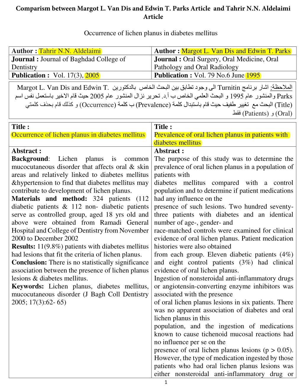
Comparism between Margot L. Van Dis and Edwin T. Parks Article and Tahrir N.N. Aldelaimi Article Occurrence of lichen planus in diabetes mellitus Author : Tahrir N.N. Aldelaimi Journal : Journal of Baghdad College of Dentistry Publication : Vol. 17(3), 2005 Author : Margot L. Van Dis and Edwin T. Parks Journal : Oral Surgery, Oral Medicine, Oral Pathology and Oral Radiology Publication : Vol. 79 No.6 June 1995 L. Van Dis and Edwin T. ريخلاا ماق ثيح ب مسا سفن لمعتسا يتملك فذحب ماق كلذك و ) Margot Turnitin و نيب قباطت دوجو ىلا ب صاخلا يملعلا ثحبلا ( ةملك لادبتساب ماق ثيح فيفط Prevalence ( ةملك ب ) Occurrence نيروتكدلا ماع روشنملا لازن ريرحت .د.أ صاخلا ثحبلا ب جمانرب راشا ماع 5991 رييغت Patients طقف ) ظحلاملا Parks Title ) Oral ) ( و :ة 5001 روشنملاو عم ثحبلا ( ( Title : Occurrence of lichen planus in diabetes mellitus Title : Prevalence of oral lichen planus in patients with diabetes mellitus Abstract : The purpose of this study was to determine the prevalence of oral lichen planus in a population of patients with diabetes mellitus compared with a control population and to determine if patient medications had any influence on the presence of such lesions. Two hundred seventy- three patients with diabetes and an identical number of age-, gender- and race-matched controls were examined for clinical evidence of oral lichen planus. Patient medication histories were also obtained from each group. Eleven diabetic patients (4%) and eight control patients (3%) had clinical evidence of oral lichen planus. Ingestion of nonsteroidal anti-inflammatory drugs or angiotensin-converting enzyme inhibitors was associated with the presence of oral lichen planus lesions in six patients. There was no apparent association of diabetes and oral lichen planus in this population, and the ingestion of medications known to cause tichenoid mucosal reactions had no influence per se on the presence of oral lichen planus lesions (p > 0.05). However, the type of medication ingested by those patients who had orallichen planus lesions was either nonsteroidal anti-inflammatory drug or Abstract : Background: mucocutaneous disorder that affects oral & skin areas and relatively linked to diabetes mellitus &hypertension to find that diabetes mellitus may contribute to development of lichen planus. Materials and method: 324 patients (112 diabetic patients & 112 non- diabetic patients serve as controlled group, aged 18 yrs old and above were obtained from Ramadi General Hospital and College of Dentistry from November 2000 to December 2002 Results: 11(9.8%) patients with diabetes mellitus had lesions that fit the criteria of lichen planus. Conclusion: There is no statistically significance association between the presence of lichen planus lesions & diabetes mellitus. Keywords: Lichen planus, diabetes mellitus, mucocutaneous disorder (J Bagh Coll Dentistry 2005; 17(3):62- 65) Lichen planus is common 1
Comparism between Margot L. Van Dis and Edwin T. Parks Article and Tahrir N.N. Aldelaimi Article angiotensin-converting enzyme inhibitor, which was asignificant association (t9 < 0.00). (ORAL SURG ORAL MED ORAL PATHOL ORAL RADIOt ENDOD 1995;79:696-700) 2
Comparism between Margot L. Van Dis and Edwin T. Parks Article and Tahrir N.N. Aldelaimi Article ( خسنب 21 Introduction Introduction يملعلا هثحب هقصل و افنا روكذملا يف ( نم ةنوكم لازن ريرحت .د.أ ثحبب ةصاخلا 92 ارطس ) ثحبلا نم ارطس ) ) ( ةمدقملا يف لازن ريرحت .د.أ ماق Turnitin ةمدقملا نا املع ( :ة ظحلاملا ) جمانرب راشا امك Author : Tahrir N.N. Aldelaimi Introduction : Lichen planus mucocutaneous approximately 0.1% to 2.0% of the general western population (1,2).The prevalence rates may differ among races and graphic areas. Some investigations have reported a slight female predilection (1,3,4) whereas others have suggested the condition is somewhat more common in males [5]. Lichen planus primarily affects adults, with the mean age of onset in the fourth to fifth decades of life (4). The oral lesions are frequently found on the buccal mucosa, tongue, soft palate, gingival, and lips and they may have a variety of clinical appearances. Accurate diagnosis of lichen planus may impact on a patient’s health as there have been reports that lichen planus lesions may undergo malignant transformation in a small percentage of patients (6,7,8). Lichen planus in particular, has been linked to diabetes mellitus (DM) and hypertension (Grinspan’s syndrome), and it has been antihypertensive medications that the patients take may cause a lichenoid mucosal reaction (9,10). Most of the associations between lichen planus and systemic controversial as they rely on isolated, anecdotal evidence (4,11-14). It has been proposed that the endocrine dysfunction in diabetes mellitus may be related to immunologic defect that may also contribute to the development of Lichen planus (15,16). Author : Margot L. Van Dis and Edwin T. Parks Introduction : Lichen planus (LP) is a relatively common mucocutaneous disorder approximately 0.1% to 2.0% of the general population. 1-5 The prevalence rates may differ among races and geographic areas.For example, African-Americans have been reportedto have a prevalence of 0.3%, 1 Libyans only 0.08%, 51.6% in a particular area in India, 3 whereas the prevalencein Scandinavian whites has been reported torange from 0.8% to 2.0%. 3, 5 Some investigators have reported a slight female predilection, 1, 3, 4 whereas othershave suggested the condition is somewhat morecommon in males. 2 LP primarily affects adults, with the mean age of onset in the fourth to fifth decades of life. 4 LP may affect only the skin, only the oral mucosa, or both. The oral lesions are frequently found on the buccal mucosa, tongue, soft palate, gingiva, and lips, and they may have a variety of clinical appearances.Oral lichen planus (OLP) has reticular, papular, plaque-like, atrophic, erosive, and bullous forms. The lesions may appear in only one form or in a combination of forms. The atrophic, erosive, and bullous forms may have associated symptoms ranging from mild discomfort (especially during the consumption of hot or spicy foods) to severe pain or a burning sensation. 4 OLP tends to be a dynamic condition with remission and exacerbation of symptoms; change in lesion site is commonplace. 6 Accurate diagnosis of OLP may impact on a patient's health as there have been reports that OLP lesions may undergo malignant transformation in a small percentage of patients. 4'7'8 However, not all scholars agree that OLP is a premalignant lesion. 9 Although the exact cause of OLP is unknown, experimental evidence suggests that it is an inflammatory,T-cell-mediated immune response.I, 4 Possible antigens that may serve as stimuli include restorative dental materials l~ and a variety of medications (TableI). 1 It has been suggested that increased levels of stress and mental disturbances are associated with OLP, u but other authors have not agreed with this finding.X2, 13 Because the exact cause of LP remains elusive, treatment is based on alleviating symptoms and usually consists of topical or systemic corticosteroids.4 is a relatively common affects disorder that that affects suggested that the diseases remain 3
Comparism between Margot L. Van Dis and Edwin T. Parks Article and Tahrir N.N. Aldelaimi Article There have been reports of associations between LP and a variety of systemic disorders such as ulcerative colitis, alopecia areata, vitiligo, myasthenia gravis, chronic active hepatitis, primary biliary cirrhosis, multiple sclerosis, and primitive pulmonary fibrosis. 14 OLP, in particular, has been linked to diabetes mellitus(DM) and hypertension (Grinspan's syndrome).15 However, the association of hypertensionand LP has been refuted, 15 and it has been suggestedthat the antihypertensive medications that the patientstake may cause a lichenoid mucosal reaction.16 Most of the reported associations betweenOLP and systemic diseases remain controversial asthey rely on isolated, anecdotal evidence. 4, 17The association between DM and OLP has receivedmuch attention in the literature. The prevalence ofDM in patients with OLP has been reported to rangefrom 10% to 85%. 18-21 It has been proposed thatthe endocrine dysfunction in DM may be related toan immunologic defect that may also contribute tothe development of LP. 21 However, others havereported that the prevalence of DM in OLP patientsdoes not differ from that of DM in the general population.22, 23Virtually all studies on the association of LP andDM have been conducted in patients with diagnosed cases of OLP, looking for evidence of DM. The reverse association, the prevalence of OLP in patients with DM, was not sought by investigators until recently. Albrecht et al' 24 conducted a study in Hungary investigating the occurrence of oral leukoplakia and LP in 1600 patients with DM. They found clinical evidence of OLP in 1% of the patients with DM and no cases (0%) in the control group. However, the control group consisted of 621 persons, one third of whom were less than 20 years of age. Nearly two thirds of the patients with DM were age 35 or older.No mention was made in the article of the statistical significance of the different prevalence rates or the different populations in each group. The authors also discussed the fact that antidiabetic drugs sometimes produce mucosal lesions resembling lichen planus. Nearly half of the diabetic patients were non-insulin dependent and could have been taking oral hypoglycemic medication, but the existence or nature of any "lichenoid" mucosal reactions were not specified. Borghelli et al. 25 recently found a prevalence rate of OLP of 0.55% in a group of 729 Argentine diabetic patients. Their control group was close in size to the diabetic group and was age- and gender-matched; the prevalence rate in the control group was 0.74%. The difference was not statistically significant. In the United States, people tend to take more medications as they grow older. 26 Because OLP tends to occur in persons who are middle-aged or older,there is a greater likelihood that these persons are taking at least one medication. Many medications, prescription as well as over-the-counter products, have been reported to produce lichenoid mucosal reactions.I, 13, 26 In a recent survey, 26 nearly 5% of a sample population took a medication that might cause a mucosal reaction. Potts et al. 27 found an association 4
Comparism between Margot L. Van Dis and Edwin T. Parks Article and Tahrir N.N. Aldelaimi Article between nonsteroidal anti-inflammatory drugs (NSAIDs) and OLP lesions, and Robertson and Wray 13 found that NSAIDs were associated with more erosive than nonerosive LP lesions. However, in many studies of LP, the patients were not taking any medications, and therefore the prevalence of "lichenoid" mucosal reactions is unknown. Neither of the recent investigations of the prevalence of OLP in patients with DM considered the medications their subjects may have been taking. The purpose of this study was to determine the prevalence of OLP in a U.S. population of diabetic patients compared to an age-, gender- and race-matched population and to determine if patient medications had any influence on the presence of such lesions. 5
Comparism between Margot L. Van Dis and Edwin T. Parks Article and Tahrir N.N. Aldelaimi Article Material And Methods ( خسنب ) 29 ( نا املع Material And Methods يف هقصل و افنا روكذملا ثح هثحب لازن ريرحت .د.أ بلا نم ارطس ) ) ( يف لازن ريرحت .د.أ ماق جمانرب راشا امك رييغت يا نودب يملعلا Turnitin : :ة ظحلاملا ثحبب ةصاخلا ارطس ) 33 ( نم ةنوكم Author : Tahrir N.N. Aldelaimi Material And Methods : Author : Margot L. Van Dis and Edwin T. Parks Material And Methods : Patients over the age of 18 years old with conditions that had been diagnosed as diabetes were obtained from Ramadi General Hospital, Ramadi City-Anbar from November 2000 to December 2002. Patients with diabetes mellitus of any type were excluded from the control group. To avoid any potential influence of other systemic disorders that have been reported to be associated with oral lichen planus, persons with the following conditions were excluded from the study: ulcerative colitis, alopecia areata, vitiligo, myasthenia gravis, chronic active hepatitis, primary biliary cirrhosis, multiple scelerosis, and primitive pulmonary fibrosis (14).112 patients were obtained for each group,demographic information was recorded for each patient who agreed to participate in the study. In addition, the patients at the diabetes clinic to obtain their medication histories, past and current, from their medical records granted permission. In addition, the length of time since the patient’s diabetic condition had been diagnosed was recorded, current fasting blood glucose levels were obtained from the medical record. A visual inspection of each patient’s oral cavity was conducted by two senior examiners (with double blind technique) using an artificial light, a mouth mirror, gauze, and wooden tongue blade. The diagnosis of lichen planus was based on the clinical criteria as based by Neville (4). The reliability of making a clinical diagnosis of lichen planus on the basis of clinical criteria was approved when compared with biopsy results, student “t" test used for statistical analysis. Patients over the age of 18 with conditions thathad been diagnosed as insulin-dependent diabetes were recruited from a hospital-based outpatientdiabetes clinic. Age-, gender- and race-matched persons were recruited from the population at a dental school to serve as controls. Patients with diabetes mellitus of any type were excluded from the control group. To avoid any potential influence of other systemic disorders that have been reported to be associated with OLP, persons with the following conditions were excluded from the study: ulcerative colitis, alopecia areata, vitiligo, myasthenia gravis, chronic active hepatitis, primary biliary cirrhosis, multiple sclerosis, and primitive pulmonary fibrosis. 14 Two hundred seventy-three patients were obtained for each information(age, gender, race) was recorded for each patient who agreed to participate in the study. In addition, permission was granted by the patients at the diabetes clinic to obtain their medication histories, past and current, from their medical records. In addition, the length of time since the patient's diabetic condition had been diagnosed was recorded. When available, current fasting blood glucose levels were obtained from the medical record. Medication histories were obtained verbally from the control patients. A visual inspection of each patient's oral cavity was conducted by a single examiner with advanced training in oral diagnosis and oral medicine with the use of an artificial light, a mouth mirror, gauze, and wooden tongue blade. The diagnosis of OLP was based on the following clinical criteria: white, pinhead-sized papules or distinct striae, forming linear, reticular or annular patterns; white, plaquelike lesions with papules or striae at the margins; and atrophies, ulcerations, and vesicles seen concomitantly with white striae or papules at the periphery. group.Demographic 6
Comparism between Margot L. Van Dis and Edwin T. Parks Article and Tahrir N.N. Aldelaimi Article The reliability of making a clinical diagnosis of lichen planus on the basis of these criteria can approach 97% when compared with biopsy results. 3 Results were analyzed using Chi-square and were considered significant at the p < 0.05 level. 7
Comparism between Margot L. Van Dis and Edwin T. Parks Article and Tahrir N.N. Aldelaimi Article Result ( s ارطس ) نم ثحبلا روكذملا افنا و هقصل يف 81 ( خسنب ) جئاتنلا يف لازن ريرحت أ . د . ماق :ة ظحلاملا Results Turnitin ارطس ) 02 ( نم ةنوكم لازن ريرحت أ . د . ثحب ب ةصاخلا ) جئاتنلا نا املع ( جمانرب راشا امك يملعلا هثحب Author : Margot L. Van Dis and Edwin T. Parks Results : Characteristics of each patient group are displayed in Table II. Subject age ranged from 18 years to 77 years. Although care was taken to insure that equal numbers of subjects were contained in each age range,there was a slight difference in exact ages between groups. The mean age of the diabetic group was 48.4 years, and the mean age of the control group was 50.8years. These differences were not statistically significant.The majority of subjects were not taking medications that have been reported to be associated with oral mucosal lesions. Characteristics of patients who showed clinical evidence of OLP are displayed in Tables III and IV.Eleven of the patients with DM (4.0%) had lesions that fit the diagnostic criteria for OLP; two of these patients had more than one lesion. Eight of the control patients had lesions that could be classified as OLP (2.9%);one of these patients had more than one oral lesion. None of the patients in either group was aware that they had LP lesions, and none wassymptomatic. However, none of the lesions was erosive; erosive lesions are usually the more symptomatic lesions. There was no association between the presence of lesions and the duration of diabetes in the diabetic patients. Fasting blood glucose levels were available for 257 of the 273 diabetic patients, including 10 of the 11 diabetic patients who had LP lesions. There was no significant association between glucose levels and the presence of LP in these patients (p = 0.2734). There was no overall difference between the diabetic and nondiabetic patients with LP (p = 0.3384) and no statistically significant difference in terms of gender, race, age, or whether medications other than insulin were taken or not. There was a significant difference, however, in the types of medications associated with the oral lesions. NSAIDs and angiotensinconverting enzyme (ACE) inhibitors were more likely to be implicated com Author : Tahrir N.N. Aldelaimi Results : Characteristics of each patient group are displayed in Table 1. Subject age ranged from 18 years to 78 years. The mean age of the diabetic group was 45 years, and the mean age of the control group was 47 years. Characteristics of patients who showed clinical evidence of lichen planus are displayed in Tables 2 and 3. Eleven (9.8%) of the patients with diabetes mellitus had lesions that fit the diagnostic criteria for lichen planus. Six (5.3%) of the control patients had lesions that could be classified as lichen planus. Dtudent " t "test showed that there was no association between the presence of lesions and diabetes in the diabetic patients, fasting blood glucose levels were available for all diabetic patients (table 3), including 11 diabetic patients who had lichen planus lesions, there was no significant association between glucose levels and the presence of lichen planus in these patients age, or whether medications other than insulin were taken or not. 8
Comparism between Margot L. Van Dis and Edwin T. Parks Article and Tahrir N.N. Aldelaimi Article Discussion روكذملا افنا و هقصل يف ثحبلا ارطس ) نم ( خسنب ) 82 ةشقانملا ( يف لازن ريرحت أ . د . ماق :ة ظحلاملا Discussion Turnitin ) 89 ( نم ةنوكم لازن ريرحت أ . د . ثحب ب ةصاخلا ) ةشقانملا ( نا املع جمانرب راشا امك يملعلا هثحب ارطس Author : Margot L. Van Dis and Edwin T. Parks Discussion : The prevalence rates of OLP lesions in both the diabetic and control populations were higher in this study than in other reports in the literature. 24,25 Borghelli et al. 25 reported prevalence rates of 0.55% in diabetic patients and 0.74% in their control group.Albrecht et al. 24 reported a 1% prevalence rate of LP in the diabetic patients and no cases in their control group. However, it must be pointed out that the sample of patients in this present study was not randomized.For example, African-Americans made up about 32% of the sample population. Although this sample is representative of the population of the diabetes clinic, it is not representative of the general U.S. population, which is approximately 12% African-American.Therefore the prevalence rates determined in this study cannot be applied to the general population of the United States. The diagnostic criteria for LP was not mentioned in the Borghelli and Albrecht studies. Borghelli et alY used unspecified clinical criteria plus "histologic study in special cases" in their study of OLP and DM. In addition, Albrecht et al. 24 found a prevalence of leukoplakia in 6.2% of diabetic patients and 2.2% in controls. The higher prevalence rates in this study may have been due to a sampling error (the groups are relatively small) or to a misdiagnosis of white or atrophic lesions as OLP when, in fact, they were other entities.The diagnoses of OLP were made on the basis of clinical features and were not confirmed histologically.Errors could have been made in the clinical diagnosis despite the report that use of the criteria could be 97% effective. 3 Although the prevalence rates were higher in this study, there was no difference in the prevalence rates of OLP between the diabetic group and the control group. Because the same criteria were applied to each group, the lack of difference may still be valid.Treatment of the lesions (topical steroids) was offered to the patients in whom these lesions were discovered.All but one refused treatment on the grounds that the lesions were asymptomatic. One control patient (47-year-old white woman taking no medications) with a reticular lesion on the buccal mucosa elected to receive Author : Tahrir N.N. Aldelaimi Discussion : The percentage of oral lichen planus lesions in both the diabetic and (non-diabetic) control populations was higher in this study than in other reports in the literature (16-18). Borghelli et al.(17) reported prevalence rates of 0.55% in diabetic patients and 0.74% in their control group. Albercht et al. reported a 1% prevalence rate of lichen planus in the diabetic patients and no cases in their control group. (16) Patients with lesions who were taken medications that have been associated with mucosal reactions were informed of the possible implication of their medications. However, the medication implicated with the lesions prescribed by a physician other than the physician treating the diabetes. Despite the patients who were taken these medications showed any evidence of lichen planus or lichenoid lesions. The types of medications that were associated with the oral lesions, NSAIDs (non steroidal anti inflammatory drugs) and ACE (angiotensin converting enzyme) inhibitors, were statistically significant (p<0.005), which is similar to the findings of Robertson and Wray [13] and Potts et al. (19). However, Robertson and Wray [13] reported no overall association between lichen planus and the ingestions of NSAIDs and lichen planus. Neither Potts et al. (19) norRobertson and Wray (13) found an association between oral lichen planus lesions and the ingestion of antihypertensive medication. 9
Comparism between Margot L. Van Dis and Edwin T. Parks Article and Tahrir N.N. Aldelaimi Article treatment. Fluocinonide gel (0.05%) was prescribed for her, and when she was seen again for routine dental treatment about 3 weeks later, the lesion's clinical appearance had improved, although faint striae could still be detected. She continued with the fluocinonide gel and the lesion was virtually undetectable after a total of 6 weeks' treatment. The lesion occasionally recurs and she uses the gel as needed with good results. Patients with lesions who were taking medications that have been associated with mucosal reactions were informed of the possible implication of their medications. The two control patients expressed reluctance to change medications as they felt their medications were working and their LP lesions were asymptomatic. The presence of the lesions in the four diabetic patients was discussed with the attending physicians in the diabetes clinic. However, in each instance, the medication implicated with the lesions was a medication prescribed by a physician other than the physician treating the diabetes. Therefore there was a reluctance to change the medications. Although notations concerning the presence of the lesions and possible implication of the medications were placed in the patients' charts, a review of these charts more than 8 months later showed that none of the medications had been changed or discontinued.Quite a few of the patients in the study were taking medications that have been implicated with "lichenoid" mucosal reactions (approximately 43% of the diabetic patients and 28% of the control patients). Despite these numbers of patients, only six patients (four with DM, two without) who were taking these medications showed any evidence of LP or lichenoid lesions. There was a statistical lack of association between the presence of OLP and whether or not patients were taking medications in this study. This suggests that the role of these medications in OLP lesions may be limited. However, the types of medications that were associated with the oral lesions, NSAIDS and ACE inhibitors, were statistically significant. Ten percent of the patients with OLP lesions were taking NSAIDs, which is similar to the findings of Robertson and Wray 13 and Ports et al., 27 who reported that 12% and 17% of their patients who had OLP lesions were taking NSAIDs, respectively. However, Robertson and Wray 13reported no overall association between histologically confirmed OLP and the ingestions of NSAIDs, but they did report an association between 10
Comparism between Margot L. Van Dis and Edwin T. Parks Article and Tahrir N.N. Aldelaimi Article NSAIDs and the presence of erosive versus nonerosive LP lesions. In addition, they found an association between NSAID ingestion and the presence of infiltrated oral keratoses. Potts et al. 27 did find a significant association between NSAIDs and LP, especially in the more severe (erosive) forms. None of the patients in this study had erosive LP. Neither PoLLs et al. 27 or Robertson and Wray 13 found any association between OLP lesions and the ingestion of antihypertensive medications. Four of the nineteen patients with OLP lesions in this study were taking ACE inhibitors. However, none of the other antihypertensive medications diuretics, propranolol) were implicated. None of the other authors specified which medications were included in the antihypertensive groups. It is possible that ACE inhibitors were not included in their studies.It is also possible that the finding in this study is somewhat skewed by the small samples. In summary, the findings in this study failed to associate DM and OLP lesions, which agrees with the findings of other authors. The ingestion of medications that have been implicated with lichenoid mucosal reactions played a minimal role in the prevalence of lesions in this population, although NSAID medications and ACE inhibitors may be more likely than other medications to be associated with such oral lesions. However, sample populations in this study did not represent the general U.S. population, and the number of patients in this study who had lesions was very small. Therefore the results may not be applied universally. (furosemide, thiazide 11
Comparism between Margot L. Van Dis and Edwin T. Parks Article and Tahrir N.N. Aldelaimi Article Reference ( ( خسنب اردصم ) نم ثحبلا روكذملا افنا و هقصل يف 81 ( رداص s ملا يف لازن ريرحت أ . د . ماق :ة ظحلاملا Reference ( Turnitin ) 81 ( نم ةنوكم لازن ثحبب ةصاخلا أ . د . ريرحت ( رداص s نا املع ملا جمانرب راشا امك يملعلا هثحب اردصم Author : Margot L. Van Dis and Edwin T. Parks References : 1. Scully C, E1-Kom M. Lichen planus: review and update on pathogenesis. J Oral Pathol 1985;14:431-58. 2. Bouquot JE, Gorlin RJ. Leukoplakia, lichen planus, and other keratoses in 23,616 white Americans over the age of 35 years.ORAL SURG ORAL MED ORAL PATrtOL 1986;61:373-81. 3. Axell T, Rundquist L. Oral lichen planus: a demographic study. Community Dent Oral Epidemiol 1987;15:52-6. 4. Vincent SD, Fotos PG, Baker KA, Williams TP. Oral lichen planus: the clinical, historical, and therapeutic features of 100 cases. ORAL SURG ORAL MED ORAL PATHOL 1990;70:165-71. 5. Zegarelli D J, Sabbagh E. Relative incidence of intraoral pemphigus vulgaris, mucus membrane pemphigoid, and lichen planus. Ann Dent 1989;48:5-7. 6. Vincent SD. Diagnosing and managing oral lichen planus. J Am Dent Assoc 1991;122:93-6. 7. Silverman S, Gorsky M, Lozada-Nur F. A prospective follow-up study of 570 patients with oral lichen planus: persistence, remission, and malignant association. ORAL SURG ORAL MED ORAL PATHOL 1985;60:30-4. 8. Voute ABE, de Jong WFB, Schulten EAJM, Snow GB, van der Waal I, Possible premalignant character of oral lichen planus: the Amsterdam experience. J Oral Pathol Med 1992;21:326-9. 9. Eisenberg E, Krutchkoff DJ. Lichenoid lesions of oral mucosa: diagnostic criteria and their importance in the alleged relationship to oral cancer. ORAL SURG ORAL MED ORAL PATHOL 1992;73:699-704. 10. Eversole LR, Ringer M. The role of dental restorative metal in the pathogenesis of oral lichen planus. ORAL SURG ORAL Author : Tahrir N.N. Aldelaimi References : 1.Scully C, El-Com M. Lichen planus: review and update on pathogenosis. J Oral Pathol 1985; 14: 431-58. 2.Bouquot JE, Gorlin RJ. Leukemia, lichen planus, and other keratoses in 23, 616 white Americans over the age of 35 years. Oral Surg Med Pathol 1986; 61: 373-81. 3.Axell T, Rundquist L. Oral lichen planus: a demographic study. Community Dent Oral Epidemiol 1987; 15: 52-6. 4.Brad W, Neville. Dermatological Diseases, oral and maxillofacial pathlogy, Mosby, 1996; pp 572-77. 5.Zegarelli DJ, Sabbagh E. Relative incidence of intraoral pemphegus vulgaris, mucus membrane pemphegoid, and lichen planus. ANN DENT 1989; 48: 5-7. 6.Vincent SD. Diagnosing and managing oral lichen planus. J AM DENT ASSOC 1991; 122: 93-6. 7.Silverman S, Gorsky M. Lozada-Nu F. A perspective follow-up study of 570 patients with oral lichen planus: persistence, remission, and malignant association. ORAL SURG MED PATHOL 1985; 60: 30-4. 8.Voute ABE, de Jong WFB. Schulten EAJM, Snow GB, Van der waal I. Possible premalignant character of oral lichen planus: the Amesterdam experience. J ORAL PATHOL MED 1992; 21: 326-9. 9.Eisenberg E, Krutchkov DJ. Lichenoid lesions of oral mucosa : diagnostic criteria and their importance in the alleged relationship of oral cancer. ORAL SURG MED PATHOL 1992 ; 73:699-704. 10.Eversole LR, Ringer M.Role of dental restorative metal in the pathogenesis of oral lichen 12
Comparism between Margot L. Van Dis and Edwin T. Parks Article and Tahrir N.N. Aldelaimi Article planus. ORAL SURG MED PATHOL 1984; 57: 383-7. 11.Hampf BGC, Malmstrom MJ, Aalberg VA, Hannula JA, Vikkula J. Psychiatric disturbance in patients with oral lichen planus. ORAL SURG MED PATHOL 1987; 63:429-32. 12.Allen CM, Beck FM, Rossi KM, Kaul TJ. Relation of stress and anxiety to oral lichen planus. ORAL SURG MED PATHOL 1986; 61: 44-6. 13.Robertson WD, Wray D. Ingestion of medication among patients with oral keratoses including lichen planus. ORAL SURG MED PATHOL 1992; 74: 183-5. 14.Gruppo Italiano Dermatologia. Epidemiological evidence of the association between lichen planus and two immune-related diseases: alopecia areata and ulcerative colitis. ARCH DERMATOL 1991; 127:688-91. 15.Lamey PJ, Gibson J, Barclay SC, Miller S. Grinspan’s syndrome: phenomenon? ORAL SURG MED PATHOL 1990; 70: 184-5. 16.Albercht M, Banoczy J, Dinya E, Tamas G Jr. Occurrence of oral leukemia and lichen planus in diabetes mellitus. J ORAL PATHOL MED 1992; 21: 364-6. 17.Borghelli RF, Pettinari IL, Chuchurra JA, Striparo MA. Oral lichen planus in patients with diabetes: an epidemiologic study. ORAL SURG MED PATHOL 1993; 75: 498-500. 18.Miller CS, Kaplan AL, Guest GF, Cottone JA. Documenting medication use in adult dental patients: 1987-1991. J AM DENT ASSOC 1992; 123: 41-8. 19.Potts AJC, Hamberger J, Scully C. The medication of patients with oral lichen planus and the association of nonsteroidal anti-inflammatory drugs with erosive lesions. ORAL SURG MED PATHOL 1987; 64: 541-3. MED ORAL PATHOL 1984;57:383-7. 11. Hampf BGC, Malmstrom M J, Aalberg VA, Hannula JA, Vikkula J. Psychiatric disturbance in patients with oral lichen planus. ORAL SURG ORAL MEt) ORAL PATHOL 1987;63:429-32. 12. Allen CM, Beck FM, Rossi KM, Kaul TJ. Relation of stress and anxiety to oral lichen planus. ORAL SURG ORAL MEI) ORAL PATHOL 1986;61:44-6. 13. Robertson WD, Wray D. Ingestion of medication among patients with oral keratoses including lichen planus. ORAL SURG ORAL MED ORAL PATHOL 1992;74:183-5. 14. Gruppo Italiano Studi Epidemiologici in Dermatologia. Epidemiological evidence of the association between lichen planus and two immune-related diseases: alopecia areata and ulcerative colitis. Arch Dermatol 1991;127:688- 91. 15. Lamey P-J, Gibson J, Barclay SC, Miller S. Grinspan's syndrome: phenomenon? ORAL SURG ORAL MED ORAL PATHOL 1990;70:184-5. 16. Christensen E, Holmstrup P, Wiberg-J~rgensen F, Neumann-Jensen B, Pindborg JJ. Arterial blood pressure in patients with oral lichen planus. J Oral Pathol 1977;6:139- 42. 17. Millard HD, Mason DK. Perspectives on the 1988 world workshop on oral medicine. Chicago: Year Book Medical Publishers 1989:60-2. 18. Jolly M. Lichen planus and its association with diabetes mellitus. Med J Aust 1972;1:990-2. 19. Howell FV, Rick GM. Oral lichen planus and diabetes: a potential syndrome. J Calif Dent Assoc 1973;1:58-9. 20. Lowe N J, Cudworth AG, Clough SA, Bullen MF. Carbohydrate metabolism in lichen planus. Br J Dermatol 1976;95:9-12. 21. Lundstrom IMC. Incidence of diabetes mellitus in patients with oral lichen planus. Int J Oral Surg 1983;12:147-52. 22. Bussell SN, Smales FC, Sutton RBO, Duckworth R. Glucose tolerance in patients with lesions of the oral mucosa. Br Dent J 1979;146:186-8. 23. Christensen F, Holstrup P, Wiberg-JCrgensen F, Neumann-Jensen B, Pindborg JJ. Glucose tolerance in patients with oral lichen planus. J Oral Pathol 1977;6:143- 51. Studi Epidemiologici A drug-induced A drug-induced 13
Comparism between Margot L. Van Dis and Edwin T. Parks Article and Tahrir N.N. Aldelaimi Article 24. Albrecht M, Banoczy J, Dinya E, Tamas G Jr. Occurrence of oral leukoplakia and lichen planus in diabetes mellitus. J Oral Pathol Med 1992;21:364-6. 25. Borghelli RF, Pettinari IL, Chuchurru JA, Stirparo MA. Oral lichen planus in patients with diabetes: an epidemiologic study. ORAL SURG ORAL MED ORAL PATHOL 1993;75:498-500. 26. Miller CS, Kaplan AL, Guest GF, Cottone JA. Documenting medication use in adult dental patients: 1987-1991. 1992;123:41-8. 27. PoLLs AJC, Hamburger J, Scully C. The medication of patients with oral lichen planus and the association of nonsteroidal anti- inflammatory drugs with erosive lesions. ORAL SURG ORAL MED ORAL PATHOL 1987;64:541-3.. J Am Dent Assoc 14