Download
1 / 9
90 likes | 119 Views
Direct Plagiarism

E N D
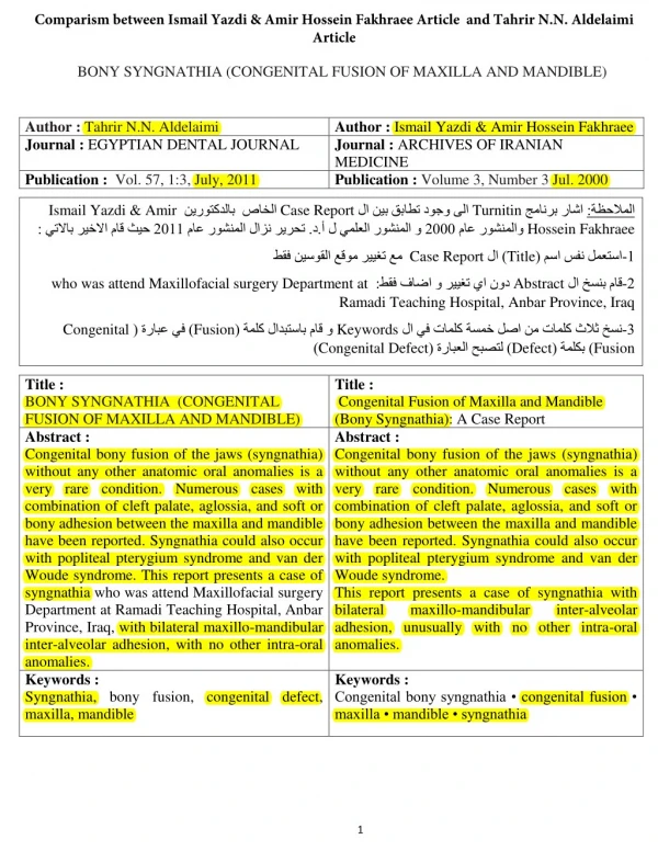
Comparism between Ismail Yazdi & Amir Hossein Fakhraee Article and Tahrir N.N. Aldelaimi Article BONY SYNGNATHIA (CONGENITAL FUSION OF MAXILLA AND MANDIBLE) Author : Tahrir N.N. Aldelaimi Journal : EGYPTIAN DENTAL JOURNAL Author : Ismail Yazdi & Amir Hossein Fakhraee Journal : ARCHIVES OF IRANIAN MEDICINE Publication : Volume 3, Number 3 Jul. 2000 Publication : Vol. 57, 1:3, July, 2011 Ismail Yazdi & Amir : يتلااب ريخلاا ماق ثيح Case Report صاخلا ب ماع روشنملا لازن ريرحت .د.أ Turnitin ماع روشنملاو نيروتكدلا نيب قباطت دوجو ىلا لا و ل يملعلا روشنملا جمانرب راشا Hossein Fakhraee :ة ظحلاملا 0222 0222 Case Report مسا سفن لمعتسا ( Title طقف نيسوقلا عقوم رييغت عم لا ) 2 - who was attend Maxillofacial surgery Department at Abstract :طقف فاضا و رييغت يا نود Ramadi Teaching Hospital, Anbar Province, Iraq لا خسنب ماق 0 - Congenital ( ةملك لادبتساب ماق و Fusion لا يف تاملك ةسمخ لصا نم تاملك ثلاث خسن Keywords ( ةرابعلا حبصتل ) Congenital Defect ( ةرابع يف ) 3 - Defect Fusion ) ( ةملكب ) Title : BONY SYNGNATHIA (CONGENITAL FUSION OF MAXILLA AND MANDIBLE) Abstract : Congenital bony fusion of the jaws (syngnathia) without any other anatomic oral anomalies is a very rare condition. Numerous cases with combination of cleft palate, aglossia, and soft or bony adhesion between the maxilla and mandible have been reported. Syngnathia could also occur with popliteal pterygium syndrome and van der Woude syndrome. This report presents a case of syngnathia who was attend Maxillofacial surgery Department at Ramadi Teaching Hospital, Anbar Province, Iraq, with bilateral maxillo-mandibular inter-alveolar adhesion, with no other intra-oral anomalies. Keywords : Syngnathia, bony fusion, congenital defect, maxilla, mandible Title : Congenital Fusion of Maxilla and Mandible (Bony Syngnathia): A Case Report Abstract : Congenital bony fusion of the jaws (syngnathia) without any other anatomic oral anomalies is a very rare condition. Numerous cases with combination of cleft palate, aglossia, and soft or bony adhesion between the maxilla and mandible have been reported. Syngnathia could also occur with popliteal pterygium syndrome and van der Woude syndrome. This report presents a case of syngnathia with bilateral maxillo-mandibular adhesion, unusually with no other intra-oral anomalies. inter-alveolar Keywords : Congenital bony syngnathia • congenital fusion • maxilla • mandible • syngnathia 1
Comparism between Ismail Yazdi & Amir Hossein Fakhraee Article and Tahrir N.N. Aldelaimi Article Case Report Introduction يف هقصل و افنا روكذملا نم ارطس ) 51 ( خسنب ) ( ةمدقملا يف لازن ريرحت .د.أ ماق جمانرب راشا امك رييغت يا نودب يملعلا هروشنم Turnitin :ة ظحلاملا Author : Tahrir N.N. Aldelaimi Introduction : Syngnathia is a rare congenital anomaly involving fusion of maxillary and mandibular bones. The fusion may be due to soft tissue adhesions between the two or a true bony fusion between maxilla and mandible1,2. The effected new born child is presenting with diffi culties in the airway protection and maintenance as well as feeding problems3. Most have other associated anomalies like popliteal pterygium syndrome, Vander Woude syndrome requiring concurrent management. Surgical management involves division of the bony fusion or break down of the adhesions in the fi rst few days of life. Depending upon the severity, these children present formidable anaesthetic challenges. The congenital bony fusion of the maxilla and mandible (bony syngnathia), especially as an isolated occurrence, is a very rare condition. Syngnathia mostly appears in association with other anatomic oral and maxillofacial anomalies. About few such cases have been reported in the literature in combination with cleft lip, cleft of hard and soft palate, aglossia, popliteal pterygium syndrome1, van der Woude syndrome, aglossia- adactylia syndrome2, oral soft tissue synechiae, hypoplasia of the proximal mandible, clefting of mandible, bifi d tongue, hemifacial microsomia, small or absent tongue, temporomandibular (zygomaticomandibular) fusion and some other regional and systemic anomalies 3-8 Author : Ismail Yazdi & Amir Hossein Fakhraee Introduction : Congenital bony fusion of the maxilla and mandible (bony syngnathia),especially as an isolated occurrence, is a very rare condition. Syngnathia mostly appears in association with other anatomic oral and maxillofacial anomalies. About 15 such cases have been reported in the literature in combination with cleft lip, cleft of hard and soft palate, aglossia, popliteal pterygium syndrom1, van der Woude syndrome, aglossia- adactylia syndrome2, oral soft tissue synechiae, hypoplasia of the proximal mandible, hemifacial microsomia, cleft of mandible, bifid tongue, small or absent tongue, (zygomaticomandibular) fusion and some other regional and systemic anomalies3-14 This condition is also seen in autosomal recessive hypomandibular craniofacial dysostosis. To our knowledge, only two isolated cases of bony syngnathia have been literature10,14,15 This report presents a case of congenital syngnathia with bilateral maxillo- mandibular inter-alveolar adhesion, unusually with no other oral or maxillofacial anomalies. temporomandibular reported in the 2
Comparism between Ismail Yazdi & Amir Hossein Fakhraee Article and Tahrir N.N. Aldelaimi Article Case Presentation لا يف Turnitin :يتلااب لازن ريرحت .د.أ مايق ىلا جمانرب راشا :ة ظحلاملا Paragraph 1 :وه فيفط رييغت عم روطس ) ( نم نوكم ) 5 ( عطقم خسنب ماق 1 - 2 ) ( مقر نع لادب ) 3 ( مقر مادختسا : لاوا OF Ramadi Teaching Hospital, Ramadi city, Anbar Province, Iraq ( نع لادب ) Pars مدختسا : ايناث ( Hospital, Tehran ) Female ( مدختسا : اثلاث Male ) ( نع لادب ) weight of 2670 g ) weight 2800g ( نع لادب ) ( مدختسا : اعبار Prohibited Prevented اهل هفدارتم يه و ) ( نع لادب ) ( ةملك مدختسا : اسماخ Paragraph 2 :وه فيفط رييغت عم روطس ) 4 ( نم نوكم ) ( عطقم خسنب ماق 2 - ( ةملك مدختسا : لاوا Clinical ( نم لادب ) Physical اهل هفدارتم يه و ) which extended posteriorly from the canine area to the molar regions, bilaterally ) which extended bilaterally from deciduous canine area to the molar regions posteriorly ) ( لا ةغايص ةداعا : ايناث ( اهلعج و Paragraph 2 :وه فيفط رييغت عم روطس ) 3 ( نم نوكم ) ( عطقم خسنب ماق 3 - of the skull and facial bones ) ( ةرابع فذح : لاوا Which ) Were ( نع لادب ) ( ةملك مادختسا : ايناث Author : Tahrir N.N. Aldelaimi Case Presentation : Author : Ismail Yazdi & Amir Hossein Fakhraee Case Presentation : A newborn female of 2 days of age was transferred to the Department of Oral and Maxillofacial Surgery of Pars Hospital, Tehran, for evaluation of the fusion of maxilla and mandible, which prohibited oral feeding. She was admitted to the neonatal intensive care unit where she was soon fed via a nasogastric tube.Physical examination revealed severe trismus due to complete adhesion of the jaws, which extended posteriorly from the canine area to the molar regions, bilaterally. The oral cavity therefore could not be seen.The child was the product of a normal vaginal delivery with a birth weight of 2670 g. Systemic examination revealed no anomalies in the rest of the body, but her general condition was poor and her rectal temperature rose to 40C.Medical consultation and pertinent laboratory tests A newborn male ( weight 2800g) of 3 days of age was referred to the Department of Maxillofacial Surgery of Ramadi Teaching Hospital, Ramadi city, Anbar Province, Iraq; for evaluation of the fusion of maxilla and mandible that prevented oral feeding. Medical consultation and laboratory tests revealed low blood glucose. Urgently thorough clinical and radiographical examinations were done and start feeding via a nasogastric (NG) tube and the patient was maintained on humidified oxygen by mask. There was no other associated local or systemic anomaly. Clinical examination revealed severe trismus due to adhesion of the jaws, which extended bilaterally from deciduous canine area to the molar regions posteriorly and revealed a bony fusion between the maxilla and the mandible involving the entire alveolar margin with a small gap about 18 mm on left side anteriorly in the canine region. 1 1 2 2 3
Comparism between Ismail Yazdi & Amir Hossein Fakhraee Article and Tahrir N.N. Aldelaimi Article 3 The radiographs were obtained with difficulty, revealed bilateral bony fusion of the maxilla and mandible, Very feeble motion was palpable over each temporomandibular joint (TMJ). Family history showed no similar affliction could be elicited in the past generations of either the parents and maternal and paternal history was negative for any facial cleft. All other siblings were normal. ( Fig. 1 and 2) failed to reveal the cause of fever. The poor condition of the baby did not allow a CT scan or MRI to be performed. The radiography of the skull and facial bones, which was obtained with difficulty, revealed bilateral bony fusion of the maxilla and mandible, normal teeth buds and presence of normally developed condyles of the temporomandibular joints.The patient was the first child of an 18-year old mother and a 23-year old father who were first cousins, healthy and normal. No similar affliction could be elicited in the past two generations of either the parents.At the age of 50 days, the patient’s systemic condition had relatively improved. She was brought to the operating theater and a planned blind awake nasal intubation was failed. Tracheostomy was performed and anesthesia was maintained with nitrous oxide and oxygen.The syngnathia was divided by means of a scalpel and fine osteotomes via an intra-oral approach which allowed the mouth to remain open. On inspection, no other anomalies of the oral cavity,including tongue, palate and pharynx were found.Both temporomandibular joints seemed to function normally. The anesthesia was without any complication and the patient was sent to the intensive care unit in fairly good condition. The baby, who primarily was unable to bottle-feed, probably due to lack of natal sucking reflex, learned to do so after appropriate nursing.The pre-existing fluctuating temperature of the patient remitted after surgery and she was discharged from the hospital at the end of second postoperative week. A few weeks later, she was repeatedly readmitted into the hospital because of high fever and pneumonia, for which she was treated and discharged in relatively good condition each time.Finally, the child died at the age of seven months. Since permission to conduct an autopsy was not given, the exact cause of death could not be determined. However, a clinical diagnosis of acute pneumonia had been made. 3 4
Comparism between Ismail Yazdi & Amir Hossein Fakhraee Article and Tahrir N.N. Aldelaimi Article Case Report روكذملا افنا و هقصل يف Discussion ارطس ) نم 05 ( خسنب ) ( ةشقانملا يف لازن ريرحت أ . د . ماق :ة ظحلاملا Discussion Turnitin ةنوكم لازن ب ةصاخلا ) أ . د . ريرحت ( ةشقانملا نا املع جمانرب راشا امك رييغت نودب يا يملعلا ارطس ) هروشنم ( نم 05 Author : Tahrir N.N. Aldelaimi Discussion : Fusion defects of the maxilla and mandible including other anatomic oral abnormalities are not common. They may be of the connecting tissue either fibrous or bony 1,2 Soft tissue fusion 9,10 (synechiae) have been extensively reviewed by Gartlan et al. (1993) and were classified as buccopharyngeal membrane remnants or as ectopic membranes on the basis of their presumed origin 11 . Bony fusion (syngnathia), particularly its isolated occurrence, is extremely rare. The very few cases reported in the literature are mostly inadequate in description, inconsistent and confusing in nomenclature and with limited useful conventional imaging 4,10. The cause of congenital bony syngnathia is not certain. In contrast, the review of five cases presented by Dawson et al (1997) and previously reported cases provide no evidence of any familial tendency, history of drug and toxin exposure or consanguinity3. Congenital bony syngnathia can be clinically recognized and diagnosed at or after the birth of the affected neonate without any exception 3,7. The adequate useful conventional radiography and / or CT scan can support the clinical recognition of this condition and its nature, which causes inability to open the jaws. The management of patients with congenital fusion of maxilla and mandible varies according to the nature and extent of the abnormalities. The condition is problematic and interferes with feeding, breathing, general health of the patient (aspiration pneumonitis), development, induction of anesthesia. The airway is the first priority to be secured in the management of any newborn with trismus. Thereafter, feeding problems should be overcome Author : Ismail Yazdi & Amir Hossein Fakhraee Discussion : Fusion defects of the maxilla and mandible including other anatomic oral abnormalities are not common. They may be of the connecting tissue either fibrous or bony1-17. Soft tissue fusion15-16 (synechiae) have been extensively reviewed by Gartlan et al. (1993) and were classified as buccopharyngeal membrane remnants or as ectopic membranes on the basis of their presumed origin.13Bony fusion (syngnathia), particularly its isolated occurrence, is extremely rare. The very few cases reported in the literature are mostly inadequate in description, inconsistent and confusing in nomenclature and with limited useful conventional imaging3,4,10,14.The cause of congenital bony syngnathia is not certain. Some of the postulated causes, from reported cases in the literature, including Goodacre and Wallace’s summarization of the various experimental studies performed on the embryological basis, include persistence of the buccopharyngeal membrane, amniotic constriction bands in the region of the developing branchial arches, environmental insults, drugs such as meclozine and large doses of vitamin A4,5,14,12.In contrast, the review of five cases presented by Dawson et al (1997) and previously reported cases provide no evidence of any familial tendency, history of drug and toxin exposure or consanguinity3.A case of a human fetus with syngnathia without any clinical history7 and also a case of vitamin A induced bony syngnathia with a cleft of the secondary palate in rats, treated with vitamin A, has also been reported17.Congenital bony syngnathia can be clinically recognized and diagnosed at or after the birth of the affected neonate without any exception3,7.Theadequate useful conventional radiography and high resolution or spiral cut CT can support the clinical recognition of this condition and its nature, which causes inability to open the growth and 5
Comparism between Ismail Yazdi & Amir Hossein Fakhraee Article and Tahrir N.N. Aldelaimi Article by placing a nasogastric or gastrotomy tubes. Since the occurrence of bony fusion of the maxilla and mandible is extremely rare, and there is high rate of association between bony syngnathia and other regional malformations3,4,6,8,12, the patient should be under the supervision of a team of clinicians skilled in the diagnosis and appropriate treatment of congenital oral and maxillofacial anomalies 3 . Surgical division of the bony fusion, under general anesthesia (blind intubation or via tracheostomy) is the optimal treatment for the simple syngnathia in isolated occurrence or cases with the presence abnormalities6,8,12 . Proper physical therapy should be commenced immediately and the infant should be encouraged to feed normally as soon as possible3. Maxillomandibular fusion is a rare group of anomalies varying in severity from simple mocusal adhesions (synechiae) to extensive bony fusion (syngnathia). Proper physical therapy and feeding should be resumed as soon as possible after the surgery. The significant points about the case reported were; it was an isolated pure bony fusion without any associated local (cleft lip/palate) or systemic anomalies; only few cases exist in the world literature so far. jaws. The management of patients with congenital fusion of maxilla and mandible varies according to the nature and extent of the abnormalities.The condition is problematic and interferes with feeding, breathing, general health of the patient (aspiration pneumonitis), growth and development, induction of anesthesia (intubation) etc.However, on the basis of the reports from the literature, (though they are relatively imprecise and not consistently well documented), functional results, especially in the cases with isolated occurrence, are likely to be good3-5,10,15. But the rarity of this condition imposes some limitations on standardization of the treatment.4In terms of jaw function and its outcome, it is more problematic in complex cases. Dawson, et al having added five new cases to the literature proposed a system of classification and elaborated on treatment recommendations3. Their proposed classification is aimed at the treatment and likely functional outcome rather than etiology or pathogenesis of the malformation3. The airway is the first priority to be secured in the management of any newborn with trismus. Thereafter, feeding problems should be overcome by placing a nasogastric or gastrotomy tubes.Since the occurrence of bony fusion of the maxilla and mandible is extremely rare, and there is high rate of association between bony syngnathia and other regional and systemic malformations3-6,8,14 the patient should be under the supervision of a team of clinicians skilled in the diagnosis and appropriate treatment of congenital oral and maxillofacial anomalies.3Surgical division of the bony fusion, under general anesthesia (blind intubation or via tracheostomy) is the optimal treatment for the simple syngnathia in isolated occurrence or cases with the presence of other anatomic abnormalities3-6,8,14. Proper physical therapy should be commenced immediately and the infant should be encouraged to feed normally as soon as possible3.There are at least two significant points about our case. First, it was an isolated case without any other regional anatomic anomalies such as cleft lip and palate. And only two such cases are reported in recent medical literature10,14.Second, there is consanguinity in the family (parents are first cousins) which suggests the and systemic of other anatomic 6
Comparism between Ismail Yazdi & Amir Hossein Fakhraee Article and Tahrir N.N. Aldelaimi Article possibility of autosomal recessive inheritance.The pathogenesis of this condition still remains obscure although some developmental defects may have occurred in the region of the first branchial arch, in which separation of the maxilla and mandible have failed to occur. The poor general condition of the infant and her high fever could be due to an aspiration pneumonitis and respiratory distress.Whether this case and two similar cases reported in literature10,14 should be considered as amild expression related to some of the syndromes of the head and neck region or categorized as an independent anomaly, is open to question. 7
Comparism between Ismail Yazdi & Amir Hossein Fakhraee Article and Tahrir N.N. Aldelaimi Article References ( لوا لسلست يف 8 رداصم ) Case Report روكذملا افنا و لا رداصم اردصم ) نم 21 ( خسنب ) رداصملا ( يف لازن نودب يا ريرحت يملعلا أ . د . هروشنم ماق :ة ظحلاملا هقصل يف رييغت Author : Tahrir N.N. Aldelaimi References : 1. Hamamoto J, Matsumoto T. A case of facio- genito-popliteal syndrome. Ann Plast Surg. 1984; 13: 224-9. 2. Johnsson GF, Robinow M. Aglossia-adactylia. Radiology. 1978; 128: 127-32. 3. Dawson KH, Gruss JS, Myall RW. Congenital bony syngnathia. A proposed classification. Cleft Palate Craniofac J. 1997; 2: 141-6. 4. Rao S, Oak S, Wagh S, Kulkarni M. Congenital midline palatomandibular bony fusion with a mandibular cleft and a bifid tongue. B J Plastic Surg. 1977; 50: 139-41. 5. Kamata S, Satoh K, Vemura T, Onizuka T. Congenital bilateral fusion with mandibular hypolasia. B J Plastic Surg. 1996; 49: 251-3. 6. Gorlin J, Cohen M, Michael, Levin L, Stefan. Syndromes of Head and Neck. 3rd ed. England: Oxford University Press; 1990: 630-1, 783. 7. Shah RM. Palatomandibular and maxillo- mandibular fusion, partial aglossia and cleft palate in a human embryo. Teratology. 1977; 15: 261-72. 8. Agrawal K, Chandra SS, Sreckumar NS. Congenital bilateral intermaxillary bony fusion. Ann Plast Surg. 1993; 30: 163-6. 9. Kamala G, Pillai V, Kamath V, Kumar GS, Nagamani N. Persistent membrane with cleft palate. Oral Surg Oral Med Oral Pathol. 1990; 69: 164-6. 10. Dinardo NM, Christion JM, Benneth JA, Shutack JG. Cleft palate lateral synechia synchrome. Oral Surg Oral Med Oral Pathol. 1989; 68: 565-6. 11. Gartlan MG, Davies J, Smith RJ. Congenital oral synechiae. Ann Otol Rhinol Laryngol. 1993; 102: 186-97. Author : Ismail Yazdi & Amir Hossein Fakhraee References : 1. Hamamoto J, Matsumoto T. A case of facio- genito-popliteal syndrome. Ann Plast Surg. 1984; 13: 224-9. 2. Johnsson GF, Robinow M. Aglossia-adactylia. Radiology. 1978; 128: 127-32. 3. Dawson KH, Gruss JS, Myall RW. Congenital bony syngnathia. A proposed classification. Cleft Palate Craniofac J. 1997; 2: 141-6. 4. Rao S, Oak S, Wagh S, Kulkarni M. Congenital midline palatomandibular bony fusion with a mandibular cleft and a bifid tongue. B J Plastic Surg. 1977; 50: 139-41. 5. Kamata S, Satoh K, Vemura T, Onizuka T. Congenital bilateral zygomaticomandibular fusion with mandibular hypolasia. B J Plastic Surg. 1996; 49: 251-3. 6. Gorlin J, Cohen M, Michael, Levin L, Stefan. Syndromes of Head and Neck. 3rd ed. England: Oxford University Press; 1990: 630-1, 783. 7. Shah RM. Palatomandibular and maxillo- mandibular fusion, partial aglossia and cleft palate in a human embryo. Teratology. 1977; 15: 261-72. 8. Agrawal K, Chandra SS, Sreckumar NS. Congenital bilateral intermaxillary bony fusion. Ann Plast Surg. 1993; 30: 163-6. 9.Salleh NM. Congenital partial fusion of the mandible and maxilla. Oral Surg Oral Med Oral Pathol. 1965; 20: 74-6. 10.Miskinyar SA. Congenital mandibulomaxillary fusion. Plast Reconstr Surg. 1979; 63: 120-1. 11.Behnia H, Shamse MG. Congenital unilateral fusion of the mandibular and maxillary alveolar ridge, tempo romandibular joint, and coronoid process. J Oral Maxillofac Surg. 1996; 54: 773-6. 12.Goodacre TE, Wallace AF. Congenital alveolar fusion. Br J Plast Surg. 1990; 43: 203-9. 13.Gartlan MG, Davies J, Smith RJ. Congenital oral synechiae. Ann Otol Rhinol Laryngol. 1993; 102: 186-97. zygomaticomandibular buccopharyngeal 8
Comparism between Ismail Yazdi & Amir Hossein Fakhraee Article and Tahrir N.N. Aldelaimi Article 14.Nwoku AL, Kekere-Ekun TA. Congenital ankylosis of the mandible. J Maxillofac Surg. 1986; 14: 150-2. 15.Kamala G, Pillai V, Kamath V, Kumar GS, Nagamani N. Persistent membrane with cleft palate. Oral Surg Oral Med Oral Pathol. 1990; 69: 164-6. 16.Dinardo NM, Christion JM, Benneth JA, Shutack JG. Cleft palate lateral synechia synchrome. Oral Surg Oral Med Oral Pathol. 1989; 68: 565-6. 17.Nada R. Maxillomandibular ankylosis and cleft palate in rat embryos. J Dent Res. 1970; 49: 1086-90. 12. mandibulomaxillary fusion. Plast Reconstr Surg. 1979; 63: 120-1. Miskinyar SA. Congenital buccopharyngeal 9