Evaluation of Abnormal Liver Function Tests
Evaluation of Abnormal Liver Function Tests. Dr Chris Hovell Consultant Gastroenterologist Dorset County Hospital. LFT’s. Markers of hepatocellular damage Cholestasis Liver synthetic function. Markers of Hepatocellular damage (Transaminases).

Evaluation of Abnormal Liver Function Tests
E N D
Presentation Transcript
Evaluation of Abnormal Liver Function Tests Dr Chris Hovell Consultant Gastroenterologist Dorset County Hospital
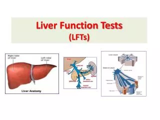
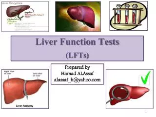
LFT’s • Markers of hepatocellular damage • Cholestasis • Liver synthetic function
Markers of Hepatocellular damage(Transaminases) • AST- liver, heart skeletal muscle, kidneys, brain, RBCs • In liver 20% activity is cytosolic and 80% mitochondrial • Clearance performed by sinusoidal cells, half-life 17hrs • ALT–more specific to liver, v.low concentrations in kidney and skeletal muscles. • In liver totally cytosolic. • Half-life 47hrs
Gamma-GT – hepatocytes and biliary epithelial cells, pancreas, renal tubules and intestine • Very sensitive but Non-specific • Raised in ANY liver discease hepatocellular or cholestatic • Usefulness limited • Confirm hepatic source for a raised ALP • Alcohol • Isolated increase does not require any further evaluation, suggest watch and rpt 3/12 only if other LFT’s become abnormal then investigate
Markers of Cholestasis • ALP – liver and bone (placenta, kidneys, intestines or WCC) • Hepatic ALP present on surface of bile duct epithelia and accumulating bile salts increase its release from cell surface. Takes time for induction of enzyme levels so may not be first enzyme to rise and half-life is 1 week. • ALP isoenzymes, 5-NT or gamma GT may be necessary to evaluate the origin of ALP
Bilirubin, Albumin and Prothrombin time (INR) • Useful indicators of liver synthetic function • In primary care when associated with liver disease abnormalities should raise concern • Thrombocytopaenia is a sensitive indicator of liver fibrosis
Patterns of liver enzyme alteration • Hepatic vs cholestatic • Magnitude of enzyme alteration (ALT >10x vs minor abnormalities) • Rate of change • Nature of the course of the abnormality (mild fluctuation vs progressive increase)
Patterns of liver enzyme alteration • Acute hepatitis –transaminase > 10x ULN • Cholestatic • Mild rise in ALT
Acute hepatitis (ALT>10xULN) • Viral • Ischaemic • Toxins • Autoimmune • Early phase of acute obstruction
Acute hepatitis (ALT>10xULN) • Viral – Hep A, B, C, E, CMV, EBV • ALT levels usually peak before jaundice appears. • Jaundice occurs in 70% Hep A, 35% acute Hep B, 25% Hep C • Check for exposure • Check Hep A IgM, Hep B core IgM and HepBsAg, Hep C IgG or Hep C RNA
Acute hepatitis (ALT>10xULN) • Ischaemic- sepsis, hypotension • ?most common cause in-patients • Often extremely high >50x • Decrease rapidly • LDH raised 80% • Rarely jaundiced
Acute hepatitis (ALT>10xULN) • Toxins - paracetamol (up to 50% of all cases of Acute Liver Failure) • Ecstasy ( 2nd most common cause in the young <35) • Any drug • herbal remedies • Alcohol – almost never, AST <7xULN in 98% • AST/ALT ratio > 1 in 92%, >2 in 70%
Acute hepatitis (ALT>10xULN) • Autoimmune • Rarely presents with acute hepatitis • Usually jaundiced and progressive liver failure • Raised IgG and autoantibodies (anti-SM, -LKM, -SLA) • Liver biopsy • Steroids and azathioprine
Acute hepatitis (ALT>10xULN) • Early phase- extrahepatic obstruction/cholangitis • Usually have history of pain • USS – dilated CBD ? ERCP or lap chole
Cholestasis • Isolated ALP 3rd trimester, adolescents • Bone – exclude by raised GGT, 5-NT or isoenzymes • May suggest biliary obstruction, chronic liver disease or hepatic mass/tumour • Liver USS/CT most important investigation-dilated ducts • Ca pancreas, CBD stones, cholangioca or liver mets
Cholestasis – non-dilated ducts • Cholestatic jaundice – Drugs- Antibiotics, Nsaids, Hormones, ACEI • PBC – anti- mitochondrial Ab, M2 fraction, IgM • PSC – associated with IBD 70%, p-ANCA, MRCP and liver biopsy • Chronic liver disease • Cholangiocarcinoma – beware fluctuating levels • Primary or Metastatic cancer, lymphoma • Infiltrative – sarcoid, inflammatory-PMR, IBD • Liver biopsy often required
“Dear Dr Hepaticus,I have just reviewed our patient data base and have identified 420 patients with persistently abnormal LFTs who are otherwise well and are not known to have liver disease. When can you see them?Yours, Dr G Practice”
COMMON CAUSES OF ABNORMAL LFTS IN THE UK • Transient mild abnormalities which are simply impossible to explain • Drugs – eg Statins • Alcohol excess • Hepatitis C • Non-Alcoholic Fatty Liver Disease (NAFLD)
Investigation of Abnormal LFTs PRINCIPLES • 2.5% of population have raised LFTs • Normal LFTs do not exclude liver disease • Interpret LFTs in clinical context • Take a careful history for risk factors, drugs (inc OTCs), alcohol, comorbidity, autoimmunity • Physical examination for liver disease • Chase likely diagnosis rather than follow algorithm unless there are no clues • If mild abnormalities and no risk factors or suggestion of serious liver disease , repeat LFTs after an interval (with lifestyle modification)
Investigation of Abnormal LFTs - ALT/AST 2-5x normal (100-200) • History and Examination • Discontinue hepatotoxic drugs • Continue statins but monitor LFTs monthly • Lifestyle modification (lose wt, reduce alcohol, diabetic control) • Repeat LFTs at 1 month and 6 months
Investigation of Abnormal LFTs- Raised ALT / AST • If still abnormal at 6 months: • Consider referral to secondary care • Hepatitis serology (B, C) • Iron studies – transferrin saturation + ferritin • Autoantibodies & immunoglobulins • Consider caeruloplasmin • Alpha-1- antitrypsin • Coeliac serology • TFTs, lipids/glucose • Consider liver biopsy esp if ALT > 100)
Hepatits C Most asymptomatic; acute hepatitis with jaundice is uncommon • 80% will have chronic / persistent infection. Of these, • 10% will develop cirrhosis of the liver 10 years after infection • 20-30% will develop cirrhosis of the liver 30 years after infection • 5% will develop hepatocellular carcinoma (liver cancer) 20 years after infection.
Hepatitis C: Factors associated with progression of liver disease • The genotype of the virus -IB • Acquiring the infection at an older age • Alcohol misuse • Male gender • Co-infection with Hepatitis B or HIV
Treatment of Hepatitis C Hep C RNA by PCR Liver biopsy for genotype I, treatment is recommended for patients with moderate to severe hepatitis Peg-interferon given by sc injection 1/ week, Ribavirin bd dose • Patients with genotypes II and III are treated with for 6 months. Response rate 70% • Patients with genotypes I, IV, V, and VI are treated with interferon and ribavirin for 12 months, if responsive on viral load at 3/12. Response rate 30%-40%.
Prevalence of Inherited Liver Diseases Leggett et al Brit J. Haem. 1990
Genetics of Haemochromatosis • Autosomal recessive • Mutations in HFE gene (C282Y and H63D) • Cause increased intestinal absorption of Fe C282Y/C282Y and C282Y/H63D are responsible for 95% of genetic haemochromatosis
Clinical Manifestations of haemochromatosis • Skin pigmentation • Liver disease • Diabetes mellitus • Arthropathy • Impotence • Fatigue • Cardiomegaly
Screening Strategy for Haemochromatosis(HFE Associated) 1.Perform transferrin saturation (or UIBC) 2. If 45% - repeat fasting 3. If still 45% - perform HFE testing 4. If C282Y +/+ or C282Y/H63D +/+: - perform serum ferritin and LFT - if SF > 1000 and/or LFT abnormal - Liver biopsy essential 5. If C282Y +/- : - Counsel re: Alcohol NASH HCV PCT 6. Venesection and family screening
Liver biopsy Findings in Abnormal LFTs Skelly et al: • 354 Asymptomatic patients • Transaminases persistently 2X normal • No risk factors for liver disease • Alcohol intake < 21 units/week • Viral and autoimmune markers negative • Iron studies normal Skelly et al. J Hepatol 2001; 35: 195-294
Liver biopsy Findings in Abnormal LFTs Skelly et al. J Hepatol 2001 • 6% Normal • 26% Fibrosis • 6% Cirrhosis • 34% NASH (11% of which had bridging fibrosis and 8% cirrhosis) • 32% Simple Fatty Liver • 18% Alteration in Management • 3 Families entered into screening programmes
Other Liver biopsy Findings in Abnormal LFTs Skelly et al. J Hepatol 2001 • Cryptogenic hepatitis 9% • Drug induced 7.6% • Alcoholic liver disease 2.8% • Autoimmune hepatitis 1.9% • PBC 1.4% • PSC 1.1% • Granulomatous disease 1.75% • Haemochromatosis 1% • Amyloid 0.3% • Glycogen storage disease 0.31%
What is the Value of Liver Biopsy in Abnormal LFTs? • The most accurate way to grade the severity of liver disease • Aminotransferase levels correlate poorly with histological activity • Narrows the diagnostic options, if not diagnostic
LIVER BIOPSY FOR SERONEGATIVE ALT < 2X NORMAL • N = 249, mean age 58, etoh < 25 units per week, 9% diabetes, 24% BMI > 27 • ALT 51-99 (over 6 m) • 72% NAFLD • 10% Normal histologically • Others: Granulomatous liver disease 4%, Autoimmune 2.7%, cryptogenic hepatitis 2.5%, ALD 1.4%, metobolic 2.1%, biliary 1.8% Ryder et al BASL 2003
LIVER BIOPSY FOR SERONEGATIVE ALT < 2X NORMAL Of those with NAFLD: • 56% had simple steatosis • 44% inflammation and/or fibrosis Risk of Severe Fibrotic Disease associated with: • BMI >27 • Gamma GT > 2x normal Ryder et al BASL 2003
Ultrasound in Liver Disease • Detects Fatty Liver • Increased echogenicity may not be specific for fat • Unable to detect Inflammation or cirrhosis (unless advanced) • Therefore unable to discriminate between NASH and simple fatty liver or identify other types of liver disease (which may include fatty change) • Liver biopsy is the only way to make an accurate diagnosis • It may be worth treating fatty liver for 6 months before considering referral for biopsy
The spectrum of Nonalcoholic Fatty Liver Disease • Type 1 Fat alone • Type 2 Fat + inflammation • Type 3 Fat + ballooning degeneration • Type 4 Fat + fibrosis and/or Mallory bodies • Only types 3 and 4 have been definitively shown to progress to advanced liver disease and can be classified as NASH
NAFLD - Classification and Causes PRIMARY Increased insulin resistance syndrome Diabetes mellitus (type II) Obesity Hyperlipidemia
NAFLD - Secondary Causes Drugs Surgical Procedures Miscellaneous Corticosteroids Gastroplexy Hepatitis C Synth oestrogens Jejunoileal bypass Abetalipoproteinaemia Amiodarone Extensive small bowel Weber-Christian Perhexiline resection disease Nifedipine Biliopancreatic diversion TPN with glucose Tamoxifen Environmental toxins Tetracycline S.bowel diverticulosis Chloroquine Wilson’s disease Salicylates Malnutrition IBD HIVinfection
Prevalence of NAFLD and NASH • No good data - histological diagnosis • Car Crash post mortem study - 24% NAFL, 2.4% NASH- Hilden et al 1977 (n=503) • USS - 16.4- 23% NAFL (Italy, and Japan)
Prevalence of NAFLD / NASHHigh risk groups • Severely obese subjects - 25% incidence of NASH at laparoscopy • Type 2 diabetes - 28-55% NAFL • Hyperlipidaemia - 20-90% NAFL • Approx 60% of NAFL occurs in females • Many patients are neither obese nor diabetic (Bacon et al 1994, George et al 1998)
Obesity and Fatty liver • Prevalence increases with weight • Up to 80% of obese individuals • Up to 10-15% of normal subjects • Correspondingly, 15-20% of morbidly obese subjects and 3% of non-obese subjects have NASH • Increasing prevalence in children AGA, Gastroenterology 2002
NAFLD - Clinical Features • Mostly an incidental finding in asymptomatic individuals • ALT 2-5x normal • AST:ALT < 1 except in severe injury • ALP, GGT 2-3x normal <50% • Bilirubin rarely raised • RUQ discomfort, fatigue and malaise in some patients
NASH - Natural History • 15-50% of NASH patients have fibrosis or cirrhosis at index biopsy James and Day 1998 • In Aetiological studies NASH is now the most common cause of cryptogenic cirrhosis Caldwell et al 1999, Poonwala et al 2000 • In a 19 year follow up study, steatosis (alone) did not progess histologically Teli et al 1995
NASH - Natural History 10 year retrospective follow up studyn = 98 11% Liver Related deaths in types 3 and 480% of those developing cirrhosis had fibrosis at index biopsy . % Developing Cirrhosis Matteoni et al 1999
NASH-natural history • Steatosis only can progress to cirrhosis 1-2 % over 5-17yrs (Danish and Italian studies) • NASH + fibrosis – cirrhosis 0% at 5yrs 12% at 8ys • Prognosis in cirrhotics poor-30% developing liver-related morbidity or mortality (liver failure + HCC) over short period • Adams et al Gastroenterology 2005
NASH - RISK FACTORS FOR FIBROSIS AND CIRRHOSIS Independent risk factors in several studies: • Age >45 • ALT > 2x normal • AST/ALT ratio > 1 • Obesity, particularly truncal • Type 2 diabetes • Insulin Resistance • Hyperlipdaemia (trigycerides > 1.7) • Hypertension • Iron overload NB: Studies are in selected groups; may not apply to all patients
NASH - Who Should Have a Liver biopsy? To Identify Patients at Risk of Progression restrict biopsy to patients with some, if not all of: • ALT > 2x normal • AST > ALT • At least moderate central obesity • NIDDM or Impaired glucose tolerance • Hypertension • Hypertriglyceridaemia Day, Gut 2002;50:5585-588
PATHOGENESIS OF NASHInsulin Resistance is the First Hit • NASH should be viewed as part of a multifactorial disease • Commonly associated with syndrome X - 85% in a retrospective study (Wilner et al 2001) • Treatment strategies may be directed at Insulin Resistance
NASH - TREATMENT • Steady Weight Loss - logical treatment • Reduces fatty infiltration • Improves LFTs • CAUTION - In some patients, inflammation and fibrosis increase especially with rapid wt loss (cf gastric and intestinal bypass) • Improved diabetic control - little histological data • Exercise - patients with NAFLD have very poor respiratory quotients. LFTs and RQ improve with exercise Elias 2001