Download
1 / 8
80 likes | 99 Views
The DynaNailu00ae TTC Fusion System, an innovative product from MedShape is designed for use in tibiotalocalcaneal (TTC) fusion (or hindfoot fusion) procedures to treat various conditions including Rheumatoid Arthritis, Osteoarthritis, Severe Deformities, Degenerative Conditions, Post-Traumatic Injury, Failed Total Ankle Arthroplasty, and Non-union from Ankle Arthrodesis.<br><br>It incorporates MedShapeu2019s patented superelastic Nickel Titanium (NiTiNOL) technology that can respond and adapt to changes in the joints and maintain active compression throughout the healing process.<br><br>In this report, Dr. Eric Giza, MD presents two cases with diabetic patients who both underwent a TTC fusion procedure after their previous treatment proved inadequate. Their successful outcomes using the DynaNailu00ae TTC Fusion System in combination with bone graft materials are reported here.<br><br>To learn more about DynaNailu00ae, its product description, instructions and directions for use, and surgical technique guide, visit www.medshape.com.

E N D
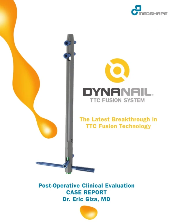
The Latest Breakthrough in TTC Fusion Technology Post-Operative Clinical Evaluation CASE REPORT Dr. Eric Giza, MD
Background DynaNail® TTC Fusion System is intended for use in tibiotalocalcaneal (TTC) fusion to treat various conditions including: • Rheumatoid Arthritis • Osteoarthritis • Severe Deformities • Degenerative Conditions • Post-Traumatic Injury • Failed Total Ankle Arthroplasty • Non-union from Ankle Arthrodesis Compressive Element DynaNail contains an internal Nickel Titanium (NiTiNOL) Compressive Element that adapts to changes in loading across the joint. The Element is stretched intra-operatively (up to 6 mm) and fixed with screws in the calcaneus and tibia. After surgery, the element can recover its stretched length in response to bone resorption or settling. This unloading allows DynaNail to sustain compression across the joint. Sliding Element Unstretched Position Element Stretched outside Outer Nail Body The figure to the left shows the axial compressive force applied by DynaNail in comparison to static intramedullary (IM) nails and an external fixation frame. Whereas static IM nails lose compression after 1 mm resorption, DynaNail maintains compression for up to 6 mm of resorption, similar to an external frame.1,2 1Yakacki et al. Pseudoelastic nailing for tibio-talo-calcaneal arthrodesis. Expert Rev Med Devices, 2011; 8(2):159-166. 2Data on File. MedShape, 2013. 2
Watch the Internal Element “In Action” The unloading of the element can be tracked by X-ray or CT scan using several methods: 1. In its fully stretched position, the Sliding Element extends plantarly from the outer body of the Nail. As resorption/settling occurs, the Sliding Element will retract back into the Nail Body. Lucent Region Immediate Post-Op: Sliding Element extending from Nail Body 2. The position of the calcaneal screws in their associated slots can be visualized under fluoroscopy and used to determine the extent the Compressive Element has unloaded. When the Compressive Element is in its fully stretched position, the calcaneal screws are located at the distal end of the slots (1) and a lucent region appears above the L-M screw (see X-ray above). As the Compressive Element unloads, the screws progressively shift upwards. The element has completely unloaded when the screws are at the proximal end of the slot and the lucent region disappears (2). 1 2 3
Introduction In many TTC fusion cases, the patient experiences severe bone loss due to a failed total ankle replacement, avascular necrosis, neuroarthropathy or infection. In these instances, a bone graft is used to fill any bony defects or replace a missing talus. However, achieving fusion using bone graft materials has proven challenging in high-risk patients with degenerative bone conditions or who are immuno-compromised. A study by Jeng et al. reported a 50% non-union rate in patients who underwent TTC fusions using a femoral head allograft and in particular, no fusions in diabetic patients.3 This report presents two cases with diabetic patients who both underwent a TTC fusion procedure after previous treatment proved inadequate. Their successful outcomes using the DynaNail TTC Fusion System in combination with bone graft materials are reported here. Performing Surgeon Dr. Eric Giza, MD University of California, Davis Medical Center Sacramento, CA Case Report #1 Background Information The patient, a 66 year old female, with a history of diabetes mellitus, spinal cord injury, ankle osteoarthritis, and flail ankle, previously underwent an attempt at posterior tibial tendon transfer and lateral ligament reconstruction. Procedure A lateral extensile approach was used to prepare the joints and perform a fibular ostectomy. An 11 mm tunnel was drilled through the calcaneus into the tibia, with a 13 mm tunnel in the calcaneus to accommodate the distal end of the implant. A 10 x 220 mm DynaNail was inserted according to the recommended surgical technique. The Compressive Element was stretched to a setting of 6 mm before fixating with screws. A 70 mm headless posterior-anterior (P-A) screw and 40 mm headed cortical L-M screw were used in the calcaneus. The proximal and distal tibial cortical screws used in the medial-lateral direction were both 25 mm. Fibular autograft and BMP-7 were used. An intra-operative X-ray image is to the right. 1 mm of Compressive Element unloading was noted, likely due to initial settling. 3Jeng CL, Campbell JT, Tang EY, Cerrato RA, Meyerson MS. Tibiotalocalcaneal arthrodesis with bulk femoral head allograft for salvage of large defects in the ankle. Foot and Ankle International, 2013. 34(9): 1256-1266. 4
Results Immediate Post-Op: No post-op complications were reported. The patient was placed in a posterior splint. 2 Weeks Post-Surgery: Week 2 P-A and M-L X-ray images are at the right. As observed on the radiograph, the position of the P-A screw in the calcaneus indicates that the Compressive Element has unloaded a total of 4 mm in response to bone resorption and settling while still maintaining compression. The wound had healed, and the patient was experiencing minimal pain with mild to moderate swelling. 6 Weeks Post-Surgery: The patient was no longer immobilized. A removable posterior splint was placed. 8 Weeks Post-Surgery: Minimal swelling was present and the patient reported no pain. 9 Weeks Post-Surgery: Ankle fusion was evident. The Compressive Element had unloaded a total of 5 mm while still maintaining compression. 12 Weeks Post-Surgery: 100% fusion was noted based on X-ray imaging. The patient was full weight-bearing in a boot with wean from boot over 1 month. 5
Case Report #2 Background Information The patient, a 68 year old female, had a history of diabetes mellitus type II with pilon ankle fracture approximately two years seven months prior to the current procedure. After initial operation, she developed Charcot neuroarthropathy with non-union and severe arthritis with deep infection. This led to a secondary operation approximately two months prior to the subject procedure, in which she had ankle incision and drainage, hardware removal and placement of antibiotic beads with placement of an external fixator/spatial frame. She received six weeks of intravenous antibiotics, but continued to experience non-union. Procedure The patient had a large resection of the tibia and talus. Femoral retrograde intra-articular aspiration of bone graft was performed. The 10 x 220 mm DynaNail was placed through a Zimmer Trabecular Metal™ knee augment implant and then coated in autograft and BMP-7. The Compressive Element was stretched to a setting of 6 mm before the nail was fixed in the calcaneus with a 70 mm P-A screw and a 45 mm L-M screw, and proximally in the tibia with two 25 mm screws in the M-L direction. An intra-operative X-ray image is shown to the right. Results Immediate Post-Op: No complications were reported, and a removable posterior splint was placed. Immediate post-op X-rays are to the right. 6
5 Weeks Post-Surgery: The Compressive Element had unloaded a total of 5 mm in response to bone resorption and settling while compression. The patient's wound had healed. still maintaining 6 Weeks Post-Surgery: Ankle fusion was evident. Minimal pain was reported by the patient and minimal swelling was observed. 10 Weeks Post-Surgery: The patient was no longer immobilized and began weight-bearing in a boot. No pain was reported by the patient. 13 Weeks Post-Surgery: The Compressive Element had continued to be unloaded a total of 5 mm in response to bone resorption and settling while still maintaining compression. consolidation of autograft around implant had occurred as observed by X-ray. There was no report of patient pain or swelling. Near-complete 7
Dr. Eric Giza, MD, is an Associate Professor in the Department of Orthopaedic Surgery at the University of California, Davis in Sacramento, CA. Dr. Giza is also Chief of the Orthopaedics Department's foot and ankle service, where he specializes in reconstruction and minimally invasive arthroscopy of the foot and ankle complex. Dr. Giza received his Doctor of Medicine degree from Temple University in Philadelphia, PA. He then completed a Combined Orthopaedic Residency at Harvard University followed by a Fellowship at St. Vincent's and North Shore Private Hospitals in Sydney, Australia, and a Fellowship at the Santa Monica Orthopaedic and Sports Medicine Group In Santa Monica, CA. Dr. Giza is Board Certifed by the American Board of Orthopaedic Surgery and is a member of the American Academy of Orthopaedic Surgeons and a member of the American Orthopaedic Foot and Ankle Society. This report is presented to demonstrate the clinical outcomes shown by Dr. Eric Giza. MedShape, as the manufacturer of this device, does not practice medicine and is not responsible for selection of the appropriate surgical technique to be utilized for an individual patient. Always refer to the package insert, product label, and/or product instructions prior to using a MedShape product. For further product information or to arrange a product demonstration, please contact your local MedShape representative or call Customer Service at 877-343-7016. You can also visit www.medshape.com. MedShape, Inc. 1575 Northside Drive, NW Suite 440 Atlanta, GA 30318 T: 877-343-7016 F: 877-343-7017 CAUTION: Federal (USA) law restricts this device to sale by or on the order of a physician. ©MedShape, Inc., 2014. All rights reserved. Printed in the USA. Protected by Patent No.: US 7,985,222. Other U.S. and International Patents Pending. MK-10110 Rev 00. Issued 03/2014. DynaNail is a registered trade- mark of MedShape, Inc. All other trademarks are trademarks of their respective owners or holders. 8