1 / 20
200 likes | 204 Views
Students should be able to: <br>- identify, with the aid of diagrams, the main stages of mitosis

E N D
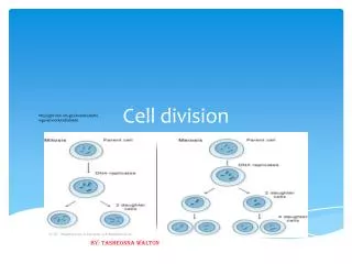
I nterphase • Is the non-dividing stage of a cell (resting stage) • During interphase, 1. Cells carry out activities like absorbing nutrients and building up protoplasm 2. The chromatin threads replicate, producing 2 identical chromatin threads (DNA molecules) 3. Centrioles divide in an animal cell (Note: Centrioles are absent in plant cells)
How a chromosome is formed - Chromosomes appear as long thin threads called chromatin (chromatin threads) Before cell division, - The chromatin threads replicate, producing 2 identical chromatin threads (DNA molecules) - The 2 chromatin threads coil and shorten to become chromosomes -Each chromosome consists of 2 identical DNA molecules (sister chromatids) -The chromatids are joined at a point (centromere)
How a chromosome is formed prophase interphase
A chromosome is formed from the condensation, coiling and shortening of chromatin threads
Structure of a chromosome - the point where the two chromatids touch, and where the microtubules attach - one of the two identical parts of the chromosome after S phase
star-shaped structure formed in the cytoplasm of a cell having fibers like rays that surround the centrosome during mitosis Prophase 1. The nucleolus disappears 2. The nuclear envelope disappears 3. The chromatin threads condense, coil and shorten to become chromosomes 4. Asters (made up of microtubules) form around centrioles 5. The 2 pairs of centrioles move apart to opposite ends of the cell 6. A spindle forms with the spindle fibers extending from one pole to the other
Metaphase 1. Chromosomes line up around the equator of the spindle 2. The centromere of each chromosome is attached to a spindle fibre
Attachment of microtubules to kinetochore - is the protein structure at the centromere of the chromosome where the spindle fibers attach during division to pull the chromosomes apart
Anaphase 1. Each centromere splits 2. The spindle fibres pull the chromatids apart to opposite poles of the cell 3. Once the chromatids are separated, they are called daughter chromosomes
Telophase 1. Spindle fibres breaks down 2. A nuclear envelope forms around the chromosomes at each pole of the cell 3. A nucleolus forms and the chromosome uncoil and lengthen to become thin chromatin threads
Cytokinesis = division of the cytoplasm 1. Furrows (cleavage) appear in the cytoplasm between the two nuclei 2. The furrows deepen and two identical daughter cells are produced
3. 1. 2. Test yourself 6. 7. 8. 9. 4. 5. ______________ Anaphase