Endoscopic Sinus Surgery
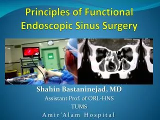
Endoscopic Sinus Surgery. Section 7 ( قسمت هفتم فایل ). Bakhshaee M, MD Rhinologist, Assistant Prof. MUMS. When a high-resolution CT scan has shown the skull base defect, MRI can help define any pathology associated with a CSF leak, e.g., brain, hematoma, CSF. Inverted Papilloma.


Endoscopic Sinus Surgery
E N D
Presentation Transcript
Endoscopic Sinus Surgery Section 7 (قسمت هفتم فایل) Bakhshaee M, MD Rhinologist, Assistant Prof. MUMS
When a high-resolution CT scan has shown the skull base defect, MRI can help define any pathology associated with a CSF leak, e.g., brain, hematoma, CSF.
Inverted Papilloma • MRI can complement CT in determining how extensive an inverted papilloma is. • It defines how much of the opacification shown on CT is due to secretions and how much is due to the tumor. This is important in planning surgery if it involves the frontal or maxillary sinus.
MR Angiography • MR angiography (MRA) delineates flow within vessels by suppressing the signal from stationary tissue. • It is performed as part of an MR examination and does not necessarily require contrast injection. • MRA provides information with respect to the principal feeding arteries in vascular tumors (such as angiofibromas, hemangiopericytomas, or paragangliomas, and certain metastases) and in vascular lesions (such as angiomas or aneurysms located at the skull base).
A negative result on MRA does not definitively rule out a vascular lesion. • A minor amount of flow or the presence of slow flow such as in a capillary hemangioma, esthesioneuroblastoma, and even in an angiofibroma may be invisible to MRA.
Digital Subtraction Angiography • Digital subtraction angiography (DSA) is the best method to delineate the vascular supply of a specific anatomical area or lesion • In a lesion (e.g., chordoma or neoplasm) that abuts the internal carotid artery at the foramen lacerum or within the cavernous sinus. • In recurrent epistaxis, DSA may be required as a means to verify the source of repeated hemorrhage.
Points to Mention on a CT Request • Write down the provisional diagnosis, e.g., “severe polyposis unresponsive to medical treatment.” • Say why you want the scan, e.g., to define the anatomy before surgery. • Detail what surgery has been done. • Ask for fine cuts if indicated, e.g., in case of a CSF leak or when sagittal reconstruction is needed. • Name the area you want examined, e.g., the frontal recess. • If you suspect a tumor, say so and ask for a contrast study.
what should you do before operating? • Minimize the amount of surgical manipulation required. • Preserve as much olfactory mucosa as possible. • Reduce peroperative bleeding to reduce the likelihood of complications. • Work out the surgical anatomy in order to minimize the chance of entering the orbit or skull base. • Set clear goals for yourself and your patient
The Preoperative Checklist • Confirm the diagnosis. • Review previous medical treatment. • Optimize the immediate preoperative condition. • Check that relevant investigations have been done ( Allergy tests, Immune status, Hematological parameters, Olfaction, Vision) • Review the relevant medical history, e.g., drug allergies, medication. • Preoperative CT checklist. • Planning and staging the procedure. • Informed consent.
Vision • Left enophthalmos due to silent sinus syndrome—involution of the maxillary sinus with collapse of its roof
The loss of color discrimination, particularly of red, is a worrying symptom of pressure on the optic nerve, and this requires urgent treatment. • For any orbital surgery, e.g., orbital decompression, an ophthalmological assessment is required. • It is alarming if a unilaterally enlarged is noticed during or after surgery.
Preoperative CT Checklist • Like an airline pilot before preparing for take-off, you must go through a systematic check of the CT scanvbefore surgery so as to avoid the surgical equivalent ofva crash.
Step 1. • When placing the scans on the viewing box, orientate the scan sequence from anterior to posterior and ensure that the sides are marked and placed as though as you are looking at the patient. • Follow the cuts anterior to posterior; follow the septum, note any deviation, and look for the size and extent of the ethmoidal bulla, which is a relatively consistent landmark.
Step 2. • Examine the lamina papyracea, uncinate process, and middle turbinate.
Localize the uncinate process (arrow) from its free margin posteriorly and follow it anteriorly and upward
A key aspect of frontal recess surgery is to define the insertion of the uncinate process as this may also “guard” anterior access to the frontal recess by forming a web if it attaches to the skull base or middle turbinate
Step 3. • Examine the area of the frontal recess. The frontal recess lies anterior and superior to the ethmoid bulla.
Step 4. • Determine the height of the skull base
Step 5. • Examine the sphenoid sinus
Informed Consent • The following issues need to be addressed. • What are the options available to the patient? • Specifically what would happen if no surgery were undertaken? • What is the patient’s prognosis with the various treatment strategies? • What does the surgery involve? • What are the complications of surgery? • This should include complications occurring more frequently than 1 in 100, and severe complications even if they are rare.
How much do we need to explain to our patients to properly gain their consent? • The surgeon may feel that mentioning complications to the patient will frighten them unnecessarily, but it is possible to mention even serious complications in the right context without causing alarm, and it is our duty to do so.
Patients need to: • Understand their diagnosis • Understand the context of their symptoms in the light of their diagnosis • Understand the principles of the surgical procedure • Be informed about complications even if they are rare • Be informed about what they can expect in the postoperative period: the healing process, the symptoms they can expect, the medical therapy they should take, and the need for time off work
Time Off Work • Rest: • After minor surgery, such as a limited anterior ethmoidectomy: one week • If they work in dusty or smoky environment, this should be extended by a further week • Patients who have had more extensive sinus surgery are advised to take 2 weeks off work.
Advice about Flying • Fly : • Some authorities have advised that it is wise to wait up to 6 weeks after surgery, • if patients are able to do a Valsalva maneuver
Complications • Our aim is to inform the patient without alarming them unnecessarily • We say that the reported risk of any moderate or serious complication is approximately 0.5% to 1% • On reviewing the world literature on the prevalence of complications associated with endoscopic sinus surgery, it is worth mentioning that these are no more common than with conventional surgery
External Incision • When undertaking frontal recess surgery, and in particular revision surgery, or when a median drainage procedure is planned, it is worth mentioning the possibility of the need for an external incision • For vascular tumors of the lateral nasal wall, such as an angiofibroma, it is important to mention that an external approach such as a lateral rhinotomy or midfacial degloving may be required.
Inverted Papilloma • In the case of accompanied SCC; more radical procedure may be required. • The surgeon should aim to remove all the diseased mucosa there is an increased risk of stenosis, particularly in the frontal recess. • The incidence of recurrent disease is as high as 30%.
Local Osteitis • A rare complication is local osteitis caused by exposure of bone. • It produces a dull, severe nagging ache that lasts for 10 days before abating. • Major analgesics are required, and local treatment appears to provide little help.
Infection • Infection following surgery is rare and can be minimized by giving perioperative antibiotics when purulent disease is present.
Surgical emphysema • Caused by air being forced through a defect in the lamina papyracea, is avoided if the surgeon advises the patient not to blow their nose or to stifle sneezes for 4 days after surgery
Visual Complications • If a patient has significant proptosis or displacement of the axis of their pupils due to paranasal sinus disease (e.g., a mucocele), they may have adapted slowly to these changes over several weeks and not have any diplopia. • Occasionally, patients may have some temporary diplopia after surgery when this displacement is suddenly corrected, and it is worth mentioning this before surgery
Diplopia in orbital decompression • Patients who undergo orbital decompression are at an increased risk of diplopia, although maintaining the medial−inferior strut of bone between the medial wall and floor of the orbit minimizes this risk.
Recurrent Polyposis • When counseling a patient with nasal polyposis associated with late-onset asthma or aspirin sensitivity, it is wise to mention that, in spite of good surgery and postoperative medical treatment, the majority of patients will have a recurrence of their polyps