Download
1 / 18
420 likes | 3.39k Views
Intraventricular Hemorrhage in the Neonate. NICU Night Team Curriculum. Objectives. To describe the epidemiology and risk factors of IVH To understand the anatomy and pathophysiology involved in IVH To demonstrate the grading system of IVH To comprehend the sequelae of high-grade IVH. Case.

E N D
Intraventricular Hemorrhage in the Neonate NICU Night Team Curriculum
Objectives • To describe the epidemiology and risk factors of IVH • To understand the anatomy and pathophysiology involved in IVH • To demonstrate the grading system of IVH • To comprehend the sequelae of high-grade IVH
Case • Two day old 750g 25 4/7 week GA male born by vertex, vaginal delivery to a mother who received good PNC after rupture of membranes 10 hours prior to delivery and precipitous labor. Otherwise healthy pregnancy. • Medications during pregnancy: • Prenatal vitamin • Ampicillin during labor for unknown GBS status x 2 doses • Betamethasone x 2 • Delivery Course: • NSVD, nuchal cord x1, easily reduced • Apgars: 51, 85 • Initially given Positive pressure ventilation then intubated • Given one dose of surfactant in delivery room • Transferred to ICN and placed on SIMV with volume control
Case (cont.) • Vital signs remained stable through Days 0 and 1 • Fluids given through TPN at total fluids: 120 cc/kg/d • Patient started on Ampicillin and Gentamicin until blood culture negative x 48 hours • Chemistries stable, checked q12h • Admission hemoglobin was 12.4, but dropped to 8.6 at 48 hours of life WHAT IS THE DIFFERENTIAL DIAGNOSIS FOR ACUTE DROP IN HEMOGLOBIN IN A NEONATE?
Differential Diagnosis • Blood Loss • Cephalohematoma • Intraventricular Hemorrhage • Subdural Hemorrhage • Subgaleal • Liver Rupture • Spleen Rupture • Pulmonary Hemorrhage • Iatrogenic • Hemolysis Secondary to Infection • Sepsis • Viral • Bacterial • TORCH
In our patient, ultrasound revealed: Grade III IVH
Epidemiology and Risk Factors • Among extremely premature babies: • IVH rates have declined from 50-80% in 1980s to current rate of 10-15%1 • Continues to be a significant problem as improved survival of extremely premature infants has resulted in more survivors with this injury What are the major risk factors for IVH? • Volpe JJ. Neurology of the Newborn, 4th Edition, WB Saunders, Philadelphia 2001. p 428
Risk Factors for IVH • Prematurity: • Occurs most frequently in infants born <32 weeks or <1500g • Incidence is 26% for infants weighing 501-1500g2 • The highest prevalence is in the least mature infants • Mortality: ~15%2 (worse prognosis with increasing severity) 2. Timing: • Virtually all IVH in premature infants occurs in first five days2 • 50, 25, and 15% on the first, second and third day, respectively2 • By the end of the first week, 90% of hemorrhages can be detected3 • Other Risk Factors3: • Intrapartum asphyxia • Chorioamnionitis • Hypoxemia • Hypercarbia • Pnuemothorax/Pulmonary Hemorrhage • Maternal fertility treatment • Vermont Oxford Network. 2002. table 1.2 • Volpe JJ. Neurology of the Newborn, 4th Edition, WB Saunders, Philadelphia 2001. p 428 • McCrea HJ and Ment LR. 2008. Clinical Perinatolgy (35(4): 777-vii
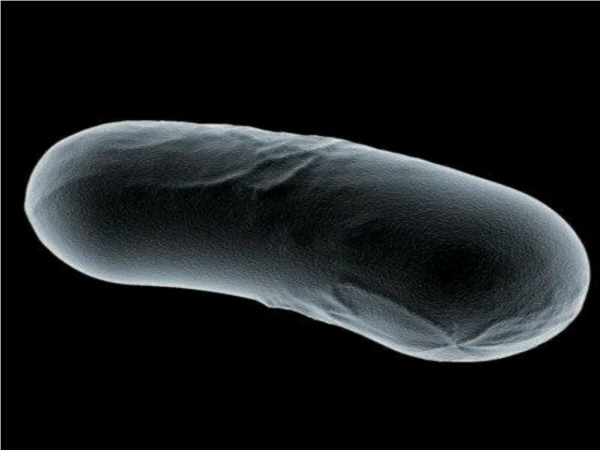
Anatomy and Pathophysiology • The Germinal Matrix in Premature Infants1 • The germinal matrix is a highly cellular layer in the subependymal, subventricular zone that gives rise to neurons and glia during development • The immature germinal matrix is highly vascularized by fragile microvessels • In response to hypertension, hypoxia, hypercapnia or acidosis, cerebral blood flow increases placing stress on vasculature • Hemorrhage in preterm babies often begins in the germinal matrix and may extend to ventricular system Arterial supply to the subependymal germinal matrix at 29 weeks gestation2 1. McCrea HJ and Ment LR. 2008. Clinical Perinatolgy (35(4): 777-vii 2. Image from Hambleton G, and Wigglesworth JS. 1976. Archives of Diseases in Childhood 51(9): 651.
How is the diagnosis of IVH made? • Cranial Ultrasound is the screening method of choice • Up to half of infants with IVH may be asymptomatic • According to the American Academy of Neurology and the Practice Committee of Child Neurology Society: • Routine ultrasound should be performed on all infants <30weeks gest age • Screening should be performed a 7-14 days and repeated at 36-40 weeks • CT or MRI offer no advantage in detection of IVH
Grading IVH Image from www.radiologyassistant.nl
Grade That Hemorrhage!! • Where is the hemorrhage? • Name the Anatomy • Grade the Hemorrhage Normal Anatomy Germinal Matrix Extension into Lateral Ventricle (no dilation) Choroid Plexus Grade II Hemorrhage Extension into lateral ventricle without dilation Image from www.pediatriceducation.org
Name the Sequelae of Intraventricular Hemorrhage 1. 2. 3. 4. 5.
Sequelae of IVH • Posthemorrhagic Hydrocephalus • Usually presents with rapid increases in head circumference • Signs and symptoms may not be evident for several weeks due to compliance of neonatal brain • Believed to be due to impaired CSF reabsorption following the inflammation related to blood in the CSF • No proven effective interventions have been described to date Serial cranial ultrasounds and MRI studies from a preterm male infant born at 24 weeks of gestation. The initial diagnosis of Grade 3 IVH at age 3 days (panel A) was followed by parenchymal involvement of hemorrhage, or Grade 4 IVH, on postnatal day 4 (arrow, panel B). A cranial ultrasound performed on day 10 because of increasing occipitofrontal head circumference and full fontanel revealed bilateral ventriculomegaly, residual intraventricular blood and a developing porencephaly (arrow, panel C). MRI study at 2 months demonstrated ventriculomegaly (panel D). Because of excessive increase in head circumference and increasing spasticity, the patient underwent third ventriculostomy following MRI scan at age 6 months (panel E). McCrea HJ and Ment LR. 2008. Clinical Perinatolgy (35(4): 777-vii
Sequelae of IVH • Periventricular Leukomalacia (PVL) • Classic white matter abnormality following IVH • Attributed to profound and long-lasting decreases in CSF • PVL may progress to porencepholy (Greek for “hole in the brain”) • Depending on severity, may lead to spastic dysplagia, visual defects, or cognitive impairment Serial cranial ultrasounds of a 30 week preterm male with Grade 3 IVH and hemorrhagic PVL at age 10 days (panel A). Repeat ultrasound 3 weeks later demonstrated unilateral ventriculomegaly and periventricular cystic cavities consistent with PVL (panel B). McCrea HJ and Ment LR. 2008. Clinical Perinatolgy (35(4): 777-vii
Sequelae of IVH • Seizures • Cerebral Palsy • Mental Retardation Risk for major neurological deficits: Grade I:5-9% Grade II: 11-15% Grade III: 30-40% Grade IV: 50-70%
Preventive Strategies Many potential pharmacologic interventions have been studied, though few are currently in wide use* *see notes section for more details Adapted from McCrea HJ and Ment LR. 2008. Clinical Perinatolgy (35(4): 777-vii
Conclusion • IVH remains a common problem of extreme prematurity • Hemorrhage often arises from vasculature of the germinal matrix • Severe neurodevelopmental sequelae can arise and is associated with increasing severity of the hemorrhage • Very few, if any, good preventive strategies are available