IHC Fixation
To succeed immunohistochemistry(IHC), fixation plays an important role. IHC fixation purposes, selection of fixing solution, methods and time are all available on immunostaining.info where you find an importance of IHC fixation and many other information related to fixation topics. Make a visit today and enhance your knowledge about IHC fixation.

IHC Fixation
E N D
Presentation Transcript
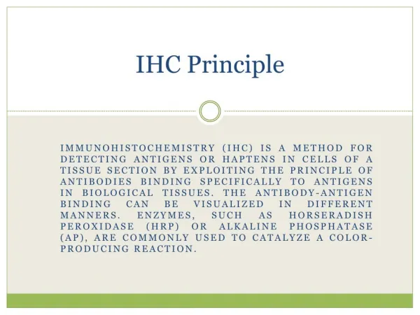
IHC Principle Immunohistochemistry (IHC) is a method for detecting antigens or haptens in cells of a tissue section by exploiting the principle of antibodies binding specifically to antigens in biological tissues. The antibody-antigen binding can be visualized in different manners. Enzymes, such as Horseradish Peroxidase (HRP) or Alkaline Phosphatase (AP), are commonly used to catalyze a color-producing reaction.
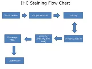
Introduction IHC is widely used in many research and clinical laboratories because this technique makes it possible to visualize the distribution and localization of specific cellular components within cells and in proper tissue context. There are numerous IHC methods that can be used to localize antigens. The method selected should include consideration of parameters such as the specimen types and assay sensitivity.
FIXATION Purposes Keep cell sharp and tissue shape to prevent postmortem autolysis, putridness, endogenic and exogenic enzyme activity Maintain cell structure and position by preventing antigen diffusion through transfer of protein, fat, sugar and enzymes of cell into insoluble substances Precipitate and curdle materials in tissue to produce different refraction Indurate tissues to enhance working with glass slides Prevent cell from shrinking and swelling Give color to clarify tissues by different affinity to coloring agent
Selection of Fixing Solution Below is a list of commonly used fixing solutions. You may need to test whether a specific type of solution is appropriate for your detected antigens because there is no standard fixing solution for different kinds of antigen immobilization. Acetone and Alcohol These two types of solutions, which are primary fixing solutions, play a role of precipitating sugars and fat as well as maintain the immunologic competence.
Alcohol is ineffective to maintain low molecular weight protein, polypeptide and cytoplasmic proteins. However, it can be mixed with glacial acetic acid, ethyl ether, chloroform and formaldehyde. Acetone is often used for frozen tissue and cytological smears because it has a strong penetrability and dehydration property.
Aldehyde It is a di-functional cross-linking agent which is widely used due to its strong penetrability, low contractibility and low background. It helps keep the cross-linking between tissues and maintain antigen. • Formalin (10% neutral buffered) is the most widely used • 4% paraformaldehyde is better than formaldehyde • Bouin’s solution (containing picric acid) is the most widely used in histology and pathology • Zamboni’s solution is applied to light and electron microscopic immunocytochemistry and is better than formaldehyde in ultrastructural organization maintenance
Non-Aldehyde Carbodiimide, dimethylacetamide, dimethyl-suberimidate, para-benzoquinone are widely used in tissue fixation of peptide hormones. These fixation agents are better mixed with glutaricdialdehyde or paraformaldehyde. In recent years, a new type of formaldehyde-free fixing solution has become available. With low toxicity and degradable chemical agent, this solution has gained a broad popularity in IHC, regular pathological examinations and molecular pathology detections due to the use of non-protein cross linking, strong DNA/RNA preservation, and absence of cell vacuole, tissue shrinkage and pyknosis.
Method and Time Method: Immersion The immersion method marinates the tissue in fixing solution (at 4℃ if needed) for a specified period which is determined by the antigen stability and type of fixing solution used. Biopsy and surgical specimens as well as other non-irrigation tissues commonly employ this fixation method.
Method: Irrigation This method has the ability to fix tissues fully and quickly, suppressing the interference of endogenous peroxidase. Therefore, it is a method of choice in animal experiments.
Fixation Time The fixation time depends on the tissue thickness, solution concentration and experimental temperature. In principle, the time is directly proportional to the tissue thickness but inversely proportional to the solution concentration.
Exercise Caution When Fixating Tissues Do not over-fix the tissues Keep the tissues fresh after fixation Use enough fixing solution and wash it off completely after fixation Use tissues of size less than 2 cm × 1.5 cm × 0.3 cm (Thickness < 0.3 cm)