Download
1 / 4
40 likes | 61 Views
Cancer and thromboembolism relationship has been acknowledged for many years. Pulmonary embolism is associated with significant morbidity and mortality in patients with cancer. A more significant line of research needs to be done to investigate thromboembolism in cancer. Ultrasonography is a reliable diagnostic bedside test for PTE in critically ill and immobile patients. In this review, we want to display a case with both obstructive and hypovolemic shock and how ultrasonography can assist us to diagnose it

E N D
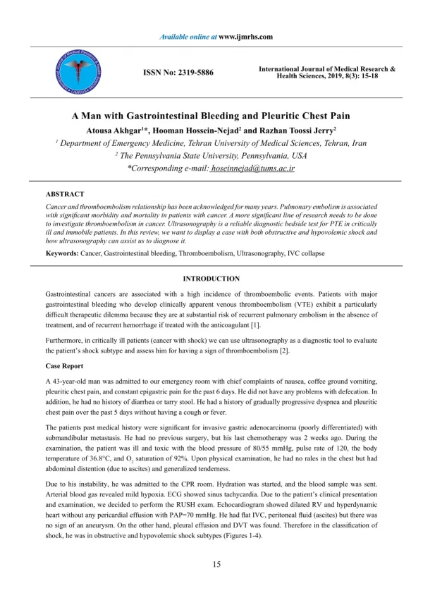
Available online at www.ijmrhs.com InternationalJournalofMedicalResearch&HealthSciences •IJMRHS • International Journal of Medical Research & Health Sciences, 2019, 8(3): 15-18 ISSN No: 2319-5886 A Man with Gastrointestinal Bleeding and Pleuritic Chest Pain Atousa Akhgar1*, Hooman Hossein-Nejad2 and Razhan Toossi Jerry2 1 Department of Emergency Medicine, Tehran University of Medical Sciences, Tehran, Iran 2 The Pennsylvania State University, Pennsylvania, USA *Corresponding e-mail: hoseinnejad@tums.ac.ir ABSTRACT Cancer and thromboembolism relationship has been acknowledged for many years. Pulmonary embolism is associated with significant morbidity and mortality in patients with cancer. A more significant line of research needs to be done to investigate thromboembolism in cancer. Ultrasonography is a reliable diagnostic bedside test for PTE in critically ill and immobile patients. In this review, we want to display a case with both obstructive and hypovolemic shock and how ultrasonography can assist us to diagnose it. Keywords: Cancer, Gastrointestinal bleeding, Thromboembolism, Ultrasonography, IVC collapse INTRODUCTION Gastrointestinal cancers are associated with a high incidence of thromboembolic events. Patients with major gastrointestinal bleeding who develop clinically apparent venous thromboembolism (VTE) exhibit a particularly difficult therapeutic dilemma because they are at substantial risk of recurrent pulmonary embolism in the absence of treatment, and of recurrent hemorrhage if treated with the anticoagulant [1]. Furthermore, in critically ill patients (cancer with shock) we can use ultrasonography as a diagnostic tool to evaluate the patient’s shock subtype and assess him for having a sign of thromboembolism [2]. Case Report A 43-year-old man was admitted to our emergency room with chief complaints of nausea, coffee ground vomiting, pleuritic chest pain, and constant epigastric pain for the past 6 days. He did not have any problems with defecation. In addition, he had no history of diarrhea or tarry stool. He had a history of gradually progressive dyspnea and pleuritic chest pain over the past 5 days without having a cough or fever. The patients past medical history were significant for invasive gastric adenocarcinoma (poorly differentiated) with submandibular metastasis. He had no previous surgery, but his last chemotherapy was 2 weeks ago. During the examination, the patient was ill and toxic with the blood pressure of 80/55 mmHg, pulse rate of 120, the body temperature of 36.8°C, and O2 saturation of 92%. Upon physical examination, he had no rales in the chest but had abdominal distention (due to ascites) and generalized tenderness. Due to his instability, he was admitted to the CPR room. Hydration was started, and the blood sample was sent. Arterial blood gas revealed mild hypoxia. ECG showed sinus tachycardia. Due to the patient’s clinical presentation and examination, we decided to perform the RUSH exam. Echocardiogram showed dilated RV and hyperdynamic heart without any pericardial effusion with PAP=70 mmHg. He had flat IVC, peritoneal fluid (ascites) but there was no sign of an aneurysm. On the other hand, pleural effusion and DVT was found. Therefore in the classification of shock, he was in obstructive and hypovolemic shock subtypes (Figures 1-4). 15
Akhgar, et al. Int J Med Res Health Sci 2019, 8(3): 15-18 Figure 1 IVC collapsibility on inspiration and expiration Figure 2 The apical 4-chamber view is displaying the right ventricular dilation Figure 3 Short axis view is showing flattening or bowing of the intraventricular septum into the LV 16
Akhgar, et al. Int J Med Res Health Sci 2019, 8(3): 15-18 Figure 4 Subxiphoid view is showing hyperdynamic heart and no pericardial effusion After an hour of admission, the patient had a cardiorespiratory arrest, and resuscitation attempts were unsuccessful. DISCUSSION This case demonstrated the application of bedside ultrasound in the diagnosis of massive PE in a patient with cancer and active gastrointestinal bleeding. Management of the critically ill patient in shock requires rapid identification of its etiology. Common mechanisms of shock are hypovolemia, cardiac, distributive, and mechanical [3]. The use of bedside ultrasound for evaluation of patients admitted to the emergency department with signs and symptoms of pulmonary embolism has been controversial [4]. There are multiple sonographic findings that support the diagnosis of acute pulmonary embolism: visualization of a free-floating thrombus in the right heart or pulmonary artery, right ventricular dilation (RV/LV ratio>0.6-1:1), abnormal septal wall motion and D shape septum in short axis, right ventricular systolic dysfunction, McConnell’s sign and IVC dilation without inspiratory collapse (Plethoric IVC) [4]. On the other side, in hypovolemic shock, we have small collapsed IVC and hyperdynamic left ventricle. We should mention here that the hyperdynamic heart can also overestimate the ejection fraction. Therefore, we may need IV fluids and blood replacement [3]. CONCLUSION Bedside ultrasound may help differentiate between the etiologies of hypotension in the unstable patient, but there are 2 things that we would like to mention. First of all, a hyper dynamic heart (tachycardia) can overestimate the ejection fraction. The other point which we would like to mention here is that in patients with pulmonary thromboembolism in the hypovolemic state, considering the IVC collapsibility index alone is not appropriate for assessing massive PTE and volume status estimation. DECLARATIONS Conflict of Interest The authors declared no potential conflicts of interest with respect to the research, authorship, and/or publication of this article. REFERENCES [1] Nieto, J. A., et al. “Acute venous thromboembolism in patients with recent major bleeding. The influence of the site of bleeding and the time elapsed on the outcome.” Journal of Thrombosis and Haemostasis, Vol. 4, No. 11, 2006, pp. 2367-72. [2] Perera, Phillips, et al. “The RUSH exam 2012: Rapid ultrasound in shock in the evaluation of the critically ill patient.” Ultrasound Clinics, Vol. 7, No. 2, 2012, pp. 255-78. 17
Akhgar, et al. Int J Med Res Health Sci 2019, 8(3): 15-18 [3] Lyon, Mathew, and Neha Verma. “Ultrasound guided volume assessment using inferior vena cava diameter.” The Open Emergency Medicine Journal, Vol. 3, 2010, pp. 22-24. [4] Patel, Adarsh N., et al. “The use of bedside ultrasound in the evaluation of patients presenting with signs and symptoms of pulmonary embolism.” Case Reports in Emergency Medicine, 2013. 18