Download

1 / 2
0 likes | 284 Views
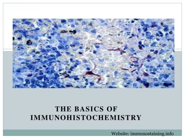
Immunohistochemistry (IHC) is a powerful technique widely employed in biological research and diagnostic pathology, allowing researchers to visualize and analyze the distribution of proteins within tissues. This article provides a comprehensive overview of an effective immunohistochemistry protocol and highlights key considerations for successful IHC staining.

E N D
Mastering Immunohistochemistry: A Comprehensive Guide to IHC Staining Protocols Immunohistochemistry (IHC) is a powerful technique widely employed in biological research and diagnostic pathology, allowing researchers to visualize and analyze the distribution of proteins within tissues. This article provides a comprehensive overview of an effective immunohistochemistry protocol and highlights key considerations for successful IHC staining. Understanding the Basics: Before diving into the protocol, it's crucial to grasp the basics of immunohistochemistry. This technique involves the use of antibodies to detect specific antigens in tissue sections. The first step is sample preparation, where tissues are fixed, embedded, and sectioned. Proper fixation is essential for preserving cellular structures and antigen city. Optimizing IHC Staining Protocols: Achieving accurate and reproducible results begins with a well-optimized staining protocol. Considerations include antigen retrieval methods, blocking steps to minimize nonspecific binding, and selecting appropriate primary and secondary antibodies. Time and temperature play pivotal roles in each stage of the protocol, influencing the sensitivity and specificity of the staining.
Antigen Retrieval: To enhance antigen accessibility, researchers often utilize heat-induced antigen retrieval methods. This step is critical for unmasking epitopes that may be masked during tissue fixation, enabling better antibody binding. Blocking and Antibody Selection: Nonspecific binding can compromise the specificity of IHC staining. Blocking agents, such as serum or bovine serum albumin (BSA), help reduce background staining. Careful selection of primary and secondary antibodies ensures accurate detection of the target antigen. Visualization and Imaging: After completing the staining protocol, the next step is visualization. DAB (3,3'-diaminobenzidine) is a commonly used chromogen, producing a brown precipitate at the site of antibody binding. Counterstaining with hematoxylin allows for better contrast and visualization of cellular structures. Troubleshooting Tips: Even with a well-established protocol, challenges may arise. Common issues include high background staining, weak signal, or inconsistent results. Regularly reviewing and adjusting the protocol can address these issues and enhance the overall reliability of the staining. In conclusion, mastering immunohistochemistry requires a thorough understanding of the protocol's intricacies. By following optimized procedures and considering key factors, researchers can unlock the full potential of IHC staining for accurate and insightful analyses of tissue samples. Address : Unit D, 3/F., Freder Centre Mok Cheong Street, Tokwawan, Hong Kong Ph No : 13808832613 Email : info@ihc-prs.com Website : https://ihc-prs.com/