Download
1 / 53
530 likes | 731 Views
Chapter 27. The Cardiovascular System. PowerPoint® presentation to accompany: Medical Assisting Third Edition Booth, Whicker, Wyman, Pugh, Thompson. Learning Outcomes. 27.1 Describe the structure of the heart and the function of each part. 27.2 Trace the flow of blood through the heart.

E N D
Chapter 27 The Cardiovascular System PowerPoint® presentation to accompany: Medical Assisting Third Edition Booth, Whicker, Wyman, Pugh, Thompson
Learning Outcomes 27.1 Describe the structure of the heart and the function of each part. 27.2 Trace the flow of blood through the heart. 27.3 List the most common heart sounds and what events produce them. 27.4 Explain how heart rate is controlled by the electrical conduction system of the heart.
Learning Outcomes (cont.) 27.5 List the different types of blood vessels and describe the functions of each. 27.6 Define blood pressure and tell how it is controlled. 27.7 Trace the flow of blood through the pulmonary and systemic circulation. 27.8 List the major arteries and veins of the body and describe their locations.
Learning Outcomes (cont.) 27.9 List and describe the components of blood. 27.10 Give the functions of red blood cells, the different types of white blood cells, and platelets. 27.11 List the substances normally found in plasma. 27.12 Explain how bleeding is controlled. 27.13 Explain the differences among blood types A, B, AB, and O.
Learning Outcomes (cont.) 27.14 Explain the difference between Rh-positive blood and Rh-negative blood. 27.15 Explain the importance of blood typing and tell which blood types are compatible. 27.16 Describe the causes, signs and symptoms, and treatments of various diseases and disorders of the cardiovascular system.
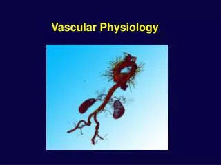
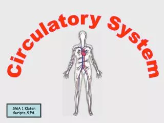
Introduction • The cardiovascular system consists of heart and blood vessels • Sends blood to • Lungs for oxygen • Digestive system for nutrients • CV system also circulates waste products to certain organ systems for removal from the blood
The Heart: Structures • Cone-shaped organ about the size of a loose fist • In the mediastinum • Extends from the level of the second rib to about the level of the sixth rib • Slightly left of the midline
The Heart: Structures (cont.) • Heart is bordered: • Laterally by the lungs • Posteriorly by the vertebral column • Anteriorly by the sternum • Rests on the diaphragm inferiorly
Heart coverings Pericardium Covers theheart and large blood vessels attached to the heart Visceral pericardium Innermost layer Directly on the heart Parietal pericardium Layer on top of the visceral pericardium Heart walls: Epicardium Outermost layer Fat to cushion heart Myocardium Middle layer Primarily cardiac muscle Endocardium Innermost layer Thin and smooth Stretches as the heart pumps The Heart: Structures (cont.) Click for Larger View
Four chambers Two atria Upper chambers Left and right Separated by interatrial septum Two ventricles Lower chambers Left and right Separated by interventricular septum The Heart: Structures (cont.) • Atrioventricular septum separates the atria from the ventricles Click for View of Heart
The Heart: Structures (cont.) • Tricuspid valve – prevents blood from flowing back into the right atrium when the right ventricle contracts • Bicuspid valve – prevents blood from flowing back into the left atrium when the left ventricle contracts • Pulmonary valve – prevents blood from flowing back into the right ventricle • Aortic valve – prevents blood from flowing back into the left ventricle Click for View of Heart
The Heart: Blood Flow Oxygenated blood out to body Deoxygenated blood in from body Oxygenated blood in lungs Deoxygenated blood out to lungs Atria Contract Ventricles Contract
Right Atrium TricuspidValve Right Ventricle PulmonaryValve Lungs Body AorticSemilunarValve Left Ventricle BicuspidValve Left Atrium PulmonarySemilunarValve The Heart: Blood Flow (cont.)
Right atrium contracts Tricuspid valve opens Blood fills right ventricle Right ventricle contracts Tricuspid valve closes Pulmonary semilunar valve opens Blood flows into pulmonary artery Left atrium contracts Bicuspid valve opens Blood fills left ventricle Left ventricle contracts Bicuspid valve closes Aortic semilunar valve opens Blood pushed into aorta The Heart: Cardiac Cycle • One heartbeat = one cardiac cycle • Atria contract and relax • Ventricles contract and relax
The Heart: Cardiac Cycle (cont.) • Influenced by • Exercise • Parasympathetic nerves • Sympathetic nerves • Cardiac control center • Body temperature • Potassium ions • Calcium ions
The Heart: Heart Sounds • One cardiac cycle – two heart sounds (lubb and dubb) when valves in the heart snap shut • Lubb – First sound • When the ventricles contract, the tricuspid and bicuspid valves snap shut • Dubb – Second sound • When the atria contract and the pulmonary and aortic valves snap shut
Group of structures that send electrical impulses through the heart Sinoatrial node (SA node) Wall of right atrium Generates impulse Natural pacemaker Sends impulse to AV node Atrioventricular node (AV node) Between atria just above ventricles Atria contract Sends impulse to the bundle of His Bundle of His Between ventricles Two branches Sends impulse to Purkinje fibers Purkinje fibers Lateral walls of ventricles Ventricles contract Link to Diagram The Heart: Cardiac Conduction System
Apply Your Knowledge ANSWER: C Match the following: __ Tricuspid valve A. Two branches; sends impulse to Purkinje fibers __ Bicuspid valve B.Covering of the heart and aorta __ Pericardium C. Between the right atrium and the right ventricle __ SA node D. In the lateral walls of ventricles __ Bundle of His E. Natural pacemaker __ Purkinje fibers F. Between the left atrium and the left ventricle F B E A D Good Job!
Strongest of the blood vessels Carry blood away from the heart Under high pressure Vasoconstriction Vasodilation Arterioles Small branches of arteries Aorta Takes blood from the heart to the body Coronary arteries Supply blood to heart muscle Blood Vessels: Arteries and Arterioles
Blood under no pressure in veins Does not move very easily Skeletal muscle contractions help move blood Sympathetic nervous system also influences pressure Valves prevent backflow Venules Small vessels formed when capillaries merge Superior and inferior vena cava Largest veins Carry blood into right atrium Blood Vessels: Veins and Venules
Blood Vessels: Capillaries • Branches of arterioles • Smallest type of blood vessel • Connect arterioles to venules • Only about one cell layer thick • Oxygen and nutrients can pass out of a capillary into a body cell • Carbon dioxide and other waste products pass out of a body cell into a capillary
Apply Your Knowledge How do arteries control blood pressure? ANSWER: The muscular walls of arteries can constrict to increase blood pressure or dilate to decrease blood pressure. Correct!
Blood Pressure Force blood exerts on the inner walls of blood vessels Highest in arteries Lowest in veins Systolic pressure Ventricles contract Blood pressure is at its greatest in the arteries Diastolic pressure Ventricles relax Blood pressure in arteries is at its lowest Reported as the systolic number over the diastolic number
Blood Pressure (cont.) • Control is based mainly on the amount of blood pumped out of the heart • The amount of blood entering should equal the amount pumped from the heart • Starling's law of the heart • Blood entering the left ventricle stretches the wall of the ventricle • The more the wall is stretched • The harder it will contract and • tTe more blood it will pump out
Blood Pressure (cont.) • Baroreceptors • Also help regulate blood pressure • Located in the aorta and carotid arteries • High blood pressure in aorta message to cardiac center in brain decreases heart rate lowers blood pressure • Low blood pressure in aorta message to cardiac center in the brain increases heart rate increases blood pressure
Apply Your Knowledge What is the difference between the systolic pressure and diastolic pressure? ANSWER: Systolic pressure is the result of the contraction of the ventricles increasing the pressure in the arteries. Diastolic pressure is the result of the relaxation of the ventricles lowering the pressure in the arteries. Good Answer!
Pulmonary circuit right atrium right ventricle pulmonary artery trunk pulmonary arteries lungs pulmonary veins heart (left atrium) Systemic circuit left atrium left ventricle aorta arteries arterioles capillaries venules veins vena cava heart (right atrium) Circulation
Circulation (cont.) Arterial system Carry oxygen-rich blood away from the heart Pulmonary arteries carry oxygen-poor blood Paired – left and right artery of the same name
Circulation (cont.) • Hepatic portal system • Collection of veins carrying blood to the liver • Venous system • Carries oxygen-poor blood toward the heart • Except pulmonary veins • Most large veins have the same names as the arteries they are next to Click for Larger View
Apply Your Knowledge Do pulmonary arteries carry blood with high levels of oxygen or low levels of oxygen? ARTERIES:Pulmonary arteries carry oxygen-poor blood. YIPPEE!
A type of connective tissue Red blood cells (erythrocytes) White blood cells (leukocytes) Platelets – cell fragments Plasma – fluid part of blood Blood Average-sized adult has 4 to 6 liters of blood Amount depends on: • Size of person • Amount of adipose tissue • Concentrations of ions • Females have less than males
Blood Components • Hematocrit • The percentage of red blood cells • Normal is about 45% • White cells and platelets = 1% • Plasma = 55%
Blood Components: Red Blood Cells • Erythrocytes • Transport oxygen throughout the body • Small biconcave-shaped cells • Hemoglobin is a pigment in RBCs • Oxyhemoglobin carries oxygen; bright red • Deoxyhemoglobin does not carry oxygen; darker red • Carries carbon dioxide, so also called carboxyhemoglobin • Anemia – low RBC count • Erythropoietin – regulates production of RBCs
Blood Components: White Blood Cells • Granulocytes • Neutrophils (55%) –destroy bacteria, viruses, and toxins in the bloodstream (phagocytes) • Eosinophils (3%) – get rid of parasitic infections such as worm infections • Basophils(1%) – control inflammation and allergic reactions • Agranulocytes • Monocytes (8%) – destroy bacteria, viruses, and toxins in blood • Lymphocytes (33%) – provide immunity for the body
Blood Components: White Blood Cells (cont.) • WBC count normally 5000 to 10,000 cells per cubic millimeter of blood • Leukocytosis • Elevated WBC count • Usually due to infection • Leukopenia • Low WBC count • Some viral infections and other conditions
Blood Components: Platelets • Fragments of cells found in the bloodstream • Also called thrombocytes • Important in the clotting process of blood • Normal count • 130,000 to 360,000 platelets per cubic millimeter of blood
Liquid portion of blood composed mostly of water Proteins Albumins Smallest plasma proteins Pull water in to help maintain blood pressure Globulins– transport lipids and fat-soluble vitamins Fibrinogen – needed for blood clotting Nutrients Amino acids Glucose Nucleotides Lipids from the digestive tract Gases – oxygen, carbon dioxide, and nitrogen Electrolytes Waste products Blood Components: Plasma
Blood: Bleeding Control • Hemostasis– the control of bleeding • Three processes of hemostasis • Blood vessel spasm • Platelet plug formation • Blood coagulation
Blood Types • Types are distinguished by antigen and antibodies • Agglutination • Clumping of red blood cells • Antigens on surface of RBCs bind to antibodies in plasma
Rh antigen – protein on RBCs Rh-positive RBCs contain the Rh antigen Rh-negative RBCs do not contain the Rh antigen Rh-positive blood is given to Rh-negative person Antibodies form If Rh-negative person receives more Rh-positive blood Antibodies bind to the donor cells Agglutination occurs Blood Types (cont.)
Apply Your Knowledge ANSWER: F RBCs pulls water into can receive any type of blood True or False: __ Hematocrit is the percentage of WBCs in the blood. __ Neutrophils destroy bacteria, viruses, and toxins in the bloodstream. __ Platelets are important to the clotting process. __ Albumin is a small plasma protein that pushes water out of the bloodstream. __ Hemostasis is the control of bleeding. __ A person with type AB blood can only receive type AB blood. __ Blood should be matched for Rh factor. T T F T F T Truely Terrific!
Cardiac Myocardial infarction Angina Pericarditis Coronary spasm Non-cardiac Heartburn Panic attacks Pleurisy Costochondritis Pulmonary embolism Sore muscles Broken ribs Chest Pain Take all complaints of chest pain seriously!
Chest Pain (cont.) • Determine cause • Electrocardiogram • Stress tests • Blood tests • Chest x-ray • Nuclear scan • Coronary catheterization • Echocardiogram • Endoscopy