Download
1 / 63
2.2k likes | 11.85k Views
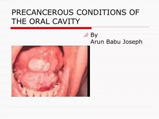
Examination of the Oral Cavity. Physical Evaluation. Oral Examination. Many diseases (systemic or local) have signs that appear on the face, head & neck or intra-orally

E N D
Examination of the Oral Cavity Physical Evaluation
Oral Examination • Many diseases (systemic or local) have signs that appear on the face, head & neck or intra-orally • Making a complete examination can help you create a differential diagnosis in cases of abnormalities and make treatment recommendations based on accurate assessment of the signs & symptoms of disease
Oral Examination • Each disease process may have individual manifestations in an individual patient • And there may be individual host reaction to the disease • Careful assessment will guide the clinician to accurate diagnosis
Scope of responsibility • Diseases of the head & neck • Diseases of the supporting hard & soft tissues • Diseases of the lips, tongue, salivary glands, oral mucosa • Diseases of the oral tissues which are a component of systemic disease
Equipment • Assure that you have all the supplies necessary to complete an oral examination • Mirror • Tissue retractor (tongue blade) • Dry gauze • You must dry some of the tissues in order to observe the nuances of any color changes
Exam of the Head & Neck; Oral Cavity • Be systematic • Consistently complete the exam in the same order • See clinic handout for a general guide
Extra-oral examination • Observe: color of skin • Examination area of head & neck • Determine: gross functioning of cranial nerves • Normal vs. abnormal • Paralysis • Stroke, trauma, Bell’s Palsy
Extra-oral examination • TMJ • Palpate upon opening • What is the maximum intermaxillary space? • Is the opening symmetrical? • Is there popping, clicking, grinding? • What do these sounds tell you about the anatomy of the joint? • When do sounds occur? • Use your stethoscope to listen to sounds
Extra-oral examination • Lymph node palpation • Refer to handout
Extra-oral examination • Thyroid Gland Palpation • Place hands over the trachea • Have the patient swallow • The thyroid gland moves upward
Exam: Lips • Observe the color & its consistency-intra-orally and externally • Is the vermillion border distinct? • Bi-digitally palpate the tissue around the lips. Check for nodules, bullae, abnormalities, mucocele, fibroma
Exam: Lips • Evert the lip and examine the tissue • Observe frenum attachment/tissue tension • Clear mucous filled pockets may be seen on the inner side of the lip (mucocele). This is a frequent, non-pathologic entity which represents a blocked minor salivary gland
Exam: Lips-palpation • Color, consistency • Area for blocked minor salivary glands • Lesions, ulcers
Exam: Lips • Frenum: • Attachment • Level of attached gingiva
Exam: Lips • Palpate in the vestibule, observe color
Examination: Buccal Mucosa • Observe color, character of the mucosa • Normal variations in color among ethnic groups • Amalgam tattoo • Palpate tissue • Observe Stenson’s duct opening for inflammation or signs of blockage • Visualize muscle attachments, hamular notch, pterygomandibular folds
Examination: Buccal Mucosa • Linea alba • Stenson’s duct
Examination: Buccal Mucosa • Lesions – white, red • Lichen Planus, Leukedema
Gingiva • Note color, tone, texture, architecture & mucogingival relationships
Gingiva • How would you describe the gingiva? • Marginal vs. generalized? • Erythematous vs. fibrous • Drug reactions: Anti-epileptic, calcium channel blockers, immunosuppressant
Exam: Hard palate • Minor salivary glands, attached gingiva • Note presence of tori: tx plan any pre-prosthetic surgery
Exam: Soft palate • How does soft palate raise upon “aah”? • Vibrating line, tonsilar pillars, tonsils, oropharynx
Exam: Oropharanyx • Color, consistency of tissue • Look to the back, beyond the soft palate • Note occasional small globlets of transparent or pink opaque tissue which are normal and may include lymphoid tissue
Exam: Tonsils • Tucked in at base of anterior & posterior tonsilar pillars • Globular tissue that has “punched out” appearing areas • Regresses after adulthood • May see white “orzo rice like” or “torpedo” shaped white concretions within the tissue
Exam: Tongue • The tongue and the floor of the mouth are the most common places for oral cancer to occur • It can occur other places; so visualize all areas • You may observe: • Circumvalate papillae, epiglottis
Exam: Tongue • Have the patient stick out their tongue • Wrap the tongue in a dry gauze and gently pull it from side to side to observe the lateral borders • Retract the tongue to view the inferior tissues
Exam: Tongue • You may observe lingual varicosities
Exam: Tongue • You may observe geographic tongue (erythema migrans)
Exam: Tongue • You may observe drug reaction
Exam: Tongue • Observe signs of nutritional deficiencies, immune dysfunction
Exam: Tongue • You may observe oral cancer
Exam: Floor of mouth • Visualize, palpate - bimanually • Wharton’s duct • Must dry to observe • Does “lesion” wipe off? • Where are the two most likely areas for oral cancer? • lateral border of the tongue • Floor of mouth
Exam: Floor of mouth • Squamous Cell Carcinoma
Exam: Floor of mouth • Squamous Cell Carcinoma
Exam: Leukoplakic area Edentulous Mandibular Ridge
Exam: Floor of mouth • Oral Cancer: • Red • White • Red and White • Does the patient have important risk factors for oral cancer? • Counseling for smoking and alcohol • Cessation
Triaging Lesions * • Describe it’s characteristics • Size, shape, color, consistency, location • How long has it been present? • Is it related to a trauma? • Fractured cusp, occlusal trauma • Has it occurred before? • Can you wipe it off? • Does the patient have specific risk factors for neoplastic lesions?
Triaging Lesions * • Any lesion that is suspicious should be re-evaluated in 2 weeks • Lesions due to infectious processes would have healed in that time frame • If it remains, the lesions should be biopsied
Exam: Maxilla & Mandible • size, shape, contour • pre-prosthetic treatment • Tori removal • tuberosity reduction • Soft or hard tissue or both
Evaluate for Epulis fissuratum If you make a new denture will the excess tissue resolve? Exam: Maxilla & Mandible
Occlusion • Orthodontic classification • Interferences