Download
1 / 23
240 likes | 495 Views
Explore the intricacies of muscle anatomy, including the three main types of muscle: skeletal, smooth, and cardiac. Skeletal muscle is voluntary, striated, and responsible for powerful contractions, covering the bony skeleton. Smooth muscle, found in hollow organs, is involuntary with spindle-shaped cells. Cardiac muscle, unique to the heart, features intercalated discs and is known for its constant contractions without fatigue. Learn about muscle attachments, organizational structures, and the cellular components that enable muscular functions in the body.

E N D
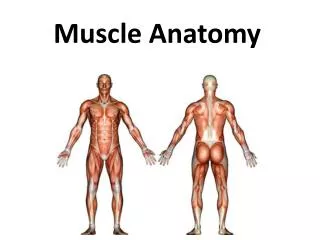
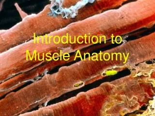
Types of Muscle 1.Skeletal • Elongated Cells • Multi nucleated • Striated – striped appearance • Voluntary • Produces powerful contractions • Tires easily, needs rest (fatigue). • Covers bony skeleton (motility) Longitudinal View Notice striations and nuclei around outside of cell. Cross Section Notice nuclei around outside of cell.
2. Smooth • Spindle-shaped Cell • Single nucleus in each cell • No Striations • Involuntary • Slow, sustained contractions • In hollow visceral organs (stomach, bladder, respiratory passages) Cross Section Nucleus is in center of cell. Cells much smaller.
3. Cardiac (Heart) • Branched cell • Contain intercalated discs • Single nucleus in each cell • Striations • Involuntary • Steady, constant contractions • Never tires
Muscle Functions • Produce movement • locomotion & manipulation • Help blood move through veins & food thru small intestines • Maintain posture • Stabilize joints • Body temp homeostasis • Shivering: movement produces heat energy
Muscle Requirements Axon of neuron Motor end plate (terminus) • Demands continuous oxygen/nutrient supply. • Lots of arteries/capillaries to muscle. • Each muscle cell w/ its own nerve ending controlling its activity. • Produce much metabolic waste due to constant activity.
Muscle Attachments • Most muscles span joints • Attaches to bone in two places: (video) 1.Insertion: the moveable bone • Bicep insertion is the radius 2.Origin: the stationary bone • bicep originates in two different places in scapula • Attachment types • Direct: attaches right onto bone - ex. intercostal muscles of ribs • Indirect: via tendon or aponeurosis (sheet-like tendon) to connect to bone - leaves bone markings such as tubercle
Muscle Organization Muscles are complex bundled structures: fibers within fibers
Muscle organization Muscle (organ) Fascicle Muscle fiber (cell) Myofibril Sarcomere Myofilaments: Actin & Myosin
Muscle Fibers • A Muscle Fiber = Muscle Cell • HUGE cell: • 10 - 100m in diameter • can be hundreds of centimeters long (created by cytoplasmic fusion of multiple embryonic cells) • extends the length of the muscle • Main content: bundles of proteins (actin and myosin) • Multinucleated • to maintain high rate of protein synthesis. • Muscle fiber nucleus = myonucleus
Insulation of Muscles • Muscle cells must be insulated from one another by specialized membranes • Muscle cells work electrically • if not insulated, nerves cannot control individual muscles.
Epimysium surrounds entire muscle • Dense CT that merges with tendon • Epi = outer • Mys = muscle • Perimysium surrounds muscle fascicles • Peri = around • Within a muscle fascicle are many muscle fibers • Endomysium surrounds muscle fiber • Endo = within
Structural Terminology Associated with Muscle Fibers Prefixes: myo, mys, and sarco all refer to muscle • Sacroplasmic Reticulum = Smooth ER of muscle (regulates calcium levels for muscle contraction) • Sarcoplasm = Cytoplasm • To maintain ATP production during cellular respiration, contains high amounts of: • mitochondria • glycosomes that store sugar • oxygen binding protein called myoglobin • Sarcolemma = Plasma Membrane • T tubules - The sarcolemma of muscle cells are not just on the outside, rather forms tubes that dive into the muscle cells • Myosin and Actin= muscle proteins that create muscle cytoskeletal filaments for contraction
myofibril Sarcoplasmic Reticulum Myosin (red) and Actin (blue) T-tubule sarcolemma
Microstructures • Each muscle fiber (muscle cell), is composed of many myofibrils. • Organized system of cytoskeleton filaments of actin and myosin proteins that do the actual contracting • Myofibrils are NOT CELLS • A sarcomere is one segment of a myofibril (muscle segments). • The series of sarcomeres produce the striated appearance of muscles
Muscle Fiber Sarcomere
Sarcomere organization • Myofibril composed of repeating series of sarcomeres with dark A and light I bands. • I bands intersected by Z discs mark the outer edges of each sarcomere. • Contraction happens within one sarcomere.
How do muscle contract? Let’s sketch the sarcomere together and discuss the sliding filament model of muscle contraction