Download

1 / 41
410 likes | 460 Views
Educative presentation for medical graduates and postgraduates

E N D
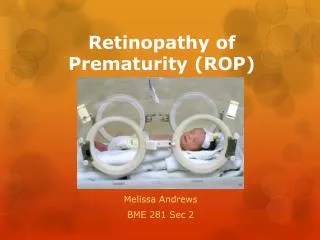
Retinopathy of Prematurity Dr.C.S.N.Vittal
Concept Map • What is ROP? • How does ROP occur? • Who should be screened for ROP? • When to screen for ROP? • How should these babies be screened for ROP? • How to diagnose ROP and recognise the severity of the disease? • What should be the frequency of screening after the initial examination? • How should they be managed • How to follow up these babies?
What is Retinopathy of Prematurity? • Retinopathy of Prematurity (ROP) is a vasoproliferative disorder of the eye that affects infants born four or more weeks preterm and have received intensive neonatal care
Epidemiology • Approximately 65% of infants with a birth weight <1,250 g and 80% of those with a birth weight <1,000 g will develop some degree of ROP. • Incidence of ROP in India is between 38-51.9% of low birth weight infants • ROP incidence is on rise in India as a result of improved neonatal care and better neonatal survival rate • 10% of the 32,300 infants affected globally (in 2010) were born in India
Normal Retinal Vascularization • Starts at the optic disc at approximately 16 weeks of gestation and proceeds peripherally. • The vessels reach ora serrata on the nasal side at around 34 to 36 weeks and temporal side by 36 to 40 weeks
Pathogenesis of ROP • Vaso-obliterative phase: • Begins when retinal vascular growth ceases after premature birth. • Vessels are vulnerable to injury and obliterated by any number of stress factors like increased O2 supply • Relatively vascular depleted retina becomes increasingly hypoxic by increasing metabolic demand of developing retina and triggers next phase.
Pathogenesis of ROP • Vaso-proliferative phase: • Formation of new vessels anarchaic and excessive resulting in invasion of vitreous and traction on retina and bleeding can occur driven by excess angiogenic factors (such as VEGF) upregulated by the hypoxic avascular retina. • These vessels are permeable; therefore, hemorrhage and edema can occur. • extraretinal fibrovascular proliferation can lead to retinal detachment and abnormal retinal function • This occurs most frequently around 33-34 weeks post conceptionally
Who should be screened for ROP? • Babies with birth weight < 2000 gm • Gestational age < 34 weeks • Gest age between 34-36 weeks but with risk factors like • Cardiopulmonay support • RDS • Fetal hemorrhage • Neonatal sepsis • Poor post natal weight gain • Prolonged oxygen therapy • Chronic lung disease • Blood transfusion • Intraventricular hemorrhage
When to screen for ROP? • The first screening is performed before discharge from the neonatal intensive care unit (NICU), but surely before 30 days of life. • All premature infants : 4 weeks of birth • Gest age < 26 weeks : 2-3 weeks of birth • For infants born weighing <1,200 g and <30 weeks gestational age, the first screening is recommended between 2 and 3 weeks of life, for aggressive posterior ROP (APROP).
How to screen for ROP? • Place: In the neonatal unit under the supervision of neonatologist • Baby swaddled and preferably fed one hour prior to exam • Incubator dependent babies – within the incubator itself • Attention to be given to possibility of bradycardia, arrhythmia, asystole, hypoventilation, apnea or aspiration
Instrument Checklist • Indirect ophthalmoscope • 20,28 and 30 D lenses • Alfonso speculum and infant scleral depressor • Dilator eye drops (one drop of phenyehrine 1% and cyclopentolate 0.2%) • Topical antibiotic eye drops (Tobramycin) • Sterile cotton and glove • ROP documentation sheet • Wide field digital camera • To reduce pain, nonpharmacologic (containment and nonnutritive sucking, oral sucrose) measures of pain relief and local anesthetic (proparacaine 0.5%) recommended.
Classification • Classification system consists of four components: • Location • Zone 1, Zone 2, Zone 3 • Severity / stage • Stage 1, 2, 3, 4 and 5 • Extent : Clock hours • Plus disease : Present or absent
Definitions • Aggressive posterior ROP (APROP) • Rapidly progressing, severe form • Posterior location (usually zone 1, but can be zone 2 posterior) • ill-defined nature of retinopathy without a classical ridge. • Ischemic loops, capillary nonperfusion, and flat intraretinal net- work neovascularization. • Untreated, it will rapidly progress to retinal detachment. • Hybrid ROP • presence of ridge tissue, characteristic of staged ROP, along with the flat neovascular syncytium • Type 1 ROP • stages 1, 2, or 3 in zone 1 with plus disease, • stage 3 in zone 1 without plus disease, and stage 2 or 3 in zone 2 with plus disease. • Type 2 ROP (older term threshold ROP) • zone 1, stage 1 or 2 without plus disease and zone 2, stage 3 without plus disease
How to diagnose ROP (revised 2005 classification of ROP) • Zones in circles • Extent of involvement in clock hours or 30 degrees
Stage 1 ROP Demarcation line • Thin structure separating the avascular retina anteriorly from the posteriorly vascularized retina • Requires close periodic examination, but may not need any treatment.
Stage 2 ROP Ridge • Ridge increases in dimensions and extends above the retina. • Blood vessel growth is moderately abnormal and in some cases may need early treatment • If not monitored, they can progress to more severe stages.
Stage 3 ROPExtraretinal fibrovascular proliferation • Neovascularization extends into the vitreous from the ridge • Blood vessel growth is severely abnormal and the newborn requires early treatment within 72 hours as this is vision threatening.
Stage 3 ROPExtraretinal fibrovascular proliferation • Schematic drawing of moderate stage III retinopathy of prematurity. • Optic nerve head is shown at the bottom, and periphery of the retina is at the top • Garner A. International classification of retinopathy of prematurity. Pediatrics. 1984;74:127.
Stage 4 ROPPartial retinal detachment • Depending on the extent • Stage 4a: extra- foveal retinal detachment • Stage 4b: involving fovea • Urgent surgical treatment is needed to diminish the chances of loss of vision.
Stage 5 ROPTotal retinal detachment • Generally tractional but may occasionally be exudative • Usually funnel-shaped • Only very few eyes get minimal vision even after advanced surgical treatment.
Plus Disease Plus disease is an ominous sign indicating severity of the disease. • Arteriolar tortuosity and venous engorgement of the posterior pole, iris vascular engorgement, pupillary rigidity, and vitreous haze. • It can be associated with any stage or zone of ROP and indicates the necessity towards treatment.
Aggressive Posterior ROP (Rush Disease) • Vary rapidly progressive severe form of ROP • If not treated timely, can progress to stage 5 skipping various classical stages • Confined to posterior location • Prominence of plus disease • Ill defined nature of retinopathy
Types of ROP Type 1 ROP ◆ Zone I, any stage with plus◆ Zone I, stage 3 without plus ◆ Zone II, stage 2 to 3 with plus Type 2 ROP ◆ Zone I, stage 1 to 2 without plus ◆ Zone II, stage 3 without plus
Management –Laser Ablative Therapy • Zone II: Plus disease with stage 2 or 3 ROP • Zone I: Plus disease with stage 1 or 2 ROP 3. • Zone I: Stage 3 ROP • Entire vasculature up to ora serrata ablated • Follow up weekly till ROP regresses and vasculature reaches ora serrate
Management – Cryotheroapy • Applied to the external surface of the sclera, and areas peripheral to the ridge of the ROP are frozen until the entire anterior avascular retina has been treated. • Approximately 35 to 75 applications are made in each eye. • Usually done under general anesthesia. • Cryotherapy causes more inflammation and requires more analgesia than laser therapy • Necessary in special cases, such as when there is poor pupillary dilation or vitreous hemorrhage, both of which prevent adequate delivery of laser therapy.
Management - Anti VEGF Intravitreal injection of VEGF inhibitor - bevacizumab Indications: • Primary therapy for aggressive posterior zone disease • Media haze due to aggressive posterior disease for laser treatment • Failed laser treatment leading to persistent neovascularizatio, tractional elements of tractional retinal detachment prior to surgery
Management – Other Medications • Vitamin E. :Maintenance of normal serum vitamin E levels is a prudent management objective • Dietary supplementation of omega-3 polyunsaturated fatty acids (PUFA): mouse model have shown a protective effect of omega-3 supplementation.
Management – Vitreoretinal surgery Stage 4 and 4 Aim: to prevent progression to retinal detachment
Management – Recent Advances • Prophylactic oral propranolol appeared to be effective in preventing severe ROP in premature infants ≤32 weeks gest age. • Ultra-wide-field retinal photography and hand- held spectral domain optical coherence tomography (SD-OCT) with and without angiography have provided a new tool for evaluating foveal growth, vascularization, and disease, and correlating with structural and functional outcomes.
Complications • Macular heterotropia • High myopia • Amblyopia • Strabismus • Anisotropia • Glaucoma • Phthisis bulbi
Prevention • Prevention of premature birth and its attendant problems • Maintaining oxygen saturation levels for severely premature infants at levels sufficiently low to minimize the risk of ROP
Dr.C.S.N.Vittal Thank you