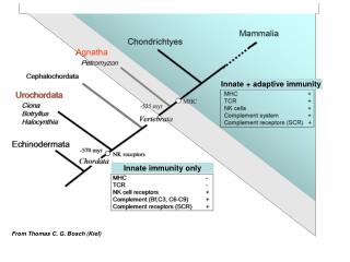
1 / 6

Lecture 02, Innate Immunity

immune system

Arslan34
Download Presentation

Lecture 02, Innate Immunity

An Image/Link below is provided (as is) to download presentation Download Policy: Content on the Website is provided to you AS IS for your information and personal use and may not be sold / licensed / shared on other websites without getting consent from its author. Content is provided to you AS IS for your information and personal use only. Download presentation by click this link. While downloading, if for some reason you are not able to download a presentation, the publisher may have deleted the file from their server. During download, if you can't get a presentation, the file might be deleted by the publisher.

E N D

Presentation Transcript


  1. 58 C H A P T E R Innate Immunity C H A P T E R C O N T E N T S The Barrier Mechanical Barrier Chemical Barrier Biological Barrier Phagocytes And Other Myeloid Cells Origin of the Myeloid Lineage Cells Pattern Recognition Receptors Of Innate Immune Cells Toll-Like Receptors C-Type Lectin Receptors NOD-Like Receptors RIG-I Helicase-Like Receptors Effector Mechanisms Of Innate Immune Cells Antigen-Presenting Cells: Macrophages & Monocytes Antigen-Presenting Cells: Dendritic Cells Granulocytes: Neutrophils Granulocytes: Eosinophils Granulocytes: Basophils & Mast Cells Natural Killer Cells Inflammatory Mediators Cytokines Chemokines The central mission of the barrier is to separate the outside world from the host, allowing various microbes to survive in niches along the surface, but preventing any from gaining a foothold where they could invade. As we will discuss later, breach of the barrier causes activation of local and systemic innate inflammation, followed by recruitment of adaptive immunity. The innate immune system is composed of physical barriers, cells, and circulating factors that are always active and ready to repel microbes. They form a boundary between your tissues and the vast majority of viruses, bacteria, and fungi that live on and inside you. Innate immune cells are also important for cleaning up debris from dying cells and repairing damaged tis- sues. In this chapter, we will discuss the mechanisms of innate immunity, including their role in host defense and in driving inflammation. Unlike the lymphocytes that make up the adap- tive immune system, which can be tuned to sense and respond in different ways to different infections, components of the innate immune system have a limited number of ways to sense and respond to infections. However, the advantage of the innate immune system is that it is activated rapidly and targeted at pat- terns that are widely shared among microbes. Mechanical Barrier The outermost layer of the barrier is formed by epithelial cells, connected to one another by tight junctions. The skin’s epidermis is covered by a layer of keratinized squamous cells that are continuously shed (“desquamated”). Along with sweat secretion, this limits the ability of microorganisms to attach and invade. Similarly, nonskin barriers are called mucous mem-branes because they are coated by mucins, a sticky mixture of glycoproteins produced by secretory epithelial cells of the respi-ratory, gastrointestinal, and genitourinary tracts. Mucus makes these surfaces more difficult to adhere to and penetrate. The cells of mucous membranes also undergo rapid divi-sion, sloughing off continuously, and the ciliated cells lining the respiratory tract, the gastrointestinal tract’s peristalsis, and the continuous flow of urine from the kidney through the bladder and urethra ensure that microbes cannot attach and invade THE BARRIER An extremely important, but often overlooked, component of host defense is the barrier formed by skin and mucous mem- branes. The epithelia covering our skin, respiratory, gastrointes- tinal, and genitourinary tracts provide the first line of defense against microbes in the external world. This occurs through a combination of mechanical, chemical, and biological means. 482 Levinson_Ch58_p482-p493.indd 482 09/03/18 4:31 PM

  2. CHAPTER 58 Innate Immunity 483 infectious diseases, including Clostridium difficile colitis, and possibly in inflammatory diseases, such as seborrheic dermatitis and inflammatory bowel disease. Additional biological immunoglobulin (Ig) A and IgG, two classes of antibody. Enormous amounts of IgA are secreted from our mucosal sur- faces, where it binds bacteria, viruses, and toxins, preventing them from adhering to the epithelial layer. Some infectious diseases are caused by pathogens that have evolved ways to bypass barrier defenses. For example, herpes-viruses and respiratory viruses attach and invade via specific target receptors on the mucosal epithelial cells. Vector-borne pathogens, such as arboviruses and malaria, have evolved to move between hosts inside insects and are released behind the barrier when we are bitten. In addition, most of the bacteria and fungi that commonly live in the environment or in our intestines are harmless, but they can cause life-threatening infections if a breakdown of the barrier allows them entry. Some examples of these include respiratory infections in people with cystic fibrosis, who have defects in mucus clearance, or infections of the bloodstream in people who require intravenous catheters. these sites. Failure of any of these mechanisms is a common cause of infection due to bacteria or fungi that otherwise colo- nize us harmlessly. In patients with severe skin burns, patients with pulmonary ciliary cell disorders, and patients with bowel or urinary obstruction, infection is the key cause of morbidity and mortality. barrier defenses are Chemical Barrier Epithelial cells also produce a number of chemicals and proteins that inhibit microbes from growing or attaching. The stomach excrete concentrated hydrochloricacid that kills bacteria. Lysozyme is an enzyme in saliva and tears that makes holes in bacterial cell walls by breaking linkages in their pepti- doglycan molecules. In addition, antimicrobial peptides, such as defensins, are produced throughout the skin and mucous membranes. Defensins are highly positively charged (i.e., cationic) peptides, primarily produced in the gastrointestinal and lower respiratory tracts, that create pores in lipid membranes of bacteria, fungi, and even some viruses. Neutrophils and Paneth cells in the intestinal crypts contain one type of defensin (α-defensins), which may have antiviral activity, whereas the respiratory tract produces dif- ferent defensins called β-defensins, which are antibacterial. Sur- factants are lipoproteins produced in the lung alveoli that bind to the surface of microbes, which can facilitate their phagocytosis (i.e., opsonin function) or can be directly bactericidal by increas- ing the bacterial cell membrane permeability. PHAGOCYTES AND OTHER MYELOID CELLS Innate immune cells respond when the barrier (first line of defense) has been breached. These are the second line of defense, and the most important of these cells are phagocytes. The name “phagocyte” derives from Greek, emphasizing these cells’ ability to “eat” foreign material and debris. (Innate immune phagocytes also play an important role in detecting, clearing, and repair- ing damaged tissue, even in the absence of a foreign infection, although this will not be covered in depth here.) There are several key phagocytic cells: tissue macrophages reside in normal tissue and are the first cells to encounter for- eign material; neutrophils migrate into damaged tissue and contribute to inflammation; and dendritic cells carry away microbial material to nearby lymphoid tissues (the lymph nodes or spleen), where they activate the adaptive immune response. A fourth cell, called a monocyte, can be recruited into inflamed tissue to take on the role played by macrophages Biological Barrier Some microorganisms have evolved ways to defend themselves against components of the chemical barrier, and this selective access attained by certain microbes over others forms ecologi- cal microenvironments throughout our bodies. We have long known that the epithelial barrier harbors a multitude of harm- less commensal microorganisms, collectively called the micro- biome, that inhabit distinct niches. These include bacteria, fungi, protozoa, and even Demodex mites that inhabit our hair follicles! Each of these species competes for nutrients, evolving antimicrobial strategies to coexist and defend the niche. When we give patients antibiotics for an infection, we are also killing off many of the commensal species, which allows the remain- ing few to proliferate, disrupting our normal microbial ecol- ogy. Microbiome disruption plays an important role in many TABLE 58–1 Components of the Barrier Anatomic Site Mechanical Chemical Biological Skin Keratinized squamous epidermis cells Fatty acids; defensins Skin flora Gastrointestinal tract Mucins; peristalsis; normal shedding of epithelial cells Gastric acid; digestive enzymes; defen- sins; lysozyme; iron-binding proteins Gut flora; IgA Genitourinary tract Urine flow Low pH Vaginal flora; IgA Respiratory tract Airflow; ciliated airway cells; coughing Surfactant proteins Nose, mouth, and pharyngeal flora; IgA Ig = immunoglobulin. Levinson_Ch58_p482-p493.indd 483 09/03/18 4:31 PM

  3. PART VII Immunology 484 Fetal Liver / Postnatal Bone Marrow Multipotent hematopoietic stem cell Common lymphoid progenitor Common myeloid progenitor Granulocyte/monocyte progenitor Natural killer cell B-cell precursor Megakaryocyte Erythroblast Granulocyte progenitor T-cell precursor Monocyte Platelets Reticulocyte Erythrocytes Macrophage Dendritic cell Eosinophil Neutrophil Mast cell Basophil FIGURE 58–1 Origin of hematopoietic cells. Stem cells in the bone marrow (or fetal liver) are the precursors of all blood cells. Stem cells differentiate into myeloid or lymphoid progenitor cells. The myeloid cells are the source of platelets, erythrocytes, granulocytes, macrophages, and dendritic cells. Monocytes are a special type of myeloid cell that can differentiate into macrophages or dendritic cells when needed. The lymphoid cells are the source of T lymphocytes, B lymphocytes, and natural killer cells. The development of lymphocytes is covered in detail in Chapter 59. a molecular pattern, called a pathogen-associated molecu- lar pattern(PAMP), that is present on the surface of many microbes but—very importantly—is not present on human cells and is difficult for those microorganisms to alter through mutation. By using this strategy, innate immune cells do not need a highly specific receptor for each individual microbe strain but can still distinguish broad classes of foreign agents from self. Table 58–2 lists examples of the four major classes of PRRs. There are two classes of receptors (Toll-like receptors and C-type lectin receptors) that recognize microbes that are outside of cells or within the cells’ vesicles. Two other classes of recep- tors in the cytoplasm of cells (NOD-like receptors and RIG-I helicase receptors) recognize microbes that have invaded the cell’s cytoplasm. Mutations in the genes encoding these pattern recep- tors result in a failure to recognize pathogens and predispose to severe bacterial, viral, and fungal infections. The most important of these PRRs are the Toll-like recep- tors(TLRs). This is a family of 10 receptors found on the sur- face of many cells, including epithelial cells and innate immune cells, such as macrophages and dendritic cells. Each of the ten TLRs recognizes a core microbial building block (e.g., endo- toxin or peptidoglycan), and the resulting signal activates tran- scription factors that enhance the synthesis of proinflammatory cytokines and cell surface molecules. The result is a rapid innate immune response, triggered by a particular microbe in a particular location. or dendritic cells. Phagocytes belong to the family of innate immune cells called myeloid cells because they originate from bone marrow “myeloid” stem cells. Origin of the Myeloid Lineage Cells By the time of birth, the stem cells that give rise to all red and white blood cells reside primarily in the bone marrow. Throughout life, these stem cells continue to produce daughter cells that become the precursor cells of all of the red and white blood cells. The immune cells, or nonerythroid cells, are often called white blood cells, or leukocytes, and include lymphoid cells and myeloid cells. In contrast to T lymphocytes and B lymphocytes, which differentiate from lymphoid stem cells, most cells of the innate immune system arise from myeloid precursors. (There are a few exceptions, such as natural killer [NK] cells, which are covered later in this chapter). PATTERN RECOGNITION RECEPTORS OF INNATE IMMUNE CELLS The first step of an immune response is that innate immune cells recognize foreign material. In order to identify what is foreign, several components of the innate immune arm detect certain carbohydrates or lipids on the surface of microorgan- isms. Components of the innate immune arm have receptors, called pattern recognition receptors (PRRs), that recognize Levinson_Ch58_p482-p493.indd 484 09/03/18 4:31 PM

  4. CHAPTER 58 Innate Immunity 485 TABLE 58–2 Pattern Recognition Receptors Location Receptor Class Examples Activating Microbial Ligands Extracellular Toll-like receptors (TLRs) TLR2 TLR4 TLR9 MBL Dectin-1 Peptidoglycan (gram-positive bacteria) Lipopolysaccharide (bacterial endotoxin) Bacterial DNA Mannose (viral, fungal, and bacterial carbohydrate) Glucans (fungal cell wall) C-type lectin receptors (CLRs) Intracellular NOD-like receptors (NLRs) NOD2 NLRP3 RIG-I Peptidoglycan (gram-positive bacteria) Staphylococcus α toxin Double-stranded RNA (viral) RIG-I helicase-like receptors (RLRs) MBL = mannan-binding lectin; NOD = nucleotide-binding oligomerization domain; RIG-I = retinoic acid-inducible gene I. pattern receptors called C-type lectin receptors (CLRs). A different CLR, calleddectin-1, recognizes beta-glucan in the cell wall of fungi suchas Candida albicans. The type of adaptive host defense that is mounted by the body differs depending on the type of organism and where it is found. For example, antibody-mediated responses are effective against extracellular (especially encapsulated) bacteria, whereas T-cell–mediated responses are required against intracellular microbes, such as viruses and mycobacteria, such as Mycobacteriumtuberculosis. NOD-Like Receptors Part of the peptidoglycan (cell wall) of bacteria is recognized by NOD-like receptors (NLRs). (NOD stands for nucleotide- binding oligomerization domain.) These receptors are located within the cytoplasm of human cells (e.g., macrophages, den- dritic cells, and epithelial cells); hence, they are important in the innate response to intracellular bacteria such as Listeria. Some important examples of pattern recognition receptors are described in the following sections. Toll-Like Receptors Endotoxin is a lipopolysaccharide (LPS) found on the surface of most gram-negative bacteria (but not on human cells).LPSstimulates a PRR called Toll-like receptor 4 (TLR4), which transmits a signal to the nucleus of the cell. This induces the production of cytokines and surface proteins that are required to activate helper T cells and to produce antibodies. Note receptor, TLR2, signals the presence of peptidoglycan from gram-positive bacteria, which has a different molecular pattern but produces the same innate cell activation. Excessive macrophage PRR activation is an important cause of septic shock and death in hospitalized patients, so drugs that modify the action of these TLRs may become important in preventing endotoxin-mediated septic shock. RIG-I Helicase-Like Receptors Finally, RIG-I helicase-like receptors, or RLRs, recognize microbial nucleic acids in the cytoplasm of infected cells. (RIG-I stands for retinoic acid–inducible gene I.) Activation of these receptors results in the synthesis of alpha- and beta-interferons that promote antiviral immune responses. that a different Toll-like EFFECTOR MECHANISMS OF INNATE IMMUNE CELLS Once recognition occurs, innate immunity is activated to increase production of proinflammatory signals that have three key effects: (1) to kill invaders and recruit other immune cells to the area, (2) to block the infection from causing disease beyond the local site of inflammation, and (3) to aid in repairing the damaged barrier. The cells that exert these functions can be categorized as antigen-presenting cells (APCs), granulocytes, and innate lymphocytes called NK cells. C-Type Lectin Receptors Many bacteria and yeasts have a polysaccharide called mannan on their surface that is not present on human cells. (Mannan is a polymer of the sugar mannose.) A PRR called mannan-binding lectin (MBL) present on the surface of macrophages and dendritic cellsis a member of a group of Levinson_Ch58_p482-p493.indd 485 09/03/18 4:31 PM

  5. CHAPTER 58 Innate Immunity 487 Antigen-Presenting Cells: Macrophages & Monocytes Interestingly, the protective function of eosinophils has not been clearly established. It seems likely that they defend against the migratory such as Strongyloides and Trichinella. These parasites become coated with IgE, and eosinophils, which have receptors for IgE, can then attach to the surface of larvae and discharge the contents of their eosinophilic granules, dam- aging the cuticle of the larvae. eosinophils also contain leukotrienes and peroxidases, which can damage tissue and cause inflammation. However, another function of eosinophils may be to reduce inflammation. The granules of eosinophils contain histaminase, an enzyme that degrades histamine, which is an important mediator of (allergic) reactions. Eosinophils can phagocytize bacteria but they do so weakly, and they do not present antigen with class II MHC. Therefore, they are not sufficient to protect against pyogenic bacterial infections in neutropenic patients. larvae of parasites, All nucleated cells express a protein called class I major histo-compatibility complex(MHC), and these cells present antigen for recognition by cytotoxic T cells. Some cells, for example, macrophages and dendritic cells, also express a different protein called class II MHCand they can only be recognized by helper T cells. The cells that are capable of presenting peptides by class II MHC proteins are referred to as “professional” APCs. The most abundant professional APCs are myeloid cells called macrophages. These are usually the first cells to encounter foreign invaders or injured tissue, and some examples include the microglial cells in the brain, the alveolar macrophages in the lung, and the Kupffer cells in the liver. The granules of the immediate hypersensitivity In addition to tissue resident macrophages, there are other cells, called monocytes, which are short-lived myeloid cells that patrol the body throughout inflammation by rapidly entering inflamed tissue and differentiating into macrophages or dendritic cells on demand. Tissue-resident macrophages and monocyte-derived macrophages have three main functions: phagocytosis, antigen presentation, and cytokine production. Granulocytes: Basophils & Mast Cells Basophils are white blood cells with cytoplasmic granules that appear blue when stained with Wright stain. Mast cells, which are similar to basophils in many ways, are fixed in tissue, especially under the skin and in the mucosa of the respiratory and gastro-intestinal tracts. Basophils and mast cells have receptors on the cell surface for the Fc portion of the heavy chain of IgE. When adjacent IgE molecules are cross-linked by antigen, the cells release preformed inflammatory mediators from their granules. Some examples of these mediators are histamine, proteolytic enzymes, and proteoglycans such as heparin. These cause inflammation and, when produced in large amounts, cause a wide range of immediate hypersensitivity reactions: the mildest form is urticaria (hives), while the most severe form is systemic anaphylaxis. Natural Killer Cells NK cells play two important roles in immunity: (1) they kill virus-infected cells and tumor cells and (2) they produce gamma interferon that activates macrophages to kill bacteria that they have ingested. NK cells are called “natural” because, unlike adaptive cells, they do not recognize their target cells by detecting antigens presented by class I or class II MHC proteins, they are not enhanced by exposure, they have no memory, and they are relatively nonspecific for any one virus or tumor. Rather, NK cells target cells to be killed by detecting other features of cell dysfunction, for example, the lack of class I MHC proteins on the cell surface. This detection process is effective because many cells lose their ability to synthesize class I MHC proteins after they have been infected by a virus. NK cells can also detect cancer cells by recognizing a protein called MICA that is found on the surface of many cancer cells but not normal cells. life, reacting to Granulocytes: Neutrophils Neutrophils are the most abundant immune cell in the blood. They are phagocytes that belong to the family of myeloid white blood cells, and in addition, they are in a subgroup called granulocytes, cytoplasmic granules visible with Wright stain. Neutrophils are a very important component of our innate host defenses, and severe bacterial and fungal infections occur if they are few in number (neutropenia) or are deficient in function (as in some of the immune disorders. Like macrophages, neutrophils have surface receptors for IgG, making it easier for them to phagocytize opsonized microbes. Note that neutrophils do not display class II MHC proteins on their surface and therefore do not present antigen to helper T cells. This is in contrast to macrophages, which are both phagocytes and APCs, as discussed earlier. named for their Granulocytes: Eosinophils Eosinophils are white blood cells with cytoplasmic granules that appear red when stained with Wright stain. The eosinophil count is elevated in two medically important types of diseases: parasitic diseases, especially those caused by tissue-invading nematodes hypersensitivity diseases, such as asthma and serum sickness. Diseases caused by protozoa are typically not characterized by eosinophilia. and trematodes and Levinson_Ch58_p482-p493.indd 487 09/03/18 4:31 PM

  6. CHAPTER 58 Innate Immunity 491 INFLAMMATORY MEDIATORS Local inflammation at the site of an infection causes the four classic symptoms of pain, redness, warmth, and swelling. These symptoms are manifestations of the immune system’s efforts to recruit leukocytes to the area and limit the infection from spreading. (3)Interleukin-6 (IL-6) is an acute-phase response cytokine released by macrophages and mast cells and probably also by nonimmune cells such as muscle and fat cells. Its primary function is to signal the liver to increase production of more acute-phase proteins, which enter the circulation and cause fever and cachexia. (4)Two other important cytokines that stimulate leukocyte migration out of the bone marrow are granulocyte colony- stimulating factor (G-CSF, or CSF1) and granulocyte-macrophage colony-stimulating factor (GM-CSF, or CSF2). (5)Interferons are glycoproteins that were originally named because they interfere with virus replication, but in fact, they are innate cytokines that have a variety of effects on cells. The type I interferons (the two types are also called alpha inter- feron or IFN-α, made by leukocytes, and beta interferon or IFN-β, made by nonhematopoietic cells) are induced when cells detect they contain a virus. Both of them signal nearby cells to make degradative enzymes that will inactivate the virus when it infects those cells. The nearby cells can prevent the virus from replicating and thereby prevent the spread of virus from cell to cell. They do this through key enzymes that (1) degrade viral messenger RNA, thereby inhibiting viral replica-tion; (2) block translation of new proteins, thereby inhibiting assembly of new virions; and (3) initiate apoptosis pathways so that the cell dies before its machinery can be used to help the virus spread. Chemokines Chemokines are a group of cytokines that attract leukocytes and help them migrate to where they are needed. Cytokines and chemokines of the innate immune system are engaged to recruit and activate leukocytes. Cytokines Cytokines are the language of the immune system, and immune cells use cytokines to communicate with each other and with the other cells of their environment. Unlike hormones, which circulate in the bloodstream, cytokines usually act over very short distances in local tissue. Cytokines can be produced and sensed by numerous cell types in different locations, and so the effects of cytokine signaling can vary depending on these factors. For example, some cytokines are both “paracrine,” meaning they act on neighboring cells, and “autocrine,” meaning they feed back to act on the same cell that produced them. Historically, cytokines were named based on a function that was originally discovered for them, such as “tumor necrosis factor” (TNF) stimulating factor” (G-CSF), but more recently, they have mostly been given the name “interleukin,” with a number assignment corresponding to the order in which they were discovered. Over time, new functions have been discovered for many cytokines, and their names have become less reflective of their function. Note that medical therapies that target many of these signaling pathways are now in use to either boost immunity (through agonism/activation) or limit excessive inflammation (through antagonism/blockade). (1)TNF-α is a proinflammatory cytokine produced primar- ily by macrophages. It has many important effects that differdepending on the concentration. At low concentrations, it increases the synthesis of adhesion molecules by endothelial cells, which allows neutrophils to adhere to blood vessel walls at the site of infection. It also activates the respiratory burst within neutrophils, thereby enhancing the killing power of these phagocytes. TNF is also an endogenous pyrogen, a cytokine that causes fever. At high concentrations, it is an important mediator of endotoxin-induced septic shock. TNF-α, as its name implies, causes the death and necrosis of certain tumors in experimental animals. It may do this by promoting intravascular coagulation that causes infarction of the tumor tissue. (2)Interleukin-1 (IL-1) is a proinflammatory cytokine that is premade in an inactive form, called pro-IL-1, which is stored, ready for use, inside macrophages and epithelial cells. Like TNF-α, IL-1 is also an endogenous pyrogen that causes fever. or “granulocyte colony- Levinson_Ch58_p482-p493.indd 491 09/03/18 4:31 PM

More Related