Download
1 / 31
320 likes | 1.2k Views
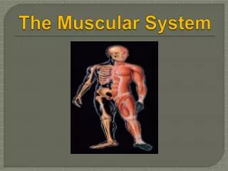
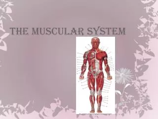
UNIT 5 – MUSCULAR SYSTEM. MUSCLE TYPES. Cardiac muscle – found only in the heart, striated, involuntary, arranged in figure-8 shaped bundles (for contraction), intercalated disks

E N D
MUSCLE TYPES • Cardiac muscle – found only in the heart, striated, involuntary, arranged in figure-8 shaped bundles (for contraction), intercalated disks • Smooth muscle – visceral (hollow organs), non-striated, involuntary, arranged in sheets or layers (contract – change shape of organ) • Skeletal muscle – where muscle connects to bone for movement, striated, voluntary
SKELETAL MUSCLE ANATOMY • Endomysium – delicate connective tissue sheeth that encloses each muscle fiber • Fasciculus – bundle of muscle fibers covered by perimysium (coarser fibrous membrane) • Epimysium – covers bundle of fasciculi (entire muscle); blends into either: • Tendon – cord of dense, fibrous tissue attaching a muscle to a bone • Aponeurosis – fibrous or membranous sheet connecting a muscle and the part is moves (usually found on torso)
MUSCLE FUNCTIONS • Produce movement • Maintains posture • Stabilizes joints • Generates heat
MICROSCOPIC ANATOMY OF SKELETAL MUSCLE • Sarcolemma – plasma membrane of muscle fiber (cell); under the endomysium • Peripheral nuclei – nuclei are pushed aside by long ribbon-like organelles called myofibrils – contain trains of tiny contractile units called sarcomeres • 2 types of myofilaments in the sarcomeres: • 1. myosin filaments – thick • 2. actin filaments – thin • Their arrangement produces a banding pattern, or striations
SKELETAL MUSCLE ACTIVITY • Stimulation and contraction of single skeletal muscle cells: • Irritability – the ability to receive and respond to a stimulus • Contractility – the ability to shorten (forcibly) when an adequate stimulus is received • Nerve stimulus and action potential – one motor neuron may stimulate a few muscle cells or hundreds of them, depending on the particular muscle and the work it does (gross motor vs. fine motor) • Motor unit – one neuron and all the skeletal muscle cells it stimulates • Neuromuscular junction – where the axon terminals for junctions with the sarcolemma • When the nerve impulse reaches the axon terminals, a neurotransmitter is released, which travels across the synaptic cleft (gap between nerve & muscle); acetylcholine (Ach) – neurotransmitter that stimulates skeletal muscle • Ach attaches to receptors which makes the membrane more permeable to Na+ • Na+ diffuses in and K+ rushes out, generating an action potential (electrical impulse), which travels over the entire surface of the sarcolemma • Muscle cell contracts • ACH is removed by acetylcholinesterase to stop contraction
SKELETAL MUSCLE ACTIVITY • steps of the action potential • Mechanism of muscle contraction: Sliding Filament Theory • AP travels down T-tubules, which causes Ca2+ to be released from the lateral sacs of the sarcoplasmic reticulum • Ca2+ binds to tropinin, causing tropomyosin to move out of the way – exposing the active site on the actin filament • Myosin heads swing back and attach to the active site on actin, forming cross-bridges • Myosin heads perform a power stroke – move toward the center of the sarcomere • Pulling actin filaments towards the center of the sarcomere • ATP is broken down to provide energy for the myosin heads to release the active site; leftover energy is stored for the next power stroke • Myosin heads grab further & further back each time • Whole muscle shortens • Whole series of events takes few thousands of a second
SKELETAL MUSCLE ACTIVITY • muscle contraction video • Contraction of skeletal muscle as a whole • Graded responses • All-or-none law – a muscle cell will contract to its fullest extent when it is stimulated adequately; it never partially contracts; is true of muscle cells only (not whole muscle) • Muscle cells react to stimuli with graded responses or different degrees of shortening • Can be produced 2 ways: • 1. By changing frequency of muscle stimulation • A single, brief, jerky contraction – muscle twitch • Nerve impulses are delivered to the muscle at a very rapid rate, so rapid that muscle does not get a chance to relax completely between stimuli; as a result, the effects of the successive contractions are “summed” (added) together and contraction gets “stronger and smoother,” with no evidence to relaxation seen – muscle is in fused, or complete, tetanus, or tetanic contractions (tetanus is normal and desirable, not to be confused with tetanus/lockjaw, which is caused by bacterium)
SKELETAL MUSCLE ACTIVITY (CONT.) • 2. by changing number of muscle cells being stimulated • How forcefully a muscle contracts depends largely on the number of muscle cells stimulated; when only a few cells are stimulated, contractions will be slight; when all cells are stimulated, contraction is strong • Providing energy for muscle contraction – as muscle contracts, ATP is broken down for energy; muscles stre a limited supply (4-6 seconds worth), so it must be regenerated continuously. Working muscles use 3 pathways for ATP regeneration: • 1. Direct phosphorylation of ADP by creatine phosphate: a phosphate group transfers from CP to ADP, regenerating more ATP; CP supplies exhaust in about 20 seconds • 2. Aerobic respiration: provides 95% of ATP at rest and during light exercise; occurs in mitochondria & involves a series of metabolic pathways that use oxygen – called oxidative phosphorylation; glucose is broken down into CO2 & H2O; some released energy is captured in ATP bonds (get 36ATP/1 glucose) • 3. Anaerobic glycolysis and lactic acid formation: initial steps of glucose breakdown occur via glycolysis which is anaerobic. • Glucose pyruvic acid with energy captured in ATP bonds (2ATP/ 1 glucose)
SKELETAL MUSCLE ACTIVITY (CONT.) • If enough oxygen is present, pyruvic acid enters aerobic pathways that occur within mitochondria • If there is not enough oxygen present (i.e. – intense muscle activity), or if oxygen or glucose delivery is inadequate, pyruvic acid is converted to lactic acid in a process called anaerobic glycolysis • Lactic acid - causes muscle soreness and fatigue (muscle fatigue occurs when the muscle can no longer contract despite still being stimulated). It results from oxygen debt which must be “paid back” (taking deep breaths) • Isotonic vs. isometric contraction: • Isotonic contractions – when myofilaments are successful in sliding movements so muscle shortens during contraction; most familiar type (i.e. – smiling, bending at knee) • Isometric contractions – when muscles do not shorten b/c muscles are pitted against some more or less immovable object, but tension keeps building (i.e. – lifting a dresser, pushing arms against a wall) • Muscle tone – state of continuous partial contraction
SKELETAL MUSCLE ACTIVITY (CONT.) • Effect of Exercise on Muscles: • Aerobic or endurance exercise • Examples – biking, jogging, swimming laps • Results in stronger more flexible muscles with greater resistance to fatigue • blood supply increases • individual muscle cells form more mitochondria and store more oxygen (makes overall body metabolism more efficient • Improves digestion and elimination of wastes • Enhances neuromuscular coordination • Makes the skeleton stronger • Heart enlarges • Fat deposits are cleared from blood vessel walls • Lungs become more efficient at gas exchange • Does NOT cause muscles to increase in size
SKELETAL MUSCLE ACTIVITY (CONT.) • Effects of Exercise on Muscles: • Resistance or isometric exercise • Examples – weightlifting, theraband or medicine ball training, bodyweight exercises like push-ups or pull-ups, plyometrics • Key is that muscles are being forced to contract with as much force as possible or as quickly as possible • Muscles increase in size and strength • Due to enlargement of individual muscle cells (more contractile filaments), not because more muscle fibers are made • Size of reinforcing connective tissue also increases to support increased muscle size
SKELETAL MUSCLE ACTIVITY (CONT.) • Attached Parts of a Muscle: • Origin – part of the muscle attached to the immovable or less movable bone • Insertion – part attached to the movable bone; insertion moves toward the origin • Types of Muscle Movement: • Flexion – decrease angle of a joint (hinge joints – knee & elbow) • Extension – increases angle of a joint (straighten knee or elbow) • Rotation – movement of a bone around its longitudinal axis (ball & socket joints – shaking your head “no”)
SKELETAL MUSCLE ACTIVITY (CONT.) • Abduction – moving a limb away from the midline (raising arm or leg out to the side) • Adduction – moving a limb toward the midline (lowering arm or leg from the side back down to the body) • Circumduction – proximal end of a limb is stationary, distal end moves in a circle, combination of flexion, extension, abduction, & adduction) • Dorsiflexion – lifting the foot so that its superior surface approaches the shin • Plantar flexion – depressing the toes (point the foot)
SKELETAL MUSCLE ACTIVITY (CONT.) • Inversion – turn the sole medially (most common type of ankle sprain) • Eversion – turn the sole laterally • Supination – forearm rotates laterally so palm faces anteriorly; radius & ulna are parallel • Pronation – forearm rotates medially so palm faces posteriorly; radius & ulna form an “X” • Opposition – movement of thumb when touching tips of other fingers on same hand
INTERACTIONS OF SKELETAL MUSCLES IN THE BODY • Prime mover – muscle that has the major responsibility for causing a particular movement • Antagonist – muscles that oppose or reverse a movement • Synergists – help prime movers by producing same movements • Fixators – hold a bone still or stabilize the origin of a prime mover so all the tension can be used to move the insertion bone (i.e. – postural muscles that stabilize the vertebrae)
NAMING SKELETAL MUSCLES • Direction of the muscle fibers – usually a reference to a midline or long axis of a limb (i.e. – rectus = straight; oblique = at a slant to) • Relative size of the muscle – maximus, minimus, longus • Location of the muscle – named for bone associated with the muscle (i.e. – temporalis, tibialis) • Number of origins – biceps brachii, triceps brachii • Location of muscle’s origin & insertion – sternocleidomastoid (originates on sternum & clavicle, inserts on mastoid process of temporal bone) • Shape of the muscle – deltoid means triangular • Action of the muscle – flexor, extensor, adductor, etc. • website tutorial
ARRANGEMENT OF FASCICLES • Circular – concentric circles around outside body opening (sphincters – eye & mouth) • Convergent – fascicles converge to single tendon (pectoralis major) • Parallel – length of fascicle runs parallel to long axis of muscle • Fusiform – spindle-shaped muscle with expanded belly (biceps brachii) • Pinnate – short fascicles attach obliquely to central tendon (uni-, bi-, or mulit-)
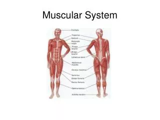
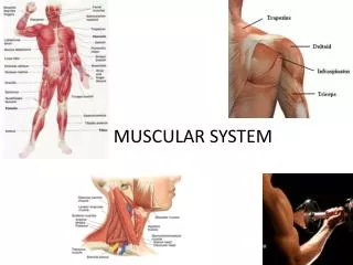
GROSS ANATOMY OF SKELETAL MUSCLES Practical 1 – head & neck • Facial Muscles • Frontalis • Orbicularis oculi • Orbicularis oris • Buccinator • Zygomaticus • Chewing Muscles • Masseter • Temporalis • Neck Muscles • Platysma • Sternocleidomastoid
GROSS ANATOMY OF SKELETAL MUSCLES Practical 2 – trunk muscles • Anterior • Pectoralis major • Intercostals (internal & external) • Muscles of the abdominal girdle: • Rectus abdominus • External oblique • Internal oblique • Transversus abdominus
GROSS ANATOMY OF SKELETAL MUSCLES Practical 2 – trunk muscles • Posterior • Trapezius • Latissimus dorsi • Erector spinae • Deltoid