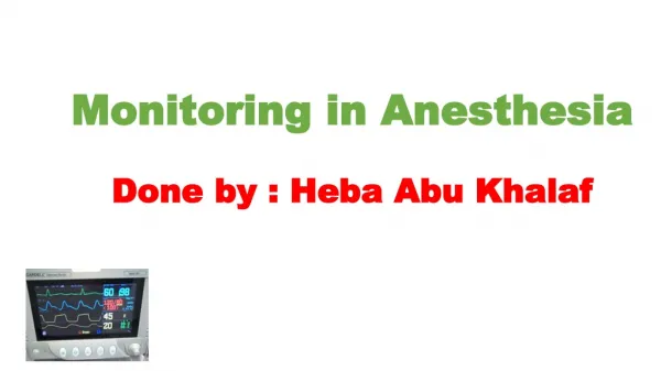
Monitoring in Anesthesia Done by : Heba Abu Khalaf
Monitoring in Anesthesia Done by : Heba Abu Khalaf. Objectives. 1. Guidelines to the practice of anesthesia and patient monitoring 2. Anesthesia depth 3 . Elements to monitor ( Oxygenation, Ventilation, Circulation, Temperature) 2.1. ECG 2.2. Pulse Oximetry 2.3. Blood Pressure

Monitoring in Anesthesia Done by : Heba Abu Khalaf
E N D
Presentation Transcript
Objectives • 1. Guidelines to the practice of anesthesia and patient monitoring • 2. Anesthesia depth • 3. Elements to monitor ( Oxygenation, Ventilation, Circulation, Temperature) • 2.1. ECG • 2.2. Pulse Oximetry • 2.3. Blood Pressure • 2.4. central venous line and pressure • 2.5. Capnography and EtCO2 • 2.6. How to identify Cyanosis • 2.7. The oxyhemoglobin dissociation curve • 3. Normal values for a healthy adult undergoing anesthesia
Guidelines to the practice of anesthesia and patient monitoring: • 1. an anesthetist present: “the only indispensable monitor”The doctor should present in the room & monitor the conduct of all general or regional anesthetics • 2. A completed pre-anesthetic checklist. • (ASA class, Hx &physical exam, investigations, NPO policy )
Con’t... • 3. perioperative anesthetic record: HR and BP every 5 min, O2 saturation, End Tidal CO2, dose and route of drugs and fluids • 4. continuous monitoring: patient’s oxygenation, ventilation, circulation and temperature .
Elements to Monitor : • I. Anesthetic Depth: • Patients with local or regional anesthesia provide verbal feedback regarding well being. • •Onset of general anesthesia signaled by lack of response to verbal commands, in addition to loss of blink reflex to light touch. • Inadequate anesthesia can be signaled by : Facial grimacing or movement of arm or leg. //blink reflex present when eyelashes lightly touched, • But with muscle relaxants ( fully paralysis), it can be signaled by : Hypertension, tachycardia, tearing or sweating. { Due to pain }
Con’t... • Excessive anesthesia can be signaled by : Cardiac depression, bradycardia, and Hypotension. also may result in hypoventilation, hypercapnia and hypoxemia when muscle relaxants is not given.
2. Oxygenation >> inspired Oxygen • we monitor it Clinically through observation of patient / skin color also by : • 1) pulse oximetry ( SaO2 ) • 2) Blood gas analysis ( Pao2 ) • 3) fraction of inspired O2 (FiO2) • Quantitavely monitored by using oxygen analyzer,equipped with an audible low oxygen concentration alarm.
• Pulse Oximetry: • ** mandatory monitor for any anesthetic ,, including cases of moderate sedation • **measure { non invasively} pt’s SpO2 ( arterial oxy saturation) • And blood flow fluctuation by plethysmograph (waveform of pulse oximeter “arterial waveform “ >> indicates that pulse oximeter is reading the arterial oxy saturation
Technique >> sensor containing light sources (Red and Infra-red light) & light detector is placed across finger tip , toe , earlobe or any other perfused tissue that can be transillumintaed • processing >>analyze amount of light absorbed by the 2 wavelengths,, then determining concentrations of oxygenated and deoxygenated forms through only arterial blood • light absorption is differ between oxyHb and deoxyHb. • analysis of oxygenation in each beat
Also it provides an indication of tissue perfusion & measure heart rate Inaccurate measurements ,, causes of oximetry artifact : 1) poor tissue perfusion (shock & hypotension) 2) movement 3)dysrhythmias 4) hypothermia ( cold extremities ) 5) cardiac arrest •Pulse oximetry (SpO2) measures oxy-, deoxy-, met-, and carboxyHb.
Pulse oximetry is never used for rapid diagnosis of hypoxia ( that may occur in unrecognized esophageal intubation ) • It used for monitoring oxygen delivery to vital organs • Also in recovery room , it helps identify post op pulmonary problems such as hypoventilation / bronchospasm / atelactasis • So timing of Spo2 monitoring >> before intubation , through the surgery ,, after extubation & recovery
Pulse oximeter tone changes with desaturation from high to low(deep) sound • So just by listening to the monitor ,,you can recognize the • 1) HR. 2) O2 saturation • Healthy patient under GA (O2= 100%) >> Spo2 96-100%
Rules : • # pay attention to the sound of pulse oximetry • # Always remember that your clinical judgment is much more superior to the monitor ,, check pt’s color for cyanosis ,, lips ,, nails
4.Temperature • ** should be monitored for patients under anesthesia • ** post op temp. >> used as quality anesthesia indicator • ** hypothermia associated with : • 1) delay drug metabolism, 2) impaired coagulation • ** hyperthermia has bad effects peri operatively leading to : • 1) tachycardia 2) vasodilation 3) neurological injury • So temp. Must be measured & recorded peri operatively
Hypothermia(<36°C) • Normal heat loss during anesthesia averages 0.5 - 1 C per hour, but usually not more that 2-3 C • Temperature below 34C may lead to significant morbidity • Hypothermia develops when thermoregulation fails to control balance of metabolic heat production and environment heat loss • Normal response to heat loss is impaired during anesthesia • • Those at high risk are elderly, burn patients , spinal cordinjuries
Causes of Hypothermia (<36°C) • **intraoperative temperature losses are common (e.g. 90% of intraoperative heat loss is transcutaneous) • >> due to: • 1) OR environment (cold room, IV fluids, instruments) • 2) open wound • ## prevented with forced air warming blanket and warmed IV fluids
Impact of Hypothermia • 1) Increased risk of wound infections >> due to impaired immune function • 2) Increases the period of hospitalization by delaying healing • 3) Reduces platelet function and impairs activation of coagulation cascade increasing blood loss and transfusion requirements • 4) Decreases the metabolism of anesthetic agents prolonging post-operative recovery
Causes of Hyperthermia (>37.5-38.3ºC) • 1) malignant hyperthermia • 2) drugs (e.g. atropine) • 3) blood transfusion reaction • 4) infection/sepsis • 5) • Increases in metabolic rate secondary to: • –Thyrotoxicosis • –Pheochromocytoma • 6) Excessive environmental warming
Continuous temperature measurements monitoring (Thermometry) is mandatory if changes in temperature are suspected. • ** intra op ,, temp measured by thermistor or thermocouple • Monitoring sites : • >> esophagus • >> tympanic membrane • >> nasopharynx. • >> Peripheral sites axilla & rectal.
2. Ventilation • Clinically, monitored through a correctly positioned endotracheal tube, also observing chest expansion and breath sounds over both lungs. • •Quantitavely by ETCO2 analysis, equipped with an audible disconnection alarm. • •Arterial blood gas analysis for assessing both oxygen and ventilation.
3. Circulation • Clinically monitored by pulse palpation, heart auscultation & monitoring intra-arterial pressure (MAP normally between 70 - 100 mmHg) or oximetry. • Quantitively using ECG & blood pressure measurements every 5 min.
Always remember that your clinical judgment is much more superior to Any monitor • monitor is present to help you not to be ignored and not to cancel you brain.