Fetal growth restriction
610 likes | 1.45k Views
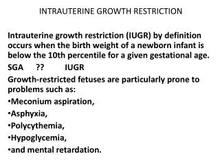
Fetal growth restriction. Joseph Breuner, md March 15, 2011. Teaching points. 16 wk rule Small vs in trouble Safer out or in: <34 wks use normal UA flow by DV to continue >34 wks deliver if Maternal htn Growth arrest x 3-4 wks Bpp low (<6/8) DV UA reversed or absent. Teaching points.

Fetal growth restriction
E N D
Presentation Transcript
Fetal growth restriction Joseph Breuner, md March 15, 2011
Teaching points • 16 wk rule • Small vs in trouble • Safer out or in: • <34 wks use normal UA flow by DV to continue • >34 wks deliver if • Maternal htn • Growth arrest x 3-4 wks • Bpp low (<6/8) • DV UA reversed or absent
Teaching points • 16 wk rule • Small vs in trouble • Safer out or in: • <34 wks use normal UA flow by DV to continue • >34 wks deliver if • Maternal htn • Growth arrest x 3-4 wks • Bpp low (<6/8) • DV UA reversed or absent
outline Risk factors Diagnosis Evaluation management
Risk factors Fetal Placental maternal
fetal • Genetic-SGA parent, sibling • Chromosomal abnormality • Congenital anomalies • Multiple gestation • infection
Placental risk factors • Ischemic placental disease=preeclampsia, FGR, abruption or combination • Small placenta- • Confined placental mosaicism
Maternal factors 1 • Uteroplacental insufficiency: htn, renal dz, diabetes, CVD, SLE, antiphospholipid syndrome, preeclampsia • Diminished caloric intake • Hypoxemia-pulm dz, cyanotic heart dz, severe anemia, high altitude
Maternal factors 2 • Hematologic d/o-sickle • Substance use-smoking, alcohol, stimulants • Toxins-meds warfarin, anticonvulsants, chemo, mtx • Caffeine • radiation
Maternal factors 3 • ART • Uterine malformations • Extremes of reproductive age • Short interpregnancy interval
outline Risk factors Diagnosis Evaluation management
16 week rule 1st 16 weeks: all cell division, uniform 2nd 16 weeks (16-32) combo cell division and growth, much more variability 32-40 primarily cell growth
16 week rule 1st 16 weeks: all cell division, uniform 2nd 16 weeks (16-32) combo cell division and growth, much more variability 32-40 primarily cell growth
definition <10%ile (50-70% will be constitutionally small) Some malnourished fetuses will be >10th%ile <5%ile Severe<3%ile
GA • Needs early US • Accuracy of Ultrasound decreases with advancing GA: • 1st TM: ± 3-5 days • 14-20 weeks: ± 7-10 days • 20-26 weeks: 2-3 weeks • Beyond 36 weeks: 3-4.5 weeks
Fundal height • Define by > 2cm discrepancy after 20 wks • Best when • Same clinician • Unmarked side of tape
Symmetric/asymmetric Symmetric-all organs decreasd proportionally due to hyperplasia(division) impairment Asymmetric-relatively greater decrease in abdominal size(liver volume/sq fat) than head circumference. Redistribution of blood flow to head
Symmetric-3 causes • Constitutionally small • Aneuploidy/congenital anomalies • Early infection
asymmetric • Always substrate deficiency
For fetuses <10th%ile 70% 20% 10%
For fetuses <5th%ile 40% 40% 20%
For fetuses <3th%ile 20% 53% 27%
ultrasound Crucial tool to Confirm/exclude dx Differentiate symmetric/asymmetric Verify fetal tolerance/well being
biometry Purpose of biometry is to detect FGR
Who gets an ultrasound? • >2cm FH discrepancy once • 2 cm FH discrepancy more than once • Or following RF’s
US Screening for IUGR • Previous IUGR fetus • Maternal medical condition • CHTN, autoimmune disorders, asthma, APS • Multiples • Uterine anomaly • Placental abnormality • Marginal/velamentous cord • Ongoing bleeding • Chronic abruption, previa • Abnormal serum screening analytes
AC most sensitive 3616 preg >25 wks gestation Single us within two wks delivery Predicted <10th%ile for GA with Sens 61% Spec 95% Ppv 86% Npv 83%
AC • More predictive of FGR than • HC • BPD • Combination
AC • More sensitive in asymmetric than symmetric GR (73%vs59%) • GA-more sensitive later in gestation • Sn/PPV at 29-31 wks 41/51%, at term 88/71% • More sensitive when interval between measurements >2wks. FP rates for interexam intervals 1,2,4 wks 31,17,3%
EFW Best algorithm uses ac,bpd,fl EFW within 10% of actual bw 75% of time Sn/sp/ppv/npv 90/85/80/90 Sens increases with worsening GR Log10 BW = 1.335 - 0.0034(AC)(FL) + 0.0316(BPD) +0.0457(AC) + 0.1623(FL)
Growth velocity • Small vs in trouble • Falling %iles shd raise alarm
ratios • HC/AC • FL/AC • TCD/AC
Diagnosis summary • Of four entities described-AC, EFW, growth velocity, and ratios • Dx depends on EFW • Log10 BW = 1.335 - 0.0034(AC)(FL) + 0.0316(BPD) +0.0457(AC) + 0.1623(FL)
Diagnosis summary • 10%ile-at risk • 5th%ile act • 3rd%ile
Evaluation and management • H+P-etoh,tobacco, maternal vasc dz • Anatomic survey/fetal echo • Karyotype for <32 wks, <3rd %ile, polyhydramnios or structural anomalies • Ab testing for cmv/rubella/vzv only if maternal symptoms suggestive or US shows echo/calcification of liver, brain, or hydrops
follow • Fetal wt for growth velocity q 2-4 wks • BPP 1-2x/wk • Amniotic fluid volume weekly (part of bpp) • doppler
doppler • Cochrane review • Doppler vs no doppler in FGR pregnancies • Reduced perinatal deaths by 29% (OR 0.71, CI 0.5-1.01)
Umbilical artery • Increasing systolic/diastolic ratio, f/b • Absent or reversed end-diastolic flow
Middle cerebral artery • Reported as umbilical/intracranial ratio, or • Peak velocity of middle cerebral artery
Umbilical vein or ductus venosus • Ductus venosus flow reverses • Umbilical vein develops pulsatile flow
Sample us report from inpt • Indication: IUGR with estimated weight on the 3rd percentile on 3/11/2011. Admitted for treatment. Comparison: 12/3/2010 through 3/11/2011. Expected menstrual age of 32 weeks 5 days with EDD 5/3/2011. The fetus is vertex with an anterior normal appearing placenta of normal thickness. The amniotic fluid is decreasing. AFI is 6 to 7 today done 3 times. The AFI was 10 on 3/11/2011. Fetal Doppler: Performed because of IUGR The umbilical arterial systolic diastolic ratio of 2.5 is normal. The umbilical/intracranial Doppler ratio of 0.5 is normal. The ductus venosus waveform was technically difficult to obtain because of fetal position, but limited adequate samples appear normal.
Sample us report • Based on EDD 5/13/2011, the expected menstrual age is 31 weeks. Twin live intrauterine fetuses identified. The presenting twin is in breech position and the nonpresenting twin is in cephalic position. Confluent posterior placenta is identified. Separating membrane is visualized. • The amount of amniotic fluid appears normal for twin A with the amniotic fluid index of 11. The amount of amniotic fluid appears normal for twin B with the amniotic fluid index of 12. There is no evidence for hydrops fetalis for either twin. • OB DOPPLER performed, per request, because of monochorionicity. Twin A: Umbilical artery Doppler shows normal systolic to diastolic ratio of 2.5. Umbilical/intracranial Doppler ratio of 0.4 is normal. Middle cerebral artery peak systolic velocity 55 cm/sec is 1.3 multiples of the median and normal. Twin B: Umbilical artery Doppler shows normal systolic to diastolic ratio of 2.9. Umbilical/intracranial Doppler ratio of 0.4 is normal. Middle cerebral artery peak systolic velocity 42 cm/sec is 1.0 multiples of the median and normal.
steroids • Two large studies conflict • Reasonable to administer • Likely the very impaired fetus can’t respond
Delivery timing • Growth Restriction Intervention Trial (GRIT) • 580 women 24-34 wks • Randomly assigned to immediate or delayed delivery groups if ob uncertain when to intervene • 90%FGR • 40% absent or reversed end diast UA flow
GRIT • Immediate delivery: when ob uncertain • Delayed delivery: when ob no longer uncertain (average delay 4.9d) • Deaths prior to hospital d/c same 29/27 • Immediate fewer stillbirths (2 v 9) but more neonatal/infant deaths (27 v 18)
Recommended management • Remote from term <34 wks • Follow doppler. Prolong pregnancy if UA flow normal. Deliver if absent/reversed • Some experts await abnormal venous flow in very preterm-investigational