Download
1 / 3
30 likes | 41 Views
Case reports are characterized as the logical documentation of a solitary clinical perception and have a long-established and rich custom in medication and logical distribution

E N D
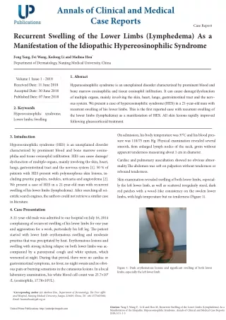
Annals of Clinical and Medical Case Reports Case Report Recurrent Swelling of the Lower Limbs (Lymphedema) As a Manifestation of the Idiopathic Hypereosinophilic Syndrome Feng Yang, Fei Wang, Kedong Li and Maihua Hou* Department of Dermatology, Nanjing Medical University, China 1. Absract Volume 1 Issue 1 - 2018 Received Date: 11 June 2018 Accepted Date: 30 June 2018 Published Date: 07 June 2018 Hypereosinophilic syndrome is an unexplained disorder characterized by prominent blood and bone marrow eosinophilia and tissue eosinophil infiltration. It can cause damage/dysfunction of multiple organs, mainly involving the skin, heart, lungs, gastrointestinal tract and the nerv- ous system. We present a case of hypereosinophilic syndrome (HES) in a 21-year-old man with recurrent swelling of his lower limbs. This is the first reported case with recurrent swelling of the lower limbs (lymphedema) as a manifestation of HES. All skin lesions rapidly improved following glucocorticoid treatment. 2. Keywords Hypereosinophilic syndrome; Lower limbs; Swelling On admission, his body temperature was 37°C and his blood pres- sure was 110/75 mm Hg. Physical examination revealed several smooth, firm enlarged lymph nodes of the neck, groin without apparent tenderness measuring about 1 cm in diameter. 3. Intuduction Hypereosinophilic syndrome (HES) is an unexplained disorder characterized by prominent blood and bone marrow eosino- philia and tissue eosinophil infiltration. HES can cause damage/ dysfunction of multiple organs, mainly involving the skin, heart, lungs, gastrointestinal tract and the nervous system [1]. 50 % of patients with HES present with polymorphous skin lesions, in- cluding pruritic papules, nodules, urticaria and angioedema [2]. We present a case of HES in a 21-year-old man with recurrent swelling of his lower limbs (lymphedema). After searching all sci- entific search engines, the authors could not retrieve a similar case in literature. Cardiac and pulmonary auscultation showed no obvious abnor- mality.The abdomen was soft on palpation without tenderness or rebound tenderness. Skin examination revealed swelling of both lower limbs, especial- ly the left lower limb, as well as scattered irregularly sized, dark red patches with a wood-1ike consistency on the swolen lower limbs, with high temperature but no tenderness (Figure 1). 4. Case Presentation A 21-year-old male was admitted to our hospital on July 16, 2014 complaining of recurrent swelling of his lower limbs for one year and aggravation for a week, particularly his left leg. The patient started with lower limb erythematous swelling and moderate pruritus that was precipitated by heat. Erythematous lesions and swelling with strong itching relapse on both lower limbs was ac- companied by a paroxysmal cough and white sputum, which worsened at night. During that period, there were no cardiac or gastrointestinal symptoms, no fever, no night sweats and no obvi- ous pain or burning sensations in the cutaneous lesions. In a local laboratory examination, his white blood cell count was 25.7×10⁹ /L (eosinophils, 17.78×10⁹/L). Figure 1: Dark erythematous lesions and significant swelling of both lower limbs, especially the left lower limb. *Corresponding Author (s): Maihua Hou, Department of Dermatology, The First Affili- ated Hospital, Nanjing Medical University, Jiangsu 210029, China, Tel: +86 13776635881; Email: houmaihua@jsph.org.cn Citation: Yang F, Wang F, Li K and Hou M, Recurrent Swelling of the Lower Limbs (Lymphedema) As a Manifestation of the Idiopathic Hypereosinophilic Syndrome. Annals of Clinical and Medical Case Reports 2018; 1(1): 1-3 United Prime Publications: http://unitedprimepub.com
Volume 1 Issue 1 -2018 Case Report Routine blood test results showed a white blood cell count of 29.95×10⁹/L with over 70% eosinophils at the absolute count of 21.54×10⁹/L. Biochemistry tests revealed uric acid(459umol/L), lipoprotein a (1107 mg/L), alpha hydroxy butyric acid dehydro- genase (230 U/L), resistance of Streptococcus hemolysin “O” (558.00 IU/mL), and total IgE (577.50 KU/L), with high-density lipoprotein cholesterol (0.77 mmol/L) and low density lipopro- tein cholesterol (2.39 mmol/L) declining mildly. No abnormality was found in his urine, coagulation routine, liver or kidneys. The results of sputum culture for several times were negative. The pa- tient was negative for antibodies to tuberculosis, filaria, trepone- ma pallidum and Human Immunodeficiency Virus (HIV). Tests for immunoglobulin (IgA, IgG, IgM), antinuclear antibody se- ries, IFN-γ and complement (C3, C4) revealed no abnormalities. Quantitative DNA of EB virus and cytomegalovirus in the blood was normal. The results of immunoglobulin gene rearrangement and TCR gene rearrangement were negative. Histology of a lesional specimen showed dense eosinophilic in- filtration, a lymphatic dilatation in the dermis, eosinophil emboli in the lumens (Figure 4), and positive staining for D2-40 in lym- phatic vessels. The FIPILT-PDGFRA fusion gene was negative. Based on the sustained eosinophilia, cutaneous manifestations and exclusion of secondary causes, a diagnosis of HES was made. Figure 4: Histology of lesional specimen: Massive eosinophilic infiltration in the dermis, around subcutaneous fat, blood vessels and collagen. Eosinophil emboli in the lumens HE×200. The patient was then treated with 40 mg methylprednisolone along with anticoagulants (warfarin, low molecular heparin) and an antiplatelet (aspirin), which led to a dramatic reduction in his peripheral blood eosinophilia (at the absolute count of 0.63×10⁹/L) and clearance of his skin rash in the first week of treatment. Thus far, follow-up has been consistent. An electrocardiogram (ECG) indicated sinus arrhythmia. Ultra- sound examination of the liver, gallbladder, pancreas and spleen showed no abnormalities. Echocardiography showed mild mitral and tricuspid regurgitation. Vascular ultrasound showed throm- bosis in his right anterior tibial artery, his left foot dorsal artery and his lower left saphenous vein. CT scans revealed multiple en- larged lymph nodes in the mediastinum, the armpit and the sur- rounding retroperitoneal and abdominal aorta. A bone marrow aspirate (Figure 2), 5. Discussion Hypereosinophilic syndrome (HES) is a disease characterized by the following: (a) a persistent absolute eosinophil count (AEC) of >1500 cells/uL documented on two occasions at least 1 month apart and/or pathologic confirmation of tissue hypereosinophil- ia, (b) evidence of eosinophil-mediated organ damage or dys- function, and (c) other potential causes of the damage have been ruled out [3-8]. In our case, bone marrow biopsy and lymph node biopsy both indicate hypereosinophilia. Owing to dense eosino- philic infiltration and lymphatic dilation in the dermis, eerythe- matous lesions and swelling relapse on both lower limbs. Mean- while, we have excluded potential causes of the damage such as maligant lymphadenoma. Accordingly, our case fulfilled those di- agnostic criteria of HES. Treatment of HES is generally aimed at long-term reduction of eosinophil levels in the blood and tissues to avoid end-organ damage, minimizing damage from the end products of eosinophil metabolism.Corticosteroid is the first-line therapy for FIP1L1-PDGFRA-negative HES, and is very effective for reducing levels of peripheral eosinophils. In this case, our patient presented recurrent nonpitting edema of lower limbs as well as scattered irregularly sized, dark red patches with a wood- 1ike consistency on the swolen lower limbs. The skin lesions were tough, and the biopsy was difficult to conduct. This lesion should be differentiated with venous thrombosis. The latter often pre- Figure 2: Bone marrow aspirate: A significant proliferation of granulocytes and increased eosinophils. Wright's staining×1000. a bone marrow biopsy and a lymph node biopsy (Figure 3) showed a great deal of eosinophilic hyperplasia. Figure 3: Lymph node biopsy: Reactive hyperplasia accompanied by eosino- philia. HE×400. Copyright ©2018 Hou M. This is an open access article distributed under the terms of the Creative Commons Attribution License, which permits unrestricted use, distribution, and build upon your work non-commercially. 2
Volume 1 Issue 1 -2018 Case Report sents acute painful edema with soft skin and could subside after prolonged elevation of the affected limb. After the treatment with glucocorticoid, our patient’s eosinophil counts normalized within 3 days, the swelling of the lower extremities and his skin rash dis- appeared in the first week. The clinical improvement strongly sug- gests that our patient had an excellent response to glucocorticoid and that the lymphatic emboli resulted from mechanical obstruc- tions by increased eosinophil counts. When the eosinophil counts rapidly returned to normal, the emboli disappeared quickly. Thus, we assume that eosinophil emboli can be relieved spontaneously and are sensitive to glucocorticoid, which is associated with the characteristics of the lymphatic system, such as a higher water content than the plasma, without platelets or other blood coagu- lation factors. Further, the lymphatic system is a regulator of tis- sue fluid. A great number of eosinophils appeared in the patient’s blood, bone marrow and lymph nodes, resulting in hematological and lymphatic system embolism. Therefore, the patient still re- quires a long-term follow-up to monitor the risk of progressing to eosinophilic leukemia. 3. Valent P, Klion AD, Horny HP, Roufosse F, Gotlib J, Weller PF, et al. Contemporary consensus proposal on criteria and classification of eo- sinophilic disorders and related syndromes. J Allergy Clin Immunol. 2012;130(3):607-12. 4. Park SM, Park JW, Kim SM, Koo EH, Lee JY, Lee CS, et al. A case of hypereosinophilic syndrome presenting with multiorgan infarctions as- sociated with disseminated intravascular coagulation. Allergy Asthma Immunol Res. 2012;4(3):161-4. 5. Rothenberg ME, Klion AD, Roufosse FE, Emmanuel Kahn J, Weller PF, Uwe Simon H, et al. Treatment of patients with the hypereosinophil- ic syndrome with mepolizumab. N Engl J Med. 2008;358(12):1215-28. 6. Park YM, Bochner BS. Eosinophil survival and apoptosis in health and disease. Allergy Asthma Immunol Res. 2010;2(2):87-101. 7. Kobayashi M, Komatsu N, Kuwayama Y, Bandobashi K, Kubota T, Ue- mura Y, et al. Idiopathic hypereosinophilic syndrome presenting acute abdomen. Intern Med. 2007;46(10):675-8. 8. Ferguson GT, Starkebaum G. Thromboangiitis obliterans associated with idiopathic hypereosinophilia. Arch Intern Med. 1985;145(9):1726- 8. References 1. Klion A. Hypereosinophilic syndrome: current approach to diagnosis and treatment. Annu Rev Med. 2009;60:293-306. 2. Carlesimo M, Fidanza L, Mari E, Feliziani G, Narcisi A, De Marco G, et al. Wells syndrome with multiorgan involvement mimicking hypereo- sinophilic syndrome. Case Rep Dermatol. 2009;1:44-8. United Prime Publications: http://unitedprimepub.com 3