Download
1 / 3
0 likes | 77 Views
In the realm of cardiovascular health assessment, the 2D echo test has long been a cornerstone. The non-invasive imaging method has completely changed how we see and comprehend the heart. As technology continues to evolve, so too does the landscape of 2D echocardiography research, unveiling new dimensions of knowledge and diagnostic capabilities

E N D
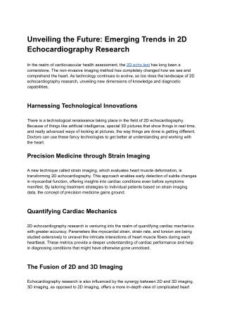
Unveiling the Future: Emerging Trends in 2D Echocardiography Research In the realm of cardiovascular health assessment, the 2D echo test has long been a cornerstone. The non-invasive imaging method has completely changed how we see and comprehend the heart. As technology continues to evolve, so too does the landscape of 2D echocardiography research, unveiling new dimensions of knowledge and diagnostic capabilities. Harnessing Technological Innovations There is a technological renaissance taking place in the field of 2D echocardiography. Because of things like artificial intelligence, special 3D pictures that show things in real time, and really advanced ways of looking at pictures, the way things are done is getting different. Doctors can use these fancy technologies to get better at understanding and working with the heart. Precision Medicine through Strain Imaging A new technique called strain imaging, which evaluates heart muscle deformation, is transforming 2D echocardiography. This approach enables early detection of subtle changes in myocardial function, offering insights into cardiac conditions even before symptoms manifest. By tailoring treatment strategies to individual patients based on strain imaging data, the concept of precision medicine gains ground. Quantifying Cardiac Mechanics 2D echocardiography research is venturing into the realm of quantifying cardiac mechanics with greater accuracy. Parameters like myocardial strain, strain rate, and torsion are being studied extensively to unravel the intricate interactions of heart muscle fibers during each heartbeat. These metrics provide a deeper understanding of cardiac performance and help in diagnosing conditions that might have otherwise gone unnoticed. The Fusion of 2D and 3D Imaging Echocardiography research is also influenced by the synergy between 2D and 3D imaging. 3D imaging, as opposed to 2D imaging, offers a more in-depth view of complicated heart
structure and function. The combination of these methods produces a complete diagnostic toolset that improves clinical judgment. Advancements in Transesophageal Echocardiography (TEE) Transesophageal echocardiography, commonly known as TEE, is experiencing significant advancements. In this technique, a probe is inserted into the esophagus to obtain a clearer image of the heart. Recent research focuses on improving TEE image quality, enhancing its diagnostic potential for conditions like atrial fibrillation, valvular diseases, and intracardiac masses. Point-of-Care Ultrasound (POCUS) With point-of-care ultrasound, clinicians can now use diagnostic tools directly at the point of care. In emergency settings and critical care scenarios, the 2D echo test is now being performed at the bedside, providing real-time insights into cardiac function. This trend not only expedites diagnoses but also improves patient outcomes through prompt interventions. Virtual Reality for Training and Education 2D echocardiography research is also branching into the realm of education and training. VR platforms are being developed to provide healthcare professionals with immersive learning experiences. These platforms allow practitioners to simulate 2D echo tests and interact with virtual cardiac anatomy, enhancing their proficiency and confidence. Enhanced Visualization with Contrast Echocardiography A new way of looking at the heart called contrast echocardiography is becoming more popular in research and medical care. They use tiny bubble-like things to make the heart's pictures clearer, especially when it's hard to see or the pictures aren't very clear. This helps doctors make better diagnoses and decide on better treatments. Big hospitals like Wockhardt now have this fancy and high-tech equipment. Conclusion: A Vision of Discovery As the canvas of 2D echocardiography research expands, new brushstrokes of knowledge are being added with each discovery. The special heart test called the 2D echo has
improved a lot over time. Using this special test, we can look at the heart more closely and understand it better. With new tools and ways of looking, we can be hopeful about finding issues and helping patients in a better way. Doctors and scientists are teaming up to learn more about the heart using this test. It's like an adventure that helps us find out secrets about the heart and learn how to keep it in good shape. It's not only about looking at pictures on a screen, but also about really understanding and taking care of hearts with care and determination.