Download
1 / 13
130 likes | 523 Views
Bone Remodeling of the Equine Distal Limb. Mark D. Fischer M.D. Orthopedic Surgeon University of Minnesota, USA Sheri L. Fischer R.N., B.S.N. Fischer Equine Lameness Foundation www.Healthehoof.com. Coffin Bone. Lateral view of coffin bone (terminal phalanx). Cortical and cancellous bone.

E N D
Bone Remodeling of the Equine Distal Limb Mark D. Fischer M.D. Orthopedic Surgeon University of Minnesota, USA Sheri L. Fischer R.N., B.S.N. Fischer Equine Lameness Foundation www.Healthehoof.com
Coffin Bone • Lateral view of coffin bone (terminal phalanx)
Cortical and cancellous bone • On the left-side is a model illustrating cancellous bone, circled in blue; on the right is a human tibia with the cancellous portion circled in blue.
Bone Blood Supply • Cancellous is circled in blue; cortical circled in red; tibia demonstrated • Blood supply is illustrated; there are both internal and external arterial sources.
Cells of bone • Osteocyte • Osteoblast • Osteoclast
Cells of Bone in Action • The large cell on the left, circled in blue, is an osteoclast. • The row of cells circled in red on the right, producing the pink material, are osteoblasts.
Osteocyte in Mature Bone • The osteocyte, identified by the large arrrow, are the small cells within the bone. • The small arrow identifies blood vessels within bone.
Osteoclast resorbing bone • Osteoclast is seen on the surface of bone, presumably reabsorbing bone.
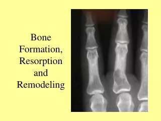
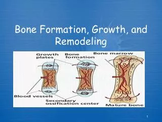
Bone formation from cartilage • Ossification of cartilage due to vascular ingrowth
Bone Marrow and Spongy Bone • Microstructure of spongy bone • Illustrated are three cells. The first is the osteoblast (light blue) producing new bone. The second is the osteoclast (pink) reabsorbing bone. The third is the osteocytes between the layers nourishing bone.
Localized Bone Loss • Loss of blood supply due to arterial occlusion has resulted in necrosis and bone loss.
Side bone • Side bone is the ossification of the lateral cartilages due to alterations in weight bearing stress forces.
Ring Bone • High and low ring bone due to increased traction forces at insertions of ligaments.