Download

1 / 36
360 likes | 642 Views
MEDICAL SCANS. How they work, what they measure, and how to read them Hosted by MEDSOC – January 17, 2007. Computerized Tomography. Overview. Simply a series of 2-D X-Rays taken to create a 3-D picture Because of many X-Rays, dangerous exposure to radiation

E N D
MEDICAL SCANS How they work, what they measure, and how to read them Hosted by MEDSOC – January 17, 2007
Overview • Simply a series of 2-D X-Rays taken to create a 3-D picture • Because of many X-Rays, dangerous exposure to radiation • CT scan measures differences in density of the tissues • 3-D nature extremely effective
How to Read a CT Scan • Lighter areas have a higher density
When are CT Scans Used? • CT Scans are most commonly used with the skeletal system • Broad-spectrum analysis of anatomical changes/insults • 3-D nature allows analysis of breaks or other trauma from more than one perspective
The CHEM-7 Test • Analysis of 7 different chemicals in the blood • Determines chemical level and concentration • Indicates abnormalities in tissues
Blood Urea Nitrogen (BUN) • Urea Nitrogen: present when amino acids break down and NH4+ (ammonium) combines with other substances • Usually indicates kidney and liver function • Normal levels: 7 to 20 mg/dl • Too low: liver failure, malnutrition, over-hydration • Too high: starvation, low protein, kidney disease/failure, heart attack, urinary track obstruction
Serum Chloride • Chloride maintains electric neutrality by counter-ion to sodium • Normal Range: 101 to 111 mmol/L • Too low: blood is acidic (acidosis), low bicarbonate levels, malfunctioning kidney • Too high: hormone deficiency (Addison’s disease, respiratory acidosis)
Carbon Dioxide • Measures CO2 and HCO3 (bicarbonate) • Kidney and Respiratory function • Normal: 20 to 29 mmol/L • Too low: problem with kidney • Too high: liver dysfunction
Creatinine • Breakdown product of creatine, an important part of muscle • Evaluates kidney function • Normal value: 0.8 to 1.4 mg/dl • Too low: muscular dystrophy • Too high: kidney disease/failure
Glucose • Blood sugar levels • Can be used to diagnose diabetes • Normal level: 100 mg/dL • Too high: most likely at risk of diabetes (greater than 100 mg/dL), diabetes diagnosed if greater than 100 mg/dL • Too low: too much insulin (rare)
Potassium • Positive ion in maintaining electric charge • Normal range: 3.7 to 5.2 mEq/L • Too high: kidney failure, respiratory acidosis • Too low: improper diet, narrowing of major blood vessels to kidney
Sodium • Sodium content in blood • Normal level: 135 to 145 mEq/L • Too high: Excessive sweating, too much aldosterone or cortisol • Too low: dehydration, heart failure, kidney disease, cirrhosis of liver, improper hormone levels (too high or low)
Blood Pressure • Stethoscope and sphygmomanometer • Measures pressure blood exerts on arteries • Highest pressure: systolic • Lowest pressure: diastolic • Typical adult blood pressure: 120 mmHg (systolic) and 80 mmHg (diastolic) • High blood pressure may lead to heart attacks, stroke • Low blood pressure needs urgent medical attention
Overview • Basic Design: a giant cube • Horizontal Tube Magnet- bore • Body part to be examined must be at the isocenter • Used for diagnosing various medical problems because of its flexibility in producing data.
The Physics • Rating unit: tesla or gauss • 3 Types of magnets in the MRI system: • Resistive Magnets • Permanent Magnets • Superconducting Magnets
The Physics • Resistive Magnets • Windings/coils of wire wrapped around a bore through which a current is passed • Require huge amounts of electricity
The Physics • Permanent Magnets • Magnet that suits its name- magnetic field is always present • Extremely heavy (drawback)
The Physics • Superconducting Magnets • Most commonly used in MRIs • Similar to the resistive magnet but… wire is continually bathed in liquid helium at -425.4oC. • Electricity required is substantially less
The Physics • Magnetic Fields • Homogenous- stable magnetic field; good for high quality imaging • Gradient Magnet- Three of these in every MRI machine; low strength • Main Magnet(resistive, etc.) -stable magnetic field • Gradient magnets- variable magnetic field
The Physics • Machine adds radio frequency pulse to area tested • Pulse causes protons in the area to spin • Pulse is transmitted through a coil that is specific to the body part tested • Gradient magnets alter magnetic field at local level
The Physics • As protons return to natural alignment, the coil picks the signals from the protons, and then sends the info to a computer system which then converts it the info into a picture using the fourier system.
How to Read an MRI • Black areas- where there is lesser hydrogen atoms in the tissue • White/Brighter areas- where there are many protons in the tissue- e.g.: fatty tissue • Tumors and “Diseases” are detected when they differ from the standard and when usually a conspicuous white “blob” is observed
When are MRI Scans Used? • Hydrogen atoms v. Hydrogen ions • May be used to differentiate between water and fatty tissue • Anatomical abnormalities
Overview • PET Scans involve radioactive isotopes • Radioactive isotopes are injected into bloodstream • Machine measures radioactivity • Constructs 2-D or 3-D image of body
The Physics • Radioactive isotopes are chemically incorporated into metabolically active molecule • Host cells uptake radioactivity based on metabolic rate • Water, glucose • Radioactivity generates a positron • Annihilates with electron
The Physics • Annihilation produces a pair of gamma photons • Measured by machine • Delay measured to remove data from random photons • Millions of these data points are collected and analyzed
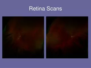
How to Read a PET Scan • Areas of high intensity (red) indicate high metabolic rate • This may correlate to high water uptake or glucose uptake, depending on which radioactive material you use • Usually used in conjunction with CT or MRI
When are PET Scans Used? • PET scans may be sensitive to changes in chemical compositions or metabolic rates even before physiological marker is present • CT, MRI for anatomical signal • Cancers almost always have different metabolic rates