Download

1 / 10
0 likes | 17 Views
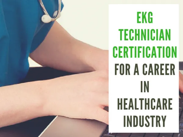
Prepare for the DHA EKG Technician exam and boost your career in Dubai's healthcare sector. Our comprehensive guide offers key insights into the DHA-EKG certification process, including eligibility, application steps, exam preparation tips, and essential study materials. Whether you're an international candidate or based in Dubai, ensure you're fully equipped to pass the DHA Electrocardiography (EKG) Technician exam and achieve certification.<br>USE 16 USD Discount Coupon Code: 9M2GK4NW<br>https://www.testsexpert.com/dha-ekg/

E N D
International DHA-EKG DHA EKG Technician (DHA-EKG) •Up to Date products, reliable and verified. •Questions and Answers in PDF Format. Full Version Features: •90 Days Free Updates •30 Days Money Back Guarantee •Instant Download Once Purchased •24 Hours Live Chat Support For More Information: https://www.testsexpert.com/ •Product Version Visit us at: https://www.testsexpert.com/dha-ekg
Latest Version: 6.0 Question: 1 The opening in the aorta that allows blood flow to the coronary arteries is called which of the following? A.Pulmonic valve B.Aortic valve C.Coronary ostium D.Right coronary artery Answer: C Explanation: The question asks about the name of the opening in the aorta that allows blood to flow to the coronary arteries. The correct answer is the "Coronary ostium." The coronary arteries are critical as they supply oxygen-rich blood to the heart muscle itself. Without this oxygenated blood, the heart tissue would not function properly, leading to possible heart damage or a heart attack. The coronary ostia (plural for ostium) are small openings in the wall of the aorta just above the aortic valve. These openings mark the beginning of the coronary arteries. There are typically two main coronary ostia: one for the right coronary artery and one for the left coronary artery. The right coronary ostium leads to the right coronary artery, which supplies the right side of the heart, the bottom portion of both ventricles, and the back of the septum. The left coronary ostium gives rise to the left coronary artery, which quickly bifurcates into the left anterior descending artery and the circumflex artery, serving the left side of the heart and much of the front of the heart and the septum. It's important to differentiate the coronary ostium from other structures such as the pulmonic valve and the aortic valve, which serve different functions. The pulmonic valve regulates blood flow from the right ventricle into the pulmonary artery leading to the lungs, while the aortic valve controls blood flow from the left ventricle into the aorta, the body's main artery. Neither of these valves is directly involved with the coronary circulation. Given this information, when considering the question, early "Coronary ostium." This term specifically refers to the openings from the aorta to the coronary arteries, enabling the delivery of oxygen-rich blood to the heart muscle. Question: 2 In some facilities, the ECG technician also does all of the following except? A.Maintains the patient files B.Cares for the equipment C.Schedules appointments D.Bills for procedures Visit us at: https://www.testsexpert.com/dha-ekg
Answer: D Explanation: An ECG (electrocardiogram) technician is a specialized healthcare professional whose primary role involves performing diagnostic tests that help in measuring a heart’s electrical activity. These tests are crucial as they assist in the detection and treatment of cardiovascular diseases. ECG technicians are trained to operate ECG machines, and their responsibilities often extend to several other duties that support the functioning of healthcare facilities. However, the scope of their responsibilities can vary depending on the specific rules and organizational structure of the facility where they are employed. Among the common responsibilities of an ECG technician are maintaining patient files, caring for the equipment, scheduling appointments, and typing doctors’ interpretations. Maintaining patient files involves organizing and managing the documentation necessary for patient care, ensuring that all records are up-to-date and available for consultations. This is crucial for maintaining continuity in patient care and for legal and compliance reasons. Caring for the equipment is another critical task. ECG machines and other diagnostic tools must be kept in optimal working condition to ensure accurate readings. This involves regular maintenance and troubleshooting any issues that arise, thereby preventing disruptions in the facility's ability to provide timely diagnostic testing. Scheduling appointments is a logistical task that ECG technicians might handle. This involves coordinating between patients and medical staff to ensure that the testing is conducted efficiently. It helps in managing the workflow of the facility, reducing waiting times, and improving overall patient satisfaction. Typing doctors’ interpretations involves transcribing the observations and conclusions made by the physicians from the diagnostic tests. This transcription must be accurate to ensure that the patient records reflect the correct medical insights, which are crucial for further treatments or interventions. However, one task that ECG technicians typically do not handle is billing for procedures. Billing involves preparing and processing details of the services provided to patients so that the facility can charge them or their insurance providers. Billing requires understanding of medical billing codes and often involves navigating complex insurance protocols and regulations. This role is usually handled by specialized billing staff or administrative professionals trained explicitly in finance and billing within the healthcare context. Therefore, while ECG technicians play a multifaceted role in supporting cardiac diagnostics and patient care, billing for procedures generally falls outside the scope of their responsibilities. This delineation helps ensure that each professional can focus on their area of expertise, thereby enhancing the efficiency and effectiveness of the healthcare facility's operations. Question: 3 What is considered to be a major component of standard precautions in a healthcare facility? A.Gloves B.Masks C.Gowns D.Hand-washing Answer: D Visit us at: https://www.testsexpert.com/dha-ekg
Explanation: Standard precautions are a set of infection control practices used to prevent transmission of diseases that can be acquired by contact with blood, body fluids, non-intact skin (including rashes), and mucous membranes. These precautions are the basic level of infection control that should be used in the care of all patients all of the time. The use of these precautions effectively reduces the risk of transmitting pathogens in healthcare settings. One major component of standard precautions is hand hygiene. Hand hygiene is considered the most important and fundamental measure for reducing the risk of transmitting infectious agents. It involves either washing hands with soap and water or using an alcohol-based hand sanitizer. Proper hand hygiene should be performed before and after every patient contact, after contact with blood, body fluids, secretions, excretions, and contaminated items; immediately after removing gloves; and between patient contacts. In addition to hand hygiene, the use of personal protective equipment (PPE) is another crucial element of standard precautions. PPE includes gloves, gowns, masks, and eye protection. Each piece of equipment serves a specific purpose: - **Gloves** protect hands from contamination with infectious materials and also help reduce the likelihood of transmitting pathogens from healthcare personnel to patients. - **Gowns** protect the skin and clothing from contamination with potentially infectious fluids and materials. - **Masks** and respirators protect mucous membranes of the nose and mouth from droplets of infectious material that might be expelled by a patient during coughing, sneezing, or certain medical procedures. - **Eye protection**, such as goggles or face shields, protect the eyes from splashes of infectious material. Another important aspect of standard precautions is the safe use and disposal of sharps. This includes needles, scalpels, and other sharp instruments or devices. Proper handling, disposal, and immediate action following a needlestick or similar injury are crucial to prevent the transmission of infections. Finally, environmental cleaning and disinfection are also key components of standard precautions. Surfaces, medical equipment, and patient care areas must be regularly cleaned and disinfected to reduce the spread of pathogens. Overall, while hand hygiene stands out as a primary and highly critical component of infection control, all elements of standard precautions are integral to preventing healthcare-associated infections. Each component contributes uniquely to the safety of both healthcare personnel and patients. Question: 4 The medulla oblongata is what control for the heart? A.Nervous control B.Size C.Thickness D.None of the above Answer: A Explanation: The medulla oblongata, a critical structure in the brain, plays a central role in regulating various involuntary functions essential for life. One of its key functions is the control over the heart, particularly in regard to the autonomic nervous system, which operates below the level of consciousness. Visit us at: https://www.testsexpert.com/dha-ekg
The autonomic nervous system is divided into the sympathetic and parasympathetic nervous systems. The medulla oblongata houses important centers that influence both these systems to maintain cardiovascular homeostasis. The cardiac control centers within the medulla oblongata can modulate heart rate and the strength of heart contractions based on continuous feedback from the body. Specifically, the medulla contains the cardiac center with two main components: the cardioacceleratory center and the cardioinhibitory center. The cardioacceleratory center activates the sympathetic neurons, which release neurotransmitters to increase heart rate and the force of heart contractions during activities requiring heightened alertness or physical exertion. Conversely, the cardioinhibitory center stimulates the parasympathetic neurons, which release acetylcholine to decrease the heart rate and conserve energy during resting or non-stressful periods. This dual control allows the body to respond rapidly to changing conditions, ensuring that oxygen and nutrient demands of tissues are met efficiently. For instance, during exercise or stress, the sympathetic influence prevails, increasing heart rate and blood pressure. During rest or sleep, parasympathetic tone dominates, slowing down the heart rate and reducing blood pressure. In summary, the medulla oblongata is essential for the nervous control of the heart, regulating its activity to adapt to both internal and external stimuli seamlessly. This makes it the central control unit for cardiovascular regulation, influencing both the rate and force of heartbeats through intricate neural pathways. Question: 5 Of the following, which would be the term for the tissue of the body that primarily functions as a source of power? A.Muscular B.Muscle C.Metabolism D.None of the above Answer: B Explanation: In the question provided, we are asked to identify the term that best describes the tissue in the body primarily functioning as a source of power. The options given are "Muscular," "Muscle," "Metabolism," and "None of the above." Let's analyze each option to determine the correct answer: "Muscular" is an adjective that describes something related to muscles or having well-developed muscles. While this term is closely related to the body's muscles, it is not a tissue itself but rather a descriptor of the characteristics or conditions related to muscles. "Muscle" is a specific type of tissue in the body composed of fibers that contract to produce movement. Muscles are integral to the body's ability to generate force and power, enabling everything from voluntary movements like walking and talking to involuntary actions such as the beating of the heart. Therefore, muscle tissue is directly responsible for generating power within the body. "Metabolism" refers to the biochemical processes that occur within a living organism, including the conversion of food to energy, the construction of proteins and nucleic acids, and the elimination of nitrogenous wastes. These processes are essential for life, supporting the body's overall energy balance and functionality, but metabolism itself is not a type of tissue. Visit us at: https://www.testsexpert.com/dha-ekg
"None of the above" would be incorrect because one of the options provided ("Muscle") correctly identifies a type of tissue that functions as a source of power. Given this analysis, the correct answer to the question is "Muscle." This term specifically refers to the tissue known for its role in generating the force and power necessary for movement and various bodily functions. Question: 6 Einthovens triangle consists of all of the following except? A.Axis of lead III B.Axis of lead I C.Axis of lead II D.Axis of V1-V6 Answer: D Explanation: Einthoven's triangle is a concept in electrocardiography that forms the basis for understanding the standard limb leads (I, II, and III) in a 12-lead ECG. The triangle is named after Willem Einthoven, who is credited with the development of the electrocardiogram. It is an imaginary formation of three leads placed around the heart which helps in visualizing the electrical activity of the heart from three different directions. Each vertex of the triangle represents one of the limb electrodes (right arm, left arm, and left leg), and the sides of the triangle represent the leads connecting these points. Specifically, Einthoven's triangle includes: - The axis of lead I, which measures the electrical difference between the left arm (negative) and the right arm (positive). - The axis of lead II, which measures the electrical difference between the left leg (positive) and the right arm (negative). - The axis of lead III, which measures the electrical difference between the left leg (positive) and the left arm (negative). Each of these leads represents a specific angle and view of the heart's electrical activity, allowing for a comprehensive analysis of the heart's rhythm and functioning. They are fundamental in diagnosing various cardiac conditions, such as arrhythmias, heart block, and myocardial infarction. The question specifically asks for an element that is NOT part of Einthoven’s triangle. The axis of V1-V6 mentioned in the question refers to the precordial or chest leads, which are not part of Einthoven's triangle. These leads (V1 to V6) are placed on the chest around the heart to give a more direct and detailed view of the heart's electrical activity from the front and the sides. They are crucial for detecting problems in different parts of the heart that may not be visible from the limb leads alone. Therefore, the correct answer to the question "Einthoven's triangle consists of all of the following except?" is the Axis of V1-V6. This axis is not part of Einthoven's triangle but part of the precordial lead system used in a standard 12-lead ECG. Question: 7 When interpreting a cardiac rhythm which is sinus, if the QRS is wide, which of the following might be considered? A.IVCD Visit us at: https://www.testsexpert.com/dha-ekg
B.Right BBB C.Left BBB D.All of the above Answer: D Explanation: *P When interpreting a sinus cardiac rhythm with a wide QRS complex, one should consider several potential underlying issues. The width of the QRS complex can indicate disruptions in the normal conductive pathways of the heart. Typically, a QRS complex should be less than 120 milliseconds in duration. A wider QRS suggests abnormal conduction. *P The following conditions are potential causes of a wide QRS complex in a sinus rhythm: 1. **Intraventricular Conduction Delay (IVCD)**: This condition occurs when there is a delay in the conduction of electrical impulses through the ventricles. It does not follow a typical right or left bundle branch block pattern and could be due to a non-specific blockage or delay within the ventricular conduction system. 2. **Right Bundle Branch Block (RBBB)**: In RBBB, there is a delay or block in the electrical impulses traveling through the right bundle branch. This leads to later depolarization of the right ventricle compared to the left ventricle. 3. **Left Bundle Branch Block (LBBB)**: Similar to RBBB, but the delay or blockage occurs in the left bundle branch. The left ventricle depolarizes later than the right ventricle, leading to a wide QRS complex. 4. **Wolff-Parkinson- White (WPW) Syndrome**: This is a pre-excitation syndrome where an accessory pathway allows electrical impulses to bypass the normal route through the AV node, leading to earlier depolarization of the ventricles. This can result in a wide QRS complex that may appear with a delta wave. *P Given these possible conditions, the answer choice "All of the above" is appropriate because each listed condition can cause a wide QRS complex in a sinus rhythm. When interpreting an electrocardiogram (ECG), identifying the pattern of the QRS complex is crucial for diagnosing the specific type of conduction abnormality. Accurate diagnosis then guides appropriate management and treatment of the underlying condition. *P Therefore, having a systematic approach to interpreting ECGs, including recognition of the characteristics of the QRS complex, is essential. Once the nature of the QRS widening is understood, further diagnostic steps and interventions can be planned to address the specific condition affecting the patient. Question: 8 Which of the following interventions would help to minimize somatic tremors in a patient? A.The patient can be covered with a blanket. B.The patient needs to exercise prior to the test. C.Both A and B D.None of the above Answer: A Explanation: **Question:** Which of the following interventions would help to minimize somatic tremors in a patient? ** Visit us at: https://www.testsexpert.com/dha-ekg
A. The patient can be covered with a blanket.** Covering the patient with a blanket can help minimize somatic tremors, which are involuntary muscle movements that can interfere with diagnostic tests like electrocardiograms (ECG). By keeping the patient warm, the blanket helps to relax muscles and reduce the shivering or trembling that might occur due to cold or anxiety. This, in turn, can lead to clearer and more accurate readings during such tests. **B. The patient needs to exercise prior to the test.** Exercising prior to a test that requires stillness and calm, such as an ECG, is generally not advisable if the goal is to minimize somatic tremors. Exercise can increase heart rate and muscle activity, which might actually enhance tremors or muscular noise in the ECG readings rather than diminish them. **C. Both A and B.** This option is incorrect because, while covering the patient with a blanket can help minimize tremors, exercising prior to the test is likely to have the opposite effect. Therefore, combining these two interventions would not be effective in achieving the goal of minimizing tremors. **D. None of the above.** This choice would be incorrect as option A, covering the patient with a blanket, is a valid and effective intervention to help minimize somatic tremors. Thus, stating that none of the interventions listed would help is inaccurate. In conclusion, the best intervention among those listed to minimize somatic tremors in a patient is to cover the patient with a blanket (option A). This approach helps stabilize the patient’s body and reduce involuntary movements that could interfere with diagnostic processes like ECGs. Question: 9 Premature ventricular contractions are often abbreviated as which of the following? A.PAC’s B.PVC’s C.PAT D.None of the above Answer: B Explanation: Premature ventricular contractions (PVCs) are commonly referred to by the abbreviation "PVC's." This term represents a specific type of cardiac arrhythmia characterized by early heartbeats originating from the ventricles, which are the lower chambers of the heart. These premature beats disrupt the regular heart rhythm, typically followed by a stronger heartbeat, and can be felt by individuals as a palpitation or skipped beat. PVCs occur when an electrical impulse within the ventricles is fired off before the next normal heartbeat would occur. Factors contributing to the occurrence of PVCs can vary widely from stress, caffeine, alcohol intake, to underlying heart conditions. In most cases, PVCs are benign and do not require treatment unless they are frequent or associated with other heart health risks. On the other hand, PAC's, which stands for premature atrial contractions, originate from the atria, the upper chambers of the heart. Similar to PVCs, PACs are early electrical impulses but are generally less concerning unless they occur frequently or lead to more serious rhythm problems. Another term mentioned is PAT, which stands for paroxysmal atrial tachycardia, a condition characterized by sudden onset and termination of rapid heartbeats originating from the atria. PAT can cause symptoms such as palpitations, fatigue, and shortness of breath. Understanding these abbreviations is crucial in the medical field, particularly in cardiology, as they help professionals quickly identify the type of arrhythmia a patient may be experiencing, which is essential for effective management and treatment. Each type of contraction or tachycardia has different Visit us at: https://www.testsexpert.com/dha-ekg
implications for patient care, and recognizing the correct abbreviation helps in documenting and communicating patient health efficiently. Question: 10 Which of the following statements regarding ECG durations would be correct? A.The QT interval should be between 0.36 and 0.44 seconds. B.The PR interval should be between 0.05 and 0.10 seconds. C.The P wave duration should be 0.36 to 0.40 seconds D.None of the above Answer: A Explanation: To answer the question correctly, we must first understand the normal durations for various components on an electrocardiogram (ECG). An ECG is a graphical representation of the electrical activity of the heart and is used widely to detect various cardiac abnormalities. The QT interval represents the total time for ventricular depolarization and repolarization. It is measured from the beginning of the Q wave to the end of the T wave. The normal range for the QT interval should be between 0.36 and 0.44 seconds. This duration can vary based on heart rate, age, and gender. An extended QT interval can indicate a risk for certain types of arrhythmias, such as Torsades de Pointes. The PR interval indicates the time taken for electrical impulse travel from the sinus node through the AV node, where it pauses, to the Purkinje fibers. It encompasses atrial depolarization (represented by the P wave) and the delay in the AV node. The normal duration for the PR interval should be between 0.12 and 0.20 seconds. A shorter PR interval might suggest pre-excitation syndromes like Wolff-Parkinson- White syndrome, while a longer PR interval may indicate first-degree heart block. The P wave represents atrial depolarization. The normal duration for the P wave should be between 0.06 and 0.12 seconds. A P wave longer than 0.12 seconds may suggest atrial enlargement. Based on the information provided in the question, the statements "The normal for the QT interval should be between 0.36 and 0.44 seconds," "The normal for the PR interval should be between 0.12 and 0.20 seconds," and "The normal for the P wave duration should be 0.06 to 0.12 seconds" are all correct. The statement "The PR interval should be between 0.05 and 0.10 seconds" and "The P wave duration should be 0.36 to 0.40 seconds" are incorrect, as they do not fall within the standard ECG duration norms. Therefore, the correct response to the question about which statement is accurate regarding ECG durations is that the statements correctly reflecting the normal ranges for the QT interval, PR interval, and P wave duration are true. Visit us at: https://www.testsexpert.com/dha-ekg
For More Information – Visit link below: https://www.testsexpert.com/ 16$ Discount Coupon: 9M2GK4NW Features: Money Back Guarantee…………..……....… 100% Course Coverage……………………… 90 Days Free Updates……………………… Instant Email Delivery after Order……………… Visit us at: https://www.testsexpert.com/dha-ekg