Download
1 / 5
50 likes | 70 Views
Foetal distress is the distress of the foetus prior to or during labour and is used to describe foetal hypoxia. As exact determination of foetal middle cerebral artery (MCA) to umbilical artery resistance index (RI) ratio has important role in the diagnosis of foetal distress, any factor that can alter this ratio can affect diagnosis of foetal distress.

E N D
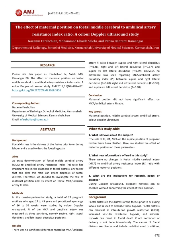
[AMJ 2018;11(10):478-482] The effect of maternal position on foetal middle cerebral to umbilical artery resistance index ratio: A colour Doppler ultrasound study Nazanin Farshchian, Mohammad Gharib Salehi, and Parisa Bahrami Kamangar Department of Radiology, School of Medicine, Kermanshah University of Medical Sciences, Kermanshah, Iran artery RI ratio between supine and right lateral decubitus (P=0.48), right and left lateral decubitus (P=0.67), and supine vs. left lateral decubitus (P=0.39). Likewise, no difference was seen regarding MCA/umbilical artery pulsatility index (PI) between supine and right lateral decubitus (P=0.20), right and left lateral decubitus (P=0.35), and supine vs. left lateral decubitus (P=0.80). Conclusion Maternal position did not have significant effect on MCA/umbilical artery RI ratio. Key Words Maternal position, middle cerebral artery, umbilical artery, colour doppler ultrasound RESEARCH Please cite this paper as: Farshchian N, Salehi MG, Kamangar PB. The effect of maternal position on foetal middle cerebral to umbilical artery resistance index ratio: A colour Doppler ultrasound study. AMJ 2018;11(10):478–482. https://doi.org/10.21767/AMJ.2018.3251 Corresponding Author: Nazanin Farshchian Department of Radiology, School of Medicine, Kermanshah University of Medical Sciences, Kermanshah, Iran Email:nfarshchian@kums.ac.ir What this study adds: ABSTRACT 1. What is known about this subject? The role of RI, UA, MCA on the supine position of pregnant mother have been clarified. Here, we studied the effect of maternal position on these parameters. 2. What new information is offered in this study? There were no changes in foetal middle cerebral artery (MCA) to umbilical artery resistance index (RI) ratio with different maternal positions. 3. What are the implications for research, policy, or practice? During Dopplerultrasound, pregnant mothers can be checked without concerning the effect of their position. Background Foetal distress is the distress of the foetus prior to or during labour and is used to describe foetal hypoxia. Aims As exact determination of foetal middle cerebral artery (MCA) to umbilical artery resistance index (RI) ratio has important role in the diagnosis of foetal distress, any factor that can alter this ratio can affect diagnosis of foetal distress. Therefore, we decided to investigate the role of maternal position and its effect on foetal MCA/umbilical artery RI ratio. Methods In this quasi-experimental study, a total of 27 pregnant mothers who aged 17 to 43 years and gestational age range of 26 to 39 weeks were studied by colour Doppler ultrasound. RI of the MCA and umbilical artery was measured at three positions, namely supine, right lateral decubitus, and left lateral decubitus positions. Results There was no significant difference regarding MCA/umbilical Background Foetal distress is the distress of the foetus prior to or during labour and is used to describe foetal hypoxia. Foetal distress can manifest as intrauterine growth restriction (IUGR), increased vascular resistance, hypoxia, and acidosis. Hypoxia can result in foetal death if not corrected or delivery is not done immediately. The causes of foetal distress are diverse and include umbilical cord conditions, 478
[AMJ 2018;11(10):478-482] foetal anomalies, drug reactions, delivery difficulties, etc.1 Colour Doppler ultrasound is a non-invasive and reliable method in the diagnosis of foetal distress. One of the methods of diagnosis of the foetal distress using Doppler ultrasound is determination of the foetal middle cerebral artery (MCA) to umbilical artery resistance index (RI) ratio. If foetal MCA/umbilical artery RI ratio lowers to less than 1, it represents foetal distress.2,3 The examination of the MCA is usually done at its proximal part. Several parameters are used in the examination of the MCA including peak systolic velocity (PSV), end-diastolic velocity (EDV), pulsatility index (PI), and RI. According to “brain sparing” theory, in any form of foetal distress decreased circulation causes decreased resistance of cerebral arteries so that avoid decreased blood flow to the brain. This change is seen as increased EDV.4-6 Physiologic responses to decreased blood flow include higher pulse rate, vascular dilation, and blood redistribution to vital organs. Therefore, in cases of decreased foetal blood flow due to uterine and foetal vascular conditions, cerebral blood flow is maintained via redistribution mechanism. To achieve this, resistance of MCA is decreased in a way that it lowers to level lower than that of the umbilical artery resistance.7,8 Regarding the importance of RI ratio between MCA and umbilical artery, any factor that can affect this ratio can have clinical significance and alter the diagnosis of foetal distress studied by colour Doppler ultrasound. Hence, recognition of responsible factors in this regard is important. In recent years, some studies have mentioned the maternal position as a responsible factor that may contribute to altered MCA/umbilical artery RI ratio.9-14 Regarding the importance of meticulous examination of MCA/umbilical artery RI ratio, and high number of pregnant patients referred for Doppler ultrasound examination during pregnancy, we decided to study the effect of maternal position on this ratio. Method In this quasi-experimental study, a total of 27 pregnant women in their third trimester of pregnancy with age range of 17–43 years (mean age=28.9 years) and gestational age range of 26–39 weeks (mean of 36 weeks) who were referred for obstetric colour examination were included. The inclusion criteria were singleton pregnancy, foetal healthiness on ultrasound study, knowing the last menstrual period (LMP) date, and gestational age within the second or third trimester. Exclusion criteria were polyhydramnios, IUGR, maternal diabetes, metabolic diseases, and drug addiction in the mother. All patients underwent colour Doppler ultrasound using iU22 (Philips) with CONVEX 3 to 5-MHz probe. The RI and PI of the MCA and umbilical artery at three positions including supine, right lateral decubitus, and left lateral decubitus positions were measured. A random number table was used for random allocation of the subjects to the three positions. Mothers were asked to keep the required position for about 5 minutes. Considering α=5 per cent and power of the study as 90 per cent, and mean (SD) of arterial resistance as 1.78 (0.27) in supine position and 1.29 (0.16) in right lateral decubitus, the required sample size was calculated as 27 subjects using the formula: - 1 2 / 1 ( 2 2 2 1 ) 2 2 Z Z ( ) a n 2 1 Statistics The data were analysed using Kolmogorov-Smirnov (KS) test to determine normal distribution of the continuous data. The data were analysed and compared between the three maternal positions using analysis of variance (ANOVA). The significance level was set at 0.05. All analyses were done using SPSS software (ver. 21.0, IBM, US). Ethics Written informed consent was obtained from the patients after explaining the details and objectives of the study. Results Table 1 presents demographic data of the studied sample. Table 1: Demographic characteristics of 27 pregnant women at second or third trimester who underwent colour Doppler ultrasound Maternal age, year Gestational age, week Parity Gravidity Fluid index Table 2 presents descriptive indices of RI and PI in the MCA and umbilical artery. There was no significant difference regarding MCA/umbilical artery RI ratio between supine and Mean (SD) 28.88 (6.04) 36.14 (3.41) 0.51 (0.84) 1.62 (0.96) 13.53 (2.1) Range 17 to 43 26 to 39 0 to 3 1 to 4 9 to 18 Doppler ultrasound 479
[AMJ 2018;11(10):478-482] right lateral decubitus (P=0.48), right and left lateral decubitus (P=0.67), and supine vs. left lateral decubitus (P=0.39) (Figure 1). Likewise, no difference was seen regarding MCA/umbilical artery pulsatility index (PI) between supine and right lateral decubitus (P=0.20), right and left lateral decubitus (P=0.35), and supine vs. left lateral decubitus (P=0.80) (Figure 2). Table 2: The resistance index (RI) and pulsatility index (PI) ratios in middle cerebral artery (MCA) to umbilical artery at the three studied positions among 27 pregnant women at the second or third trimester MCA/umbilical artery RI ratio at supine position MCA/umbilical artery RI ratio at right lateral decubitus MCA/umbilical artery RI ratio at left lateral decubitus Umbilical artery PI at supine Umbilical artery PI at right lateral decubitus Umbilical artery PI at left lateral decubitus Discussion Diagnosis of foetal complications is important and timely diagnosis of such complications can decrease complication rate. The utero-foetal circulation can be evaluated by non- invasive colour Doppler ultrasound. This ultrasound method is also helpful in determining the presence of foetal distress. Regarding the importance of measuring the RI ratio of MCA/umbilical artery as well as PI of the umbilical cord in determination of foetal distress and lack of sufficient studies, this research was carried out to investigate the effect of maternal position on this ratio and PI of the umbilical cord using colour Doppler ultrasound. The findings showed that maternal position did not have significant effect on this ratio. Likewise, maternal positions studied did not affect the umbilical cord PI. Kinsella et al. in a similar study including 20 pregnant women reported that position change from supine position to left lateral decubitus, and then supine position did not result in any change in the umbilical artery on colour Doppler ultrasound.9 These are compatible with our findings. In another study, S/D ratio, RI, and PI of MCA, umbilical artery, foetal aorta, and uterine artery among 58 pregnant women (22 patients with normal blood pressure and 36 patients with hypertension) were studied in the lateral position. Of 29 foetuses with abnormal umbilical artery indices, 11 had IUGR. Of those with reverse end-diastolic flow of the umbilical artery, one had intrauterine foetal demise (IUFD) and IUFD occurred in others in two days. These reported strong relationship between vascular indices and foetal outcome.10 In another study on 34 patients with IUGR, umbilical artery and MCA resistance had correlation with perinatal diseases and intraventricular haemorrhage.11 Another study, maternal position and its effect on foetal brain autoregulation was studied. Nineteen mothers with foetal IUGR, 25 mothers with low risk, and six mothers with small for gestational age (SGA) foetuses and low risk were selected. The umbilical artery and MCA blood flow was measured at supine and left lateral decubitus positions. The results showed that in low- risk group, MCA PI decreased from left lateral decubitus position to supine position. Likewise, MCA PI decreased in a similar manner in low risk SGA group. However, in high-risk group, MCA PI increased in supine position. The baseline MCA PI was lower in high-risk group compared to low-risk group.12 The studies show differences in vascular resistance at different maternal positions. Prior et al. evaluated foetal RI among 400 mothers before delivery. The results showed that neonates born by cesarean section had lower cerebral- to-umbilical compared to those born by vaginal delivery. This ratio can be used before foetal distress occurs.13 Another study included 1,084 foetuses within gestational age of 19–41 weeks with increased umbilical artery resistance. MCA and umbilical artery Doppler ultrasound was done. Mothers were categorized into three groups based on umbilical artery RI (<95 per cent, >95 per cent, and with diastolic blood flow). There was no significant difference in MCA RI in foetuses with preserved diastolic blood flow. But in foetuses without preserved diastolic blood flow, MCA RI was lower.14 Another study investigated the effect of maternal position on umbilical artery and MCA indices among 23 pregnant women within gestational age range of 36 to 40 weeks with low risk for complications. Their results showed that PI and PSV of the arteries decreased from left lateral decubitus to supine position.15 Another study examined the effect of maternal position on MCA and umbilical artery blood flow among 50 pregnant women within gestational age range of 36–40 weeks. The results showed that in both low-risk and high-risk groups, a significant decrease occurred 15 minutes after position change from supine to left lateral decubitus. Foetal response was seen more rapid when mothers were in left lateral decubitus position.16 Recently some concerns have raised about the possibility of late stillbirth with supine going-to-sleep position.17 In the current study, mothers were asked to be in supine positions for a short time (about 5 minutes). Therefore, the risk of stillbirth does not seem to Mean (SD) 1.34 (0.17) Range 1.02 to 1.7 1.32 (0.17) 1.06 to 1.64 1.30 (0.28) 1.04 to 2.42 0.91 (0.15) 0.87 (0.16) 0.6 to 1.3 0.61 to 1.3 0.90 (0.17) 0.6 to 1.4 480
[AMJ 2018;11(10):478-482] be significant in this research. As all mothers studied were in the third trimester, the observed changes cannot be attributed to variations in gestational age. The discrepancy among the mentioned results and our findings could be due to difference in gestational age, the method of investigation, or the sample size. A limitation that we encountered was that we were not able to recruit more subjects as some mothers did not provide consent to participate. As our findings showed that maternal position did not affect umbilical artery and MCA RI ratio, we suggest that further studies with larger sample sizes to be performed in order to reveal this mechanism of position change more accurately. Conclusion There was no difference regarding MCA/umbilical artery RI ratio and umbilical artery PI among supine, left lateral decubitus, and right lateral decubitus positions. 1990;36(1);11–17. 10.Khalid M, Wahab SH, Kumar V, et al. Doppler indices in prediction of feta outcome in hypertensive pregnant women. NJOG. 2011;6(1):28–34. 11.Marsoosi V, Bahadori F, Esfahani F, et al. The role of Doppler indices in predicting haemorrhage and perinatal mortality in fetal growth restriction. Med Ultrason. 2012;14:125–32. 12.Khatib N, Vinter D, Haberman S, et al. Postural stress can discriminate between placental insufficiency and other complications of pregnancy not associated with impaired placental function. Ultrasound Obstet Gynecol. 2012;40(suppl.1);171–310. 13.Prior T, Mullins E, Bennett P, et al. Prediction of intrapartum fetal compromise cerebroumbilical ratio: a prospective observational study. Am J Obstet Gynecol. 2013;208:124–26. 14.Morasles RJ, Hervas MD, Perales MA, et al. Doppler study of the fetal vertebral and middle cerebral arteries in fetuses with normal and increased umbilical artery resistance indices. J Clin Ultrasound. 2013;41:224–229. 15.Khatib N, Weiner Z, Beloosesky R, et al. The effect of maternal supine position on umblical and cerebral blood flow in indicies. Eur J Obstet Gynecol Reprod Biol. 2014;175:112–4. 16.El-Shahawy H, Ibrahim A, Hanafi S, et al. Effect of changing maternal position from left lateral to supine position on umbilical and fetal cerebral blood flow indices. IJOGR. 2016;3(2):240–256. 17.Heazell A, Li M, Budd J, et al. Association between maternal sleep practices and late stillbirth - findings from a stillbirth case-control 2018;125:254–262. ACKNOWLEDGEMENTS This article is the result of the thesis of No. 94330. We highly appreciate the Clinical Research Development Centre of Imam Reza Hospital for their wise advices. PEER REVIEW Not commissioned. Externally peer reviewed. CONFLICTS OF INTEREST The authors declare that they have no competing interests. FUNDING None intra ventricular using the References 1.Miller DA. Is advanced maternal age an independent risk factor for uteroplacental insufficiency. Am J Obstet Gynecol. 2005;192(6):1974–800. 2.Jugovic D, Tumbri J, Medic M, et al. New Doppler index for prediction of prediction of perinatal brain damage in growth-restricted and hypoxic fetuses. Ultrasound Obstet Gynecol. 2007;30:303–311. 3.Ertan AK, Tnriverdi HA, Meier M, et al. Perinatal risk factors for neonatal intracerebral hemorrhange in preterm infants. Eur J Obstet Gynecol Reprod Biol. 2006;127:29–34. 4.Kurmanavicius J, Florio I, Wisser J, et al. Reference resistance indices of the umbilical, fetal middle cerebral and uterine arteries at 24-42 weeks of gestation. Ultrasound Obstet Gynecol. 1997;10;112–120. 5.Yoshimura S, Masuzaki H, Gotoh H, et al. The relationship between blood flow redistribution in umbilical artery and middle cerebral artery and fetal growth in intrauterine growth retardation. Nihon Sanka Fujinka Gakkai Zasshi. 1995;47(12):1352–8. 6.McGahan JP. Goldberg BB. Diagnostic Ultrasound. 2 ed. Informa, New York, 2008, Vol 2.1169-1172. 7.Levy MN, Berne RM, Koepon BM, et al. Interply of central and peripheral factors in control of circulation. Physiology. 2004;433–42. 8.Hobbins JC. Obstetric ultrasound: artisty in practica. John Wiley & Sons; 2008. 9.Kinsella SM, Lee A, Spencer JA. Maternal and fetal effects of the supine and pelvic tilt positons in late pregnancy. Eur J Obstet Gynecol Reprod Biol. study. BJOG. 481
[AMJ 2018;11(10):478-482] Figure 1: Comparison of MCA (middle cerebral artery)/umbilical artery RI (resistance index) ratio in three positions of supine, right lateral decubitus, and left lateral decubitus among pregnant mothers Figure 2:Comparison of MCA (middle cerebral artery)/umbilical artery pulsatility index (PI) ratio in three positions of supine, right lateral decubitus, and left lateral decubitus among pregnant mothers PI mean SD 0.15 0.17 0.16 0.91 0.9 0.88 supine right left 482