Download
1 / 4
40 likes | 52 Views
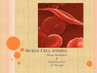
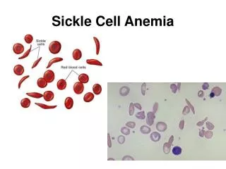
SCD develops when haemoglobin (HbS) is produced in the body as a result of a mutation occurring in haemoglobin beta chain. SLE is defined as a rare, chronic autoimmune multi-organ manifestation. SCD and SLE rarely coincide in literature due to the limited number of cases and the overlapping of symptoms of both diseases.

E N D
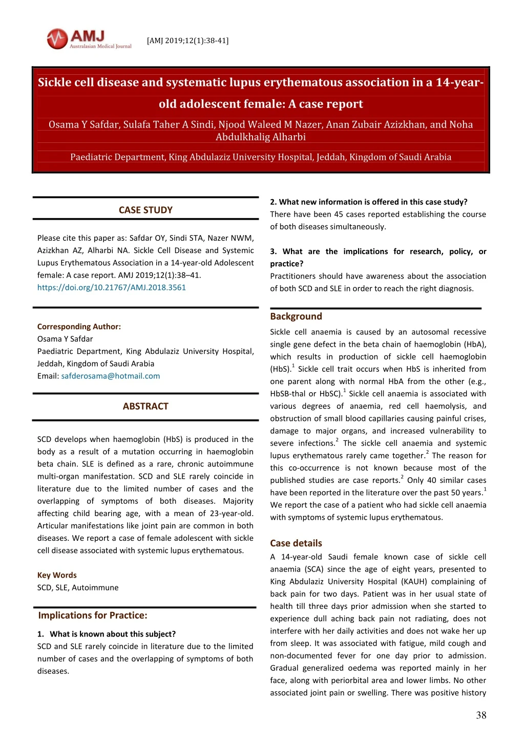
[AMJ 2019;12(1):38-41] Sickle cell disease and systematic lupus erythematous association in a 14-year- old adolescent female: A case report Osama Y Safdar, Sulafa Taher A Sindi, Njood Waleed M Nazer, Anan Zubair Azizkhan, and Noha Abdulkhalig Alharbi Paediatric Department, King Abdulaziz University Hospital, Jeddah, Kingdom of Saudi Arabia 2. What new information is offered in this case study? There have been 45 cases reported establishing the course of both diseases simultaneously. 3. What are the implications for research, policy, or practice? Practitioners should have awareness about the association of both SCD and SLE in order to reach the right diagnosis. CASE STUDY Please cite this paper as: Safdar OY, Sindi STA, Nazer NWM, Azizkhan AZ, Alharbi NA. Sickle Cell Disease and Systemic Lupus Erythematous Association in a 14-year-old Adolescent female: A case report. AMJ 2019;12(1):38–41. https://doi.org/10.21767/AMJ.2018.3561 Corresponding Author: Osama Y Safdar Paediatric Department, King Abdulaziz University Hospital, Jeddah, Kingdom of Saudi Arabia Email: safderosama@hotmail.com Background Sickle cell anaemia is caused by an autosomal recessive single gene defect in the beta chain of haemoglobin (HbA), which results in production of sickle cell haemoglobin (HbS).1 Sickle cell trait occurs when HbS is inherited from one parent along with normal HbA from the other (e.g., HbSB-thal or HbSC).1 Sickle cell anaemia is associated with various degrees of anaemia, red cell haemolysis, and obstruction of small blood capillaries causing painful crises, damage to major organs, and increased vulnerability to severe infections.2 The sickle cell anaemia and systemic lupus erythematous rarely came together.2 The reason for this co-occurrence is not known because most of the published studies are case reports.2 Only 40 similar cases have been reported in the literature over the past 50 years.1 We report the case of a patient who had sickle cell anaemia with symptoms of systemic lupus erythematous. Case details A 14-year-old Saudi female known case of sickle cell anaemia (SCA) since the age of eight years, presented to King Abdulaziz University Hospital (KAUH) complaining of back pain for two days. Patient was in her usual state of health till three days prior admission when she started to experience dull aching back pain not radiating, does not interfere with her daily activities and does not wake her up from sleep. It was associated with fatigue, mild cough and non-documented fever for one day prior to admission. Gradual generalized oedema was reported mainly in her face, along with periorbital area and lower limbs. No other associated joint pain or swelling. There was positive history ABSTRACT SCD develops when haemoglobin (HbS) is produced in the body as a result of a mutation occurring in haemoglobin beta chain. SLE is defined as a rare, chronic autoimmune multi-organ manifestation. SCD and SLE rarely coincide in literature due to the limited number of cases and the overlapping of symptoms of both diseases. Majority affecting child bearing age, with a mean of 23-year-old. Articular manifestations like joint pain are common in both diseases. We report a case of female adolescent with sickle cell disease associated with systemic lupus erythematous. Key Words SCD, SLE, Autoimmune Implications for Practice: 1.What is known about this subject? SCD and SLE rarely coincide in literature due to the limited number of cases and the overlapping of symptoms of both diseases. 38
[AMJ 2019;12(1):38-41] of dark urine without affecting the urine output. She had history of two hospital admissions in the last four months, the first one being admitted to the paediatric intensive care unit (PICU) with haemoglobin level of 2g/dl for which packed RBC transfusion was given. No history of consanguinity between the parents. On physical examination, the patient was alert, oriented, conscious, and pale, not in distress. Neurological exam revealed no motor or sensory defect, carinal nerve was intact and no cerebral deficit signs. Abdomen is soft, non- tender, hepatosplenomegaly was detected, normal bowel sounds. During the course of her admission, she had two attacks of focal convulsion involving the left leg then become generalized for 1-2 min preceded by vomiting and followed by loss of sphincter control, no loss of consciousness. During the last attack she had high blood pressure 147/90, afebrile, given two doses of diazepam and loading half loading dose of phenytoin. Magnetic resonance imaging (MRI) of the brain was done and revealed multiple cortical areas of acute/subacute infarction. No large territorial infarctions, intra or extra-axial haemorrhage. Moderate luminal narrowing the cavernous segment of the right internal carotid artery and proximal segments of the right middle cerebral artery. Complete blood count laboratory was done, results were within normal range except for low haemoglobin, mean cell volume (MCV) and mean cell haemoglobin (MCH). Readings were 4.7g/dl, 78.8FI and 27.1pg, respectively. Haemoglobin (Hb) electrophoresis test results showed Hb S at 58.1 per cent, Hb C0, and Hb A1 at 32.1 per cent, confirming a diagnosis of SCD. Our patient’s U&E showed severe renal impairment. The patient had a urea of 19mmol/l (2.5-6.4mmol/l), creatinine 676umol/l (53-115umol/l), sodium 134mmol/l (136- 145mmol/l), phosphate 1.76mmol/l (0.81-1.58mmol/l). Urine analysis showed: ++ Protein, ++++RBCs, Specific gravity is high. Immunological investigations revealed a positive titre of 1:1280, high anti-DNA antibody 485IU/mL (0–200IU/mL), lower normal C3 0.76g/L (0.75–1.65g/L) and a low C4 0.14g/L (0.2–0.6g/L. Test results for human immunodeficiency virus, hepatitis B, and hepatitis C were negative. Radiological workup including abdominal ultrasound was done. Ultrasound sound report showed the spleen is homogenous, enlarged measuring 14.5cm (median spleen size for ages 12–15 years is 10.1cm3) with no focal lesions. A diagnosis of SLE in the patient with SCD was established, with 6/11 of the diagnostic criteria of the American College of Rheumatology being met. Patient underwent renal biopsy with the following report. Histopathology report: 1.Microscopic description: Global sclerosis found in one glomerulus. The remaining intact glomeruli show glomerulonephritis with accentuated lobular configuration and endocapillary and mesangial proliferation. All the glomeruli show variable degree of segmental neutrophilic infiltration. Active cellular crescents found in two glomeruli and two fibrocelluar crescents in two glomeruli and fibrosed crescents in two glomeruli. The interstium show mild mononuclear inflammation and mild fibrosis. 2.Electron microscopy: There were 3 epithelial crescents in all three glomeruli. There is endocapillary proliferation with inflammatory infiltrate. Large numerous dense deposits are seen in mesangial and subendothelial areas, few deposits in subepithelial locations. 3.Immune fluorescence: The glomeruli demonstrate diffuse granular staining along the glomerular capillary wall and mesangium for IgG (4+), IgA (2+), IgM (2+), C3 (4+), C4 (trace) and C1q (4+). A renal biopsy revealed: class IV lupus Nephritis with high activity index 19/24, chronicity index 4/12. Patient was given monthly IV cyclophosphamide. Sodium bicarbonate 650mg tablets TID, calcium carbonate 300mg tablet QID with meals, calcium resonium 10mg powder BID. On follow up she had progressive worsening in her kidney function; test showed creatinine 766umol/L, urea 31mmol/L, haemoglobin 7.5g/dL. She had packed RBCs transfusion and she was prepared to start daily peritoneal dialysis. Renal transplant recommendation was done. Discussion Sickle cell disease (SCD) and systemic lupus erythematous (SLE) are categorized as chronic disease with multisystem organ involvement.4 SCD develops when haemoglobin (HbS) is produced in the body as a result of a mutation occurring a diffuse proliferative 39
[AMJ 2019;12(1):38-41] in haemoglobin beta chain.5It’s a hereditable disease commonly affecting women of African American descent.5,6 SLE is defined as a rare, chronic autoimmune multi-organ manifestations.7,8 Incidence of SLE in paediatric population is 0.3–0.9 per 100.000 children and prevalence 3.3–8.8 per 100.000 children.9 SCD and SLE rarely coincide in literature due to the limited number of cases and the overlapping of symptoms of both diseases.8 Females contributed to 78 per cent of cases, while only 22 per cent were male.8 Majority affecting child bearing age, with a mean of 23-year-old.10 The combination of both diseases reduces the life span of patients.11 There are many proposals regarding the conjunction of SCD and SLE.8 Encapsulated bacteria that lead to repeated infections may be a relationship between the autoimmune disease and haemoglobin S.12-14 Articular manifestations like joint pain are common in both diseases.8 SCD patients complain of joint pain and are most often diagnosed as vaso-occlusive crisis consequently detain the diagnosis of SLE.8 In our patient, back pain was the main complaint which was dull, aching and not radiating. She was diagnosed with SLE after SCD diagnosis by six years. In addition, haematological disorders have a great impact in both diseases with a derivative of anaemia, leukopenia, and thrombocytopenia.8 Our patient has anaemia with a haemoglobin value of 4.7g/dl (reference range of 14–18g/dl). White blood cell and platelet count were within normal range. In most cases of SLE, normochromic normocytic anaemia is the initial haematological manifestation followed by hypochromic microcytic anaemia.8 Thus, patients with both diseases develop a more severe type of anaemia due to the underlying pathophysiology of these two conditions.8 Neurological diseases have a prevalence of 50 per cent in SLE patients, and 25 per cent in SCD patients.15-17 Of those disorders, cerebral infarction, intracranial haemorrhage, cognitive dysfunction, and seizure disorder are widely occurring in both SCD and SLE.15 In our case, patient developed two seizure attacks that lasted for approximately two minutes. As a result, patients who manifest both diseases are prone to cerebrovascular disease.8 Renal failure is as well a result of both SCD and SLE, with lupus nephritis recorded in 29–80 per cent of paediatrics age group.8 Patient with SLE as a single disease has a risk of 47 per cent in developing nephropathy.8 Renal biopsy is widely used due to the adverse pathological impact of SCD with the coexistence of SLE on the renal function.8 Renal biopsy was done in our patient with a result of diffuse proliferative glomerulonephritis proliferation. SCD and SLE are known to affect the immunological system by producing anti-nuclear antibodies (ANA).18 It has been hypothesized that in SCD, autoantibodies could be induced by a chronic inflammatory state from chronic haemolysis.19 In an environment of rapid cell turnover, autoantibodies against self-components could be produced.19 Another hypothesis is that the dysfunctional immune complex of patients with this disease arising from functional hyposplenism, complement pathway defects and phagocytosis could impede complexes.20 Therefore when SLE is suspected in a patient with SCD, the best serologic markers appear to be SLE- specific autoantibodies such as anti-dsDNA and anti-Smith.21 Persistent hypocomplementemia presence of an immune complex-mediated disease.21 It’s positive in 100 per cent of SLE patients, however it’s positive in 20 per cent of SCD patients.18 Moreover, anti- dsDNA is commonly ordered to establish the diagnosis of the existence of both diseases.22 Central nervous system complications are appreciated in both SCD and SLE.11 Doppler carotid ultrasound usually reveals high viscosity in SCD patients.11 Those patients require continuous blood transfusion as part of medical management.11 However, in SLE patients, vasculitis and antiphospholipid antibodies play a role and hence oral anticoagulation and steroid are mandatory.11 Conclusion Awareness of the association of sickle cell disease with systemic lupus is needed in order to enable timely diagnosis and proper treatment. clearance of immune also supports the References 1.Maamar M, Tazi-Mezalek Z, Harmouche H, et al. Systemic lupus erythematosus associated with sickle-cell disease: a case report and literature review. Journal of medical case reports. 2012;6(1):366. 2.Michel M, Habibi A, Godeau B, et al. Characteristics and outcome of connective tissue diseases in patients with sickle-cell disease: report of 30 cases. Semin Arthritis Rheum. 2008;38:228–240. WB Saunders. 3.Rosenberg HK, Markowitz RI, Kolberg H, et al. Normal splenic size in infants and children: sonographic measurements. Am J Roentgen. 1991 Jul;157(1):119-21. 4.Saxena VR, Mina R, Moallem HJ, et al. Systemic lupus erythematosus in children with sickle cell disease. J Pediatric Hematology/oncology. 2003;25(8):668-71. 5.Loureiro MM, Rozenfeld S, Portugal RD. Acute clinical events in patients with sickle cell disease: epidemiology and mesangial 40
[AMJ 2019;12(1):38-41] 20.Toly-Ndour C, Rouquette AM, Obadia S, M’BAPPE PA, Lionnet F, Hagege I, Boussa-Khettab F, Tshilolo L, Girot R. High titers of autoantibodies in patients with sickle-cell disease. The Journal of rheumatology. 2011 Feb 1;38(2):302-9. 21.Khalidi NA, Ajmani H, Varga J. Coexisting systemic lupus erythematosus and sickle cell disease: a diagnostic and therapeutic challenge. J Clinical Rheuma. 2005 Apr 1;11(2):86-92. 22.White LE, Reeves JD. Polyarthritis and positive LE preparation in sickle hemoglobinopathies: a report of two cases. The J Pediatrics. 1979 Dec 1;95(6):1003-4. PEER REVIEW Not commissioned. Externally peer reviewed. CONFLICTS OF INTEREST The authors declare that they have no competing interests. FUNDING None PATIENT CONSENT The authors, Safdar OY, Sindi STA, Nazer NWM, Azizkhan AZ, Alharbi NA, declare that: 1.They have obtained written, informed consent for the publication of the details relating to the patient(s) in this report. 2.All possible steps have been taken to safeguard the identity of the patient(s). 3.This submission is compliant with the requirements of local research ethics committees. and treatment. Revista Brasileira de Hematologia e Hemoterapia. 2008;30(2):95-100. 6.Elficki Y, Rawas A, Bossei AA, et al. Coexistence of lupus nephritis and sickle cell trait, an electron microscopic assessment of renal glomerular damage: Case report of a rare association. Electronic Physician. 2017;9(9):5298. 7.Yu C, Gershwin ME, Chang C. Diagnostic criteria for systemic lupus erythematosus: a critical review. J Autoimmunity. 2014;48:10-3. 8.Robazzi TC, Alves C, Abreu L, et al. Coexisting systemic lupus erythematosus and sickle cell disease: case report and literature review. Revista brasileira de reumatologia. 2015;55(1):68-74. 9.Levy DM, Kamphuis S. Systemic lupus erythematosus in children and adolescents. Pediatric Clinics. 2012 Apr 1;59(2):345-64. 10.Manzi S. Lupus update: perspective and clinical pearls. Cleve Clin J Med. 2009;76(2):137-42. 11.Kaloterakis A, Filiotou A, Haziyannis S, et al. Sickle cell/²0-Thalassemia and systemic lupus erythematosus. Lupus. 1999;8(9):778. 12.Wilson WA, Nicholson GD, Hughes GR, et al. Systemic lupus erythematosus and sickle-cell anaemia. British Medical J. 1976;1(6013):813. 13.Wilson WA, Ceulaer KD, Morgan AG. Sickle cell anemia, complement, and systemic lupus erythematosus. Arthritis & Rheumatism: Official J the Am College of Rheumatology. 1979;22(7):803. 14.Warrier RP, Sahney S, Walker H. Hemoglobin sickle cell disease and systemic lupus erythematosus. Journal of the National Medical Association. 1984 Oct;76(10):1030. 15.Bruns A, Meyer O. Neuropsychiatric manifestations of systemic lupus erythematosus. Joint Bone Spine. 2006 Dec 1;73(6):639-45. 16.Adams RJ, Mckie VC, Hsu L, et al. Prevention of a first stroke by transfusions in children with sickle cell anemia and abnormal results ultrasonography. New England J Medicine. 1998 Jul 2;339(1):5-11. 17.Preul MC, Cendes F, Just N, et al. Intracranial aneurysms and sickle cell anemia: multiplicity and propensity for the vertebrobasilar territory. Neurosurgery. 1998 May 1;42(5):971-7. 18.Perilloux BC, Shetty AK, Leiva LE, et al. Antinuclear antibody (ANA) and ANA profile tests in children with autoimmune disorders: a retrospective study. Clinical Rheumatology. 2000 May 1;19(3):200-3. 19.Minocha V, Rana F. Lupus Nephritis in a Patient with Sickle Cell Disease. Case reports in hematology. 2013;2013. on transcranial Doppler 41