Download
1 / 1
10 likes | 139 Views
Aortic Flow Rates and Intra-Arterial Septum Mobility in Type B Aortic Dissections Quantified with Phase Contrast Magnetic Resonance Imaging C Karmonik 1,2 J Bismuth 1 , DJ Shah 1 , JE Anaya-Ayala, MG Davies 1 ,AB Lumsden 1 1 The Methodist DeBakey Heart & Vascular Center

E N D
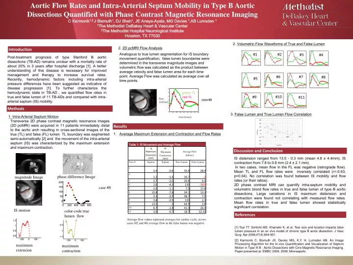
Aortic Flow Rates and Intra-Arterial Septum Mobility in Type B Aortic Dissections Quantified with Phase Contrast Magnetic Resonance ImagingC Karmonik1,2 J Bismuth1, DJ Shah1, JE Anaya-Ayala, MG Davies 1,AB Lumsden 1 1The Methodist DeBakey Heart & Vascular Center 2The Methodist Hospital Neurological Institute Houston, TX 77030 2. Volumetric Flow Waveforms of True and False Lumen 2. 2D pcMRI Flow Analysis Introduction Analogous to true lumen segmentation for IS boundary movement quantification, false lumen boundaries were determined in the transverse magnitude images and volumetric flow was calculated as the product between average velocity and false lumen area for each time point. Average Flow was calculated as average over all time points. Post-treatment prognosis of type Stanford B aortic dissections (TB-AD) remains unclear with a mortality rate of about 20% in 3 years after hospital discharge [1]. A better understanding of this disease is necessary for improved management and therapy to increase survival rates. Recently, hemodynamic factors including intra-arterial pressure differences have been suggested as indicative of disease progression [1]. To further characterize the hemodynamic state in TB-AD , we quantified flow rates in true and false lumen of 11 TB-ADs and compared with intra-arterial septum (IS) mobility. Methods 3. False Lumen and True Lumen Flow Correlation 1. Intra-Arterial Septum Motion Transverse 2D phase contrast magnetic resonance images (2D pcMRI) were acquired in 11 patients immediately distalto the aortic arch resulting in cross-sectional images of the true (TL) and false (FL) lumen. TL boundary was segmented semi-automatically [2] and the movement of the intra-arterial septum (IS) was characterized by the maximum extension and maximum contraction. Results • Average Maximum Extension and Contraction and Flow Rates Discussion and Conclusion IS distension ranged from 13.5 - 0.3 mm (mean 4.8 ± 4.4mm), IS contraction from 7.8 to 0.6 mm (2.4 ± 2.1 mm). In two cases, mean flow in the FL was negative (retrograde flow). Mean TL and FL flow rates were inversely correlated (r=-0.63, p=0.04). No correlation was found between IS mobility and flow rates (or their ratios). 2D phase contrast MRI can quantify intra-septum mobility and volumetric blood flow rates in true and false lumen of type B aortic dissections. Large variations in IS maximum distension and contraction were found not correlating with measured flow rates. Mean flow rates in true and false lumen showed statistically significant correlation. phase difference Image magnitude Image case #8 IS motion color-code true lumen flow References Average flow values represent averages for cardiac cycle, in two cases (#2 and #4) average flow in the false lumen was negative. [1] Tsai TT, Schlicht MS, Khanafer K, et al. Tear size and location impacts false lumen pressure in an ex vivo model of chronic type B aortic dissection. J Vasc Surg. Apr 2008;47(4):844-851. [2] Karmonik C, Bismuth JX, Davies MG, K.Y. H, Lumsden AB. An Image Processing Algorithm for the In-vivo Quantification and Visualization of Septum Motion in Type III B - Aortic Dissections with Cine Magnetic Resonance Imaging. Paper presented at: EMBC 2009, 2009; Minneapolis. maximum extension maximum contraction