Download
1 / 45
480 likes | 970 Views
CardioVascular Assessment Lab. C Ruckdeschel RN, BSN. Objectives. Review Anatomy of Heart Review Vascular System Review Physiologic basics for Cardiovascular System. Objectives:. Identify Skills to assess cardiovascular System: Pulse Peripheral vascular assessment Heart Sounds

E N D
CardioVascular Assessment Lab • C Ruckdeschel RN, BSN
Objectives • Review Anatomy of Heart • Review Vascular System • Review Physiologic basics for Cardiovascular System
Objectives: • Identify Skills to assess cardiovascular System: • Pulse • Peripheral vascular assessment • Heart Sounds • Blood Pressure
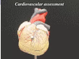
Anatomy of Heart • Right side of heart - receives deoxygenated blood from systemic circulation - LOW PRESSURE • Left Side of the heart - receives oxygenated blood from pulmonary circulation and pumps it into systemic circulation - HIGH PRESSURE
Chambers and Valves • Rt Atrium • RT AV Valve (Tricuspid) • Rt Ventricle • Rt semilunar (Pulmonic) • Left Atrium • Lft AV Valve (bicuspid, Mitral) • Left Ventricle • Left semilunar (Aortic)
Great Vessels of the Heart • Vena Cava - deoxygenated blood brought to heart • IVC (inferior vena Cava) • SVC (superior Vena Cava) • Pulmonary Artery - deoxygenated blood from rt ventricle to pulmonary capillaries • Pulmonary Veins - oxygenated blood from pulmonary capillaries to lft atrium • Aorta - • Ascending • Arch • Descending • Thoracic • Abdominal http://www.youtube.com/watch?v=PgI80Ue-AMo
Coronary Arteries • Arteries that arise from base of aorta and supply myocardium with richly oxygenated blood • LCA • LAD • Circumflex • RCA
Cardiac Conduction System • Heart is innervated by Autonomic nervous system • Sympathetic : stimulates • Parasympathetic: slows • SA Node (Sinoatrial node): located in right atria, generates impulses that travel through the conduction system & produce cardiac muscle contraction. • AV Node (atrioventricular node): located in the atrial septum • Bundle of His: right and left bundle branches • Purkinjie fibers: located in ventricular myocardium, where ventricular contraction takes place
12 Lead EKG • Chest X-ray
CommonCardiovascular Problems • CAD (Coronary Artery Disease) • HTN (Hyypertension) > 80% of US population • RHD (Rheumatic Heart Disease) - Sequelae of beta hemolytic strep infections resulting in valvular damage, more likely seen In older adults • BE (Bacterial Endocarditis) - bacteremia causes valvular damage • CHD (Congenital Heart Disease) – greatest portion diagnosed early in life
Peripheral Vascular Anatomy • Aorta • Arteries • Arterioles • Capillaries • Venules • Veins • Vena Cava
Important Vessels • Accessible arteries: • Temporal, Carotid, Aorta, Brachial, Ulnar, Radial, Femoral, Popliteal, Doraslis pedis, Posterior Tibial • Accessible veins: • Jugular, Superficial & deep arm veins, Femoral vein (deep), Popliteal vein (deep), saphenous (superficial)
Physiologic Basics • Myocardium - muscle layer of the heart that allows it to act as pump • Cardiac Output = HR x SV • Heart Rate (pulse) = beats per minute • Blood Pressure = SVR x CO • Electrical conduction of the heart
Assessing: Heart Sounds • Heart Sound Review • Location • Aortic: 2nd ICS, RSB (s2 is loudest) • Pulmonic: 2nd ICS, LSB (s2 is loudest) • Erbs Point: 3rd ICS, LSB • Tricuspid: 4th ICS, LSB (s1 is loudest) • Mitral (Apex): 5th ICS, MCL (s1 is loudest) • S1: represents ventricular contraction & ejection: S1 sound is produced by closing of AV valves (tricuspid and Mitral valves) • S2: represents ventricular relaxation & filling: S2 sound is produced by closing of semilunar valves: Aortic and Pulmonic valves http://www.youtube.com/watch?v=Ge12P7u0aQo
Assessing: Heart Sounds • Obtain History • Any medications?type • doseside effectsexpected effectstake as prescribed? • Pacemaker • Typebattery checkPresence of AICDautomated internal defibrillator • Obtain History • Risk factors/lifestyle • diet, exercise • smoking • cholesterol • stress, palpitations • dyspnea/orthopnea • edema • fatigue - relationship to exercise • chest pain • Location substernal? • Radiate precordial? • Quality crushing? • Associated N/V • Related to activity?
Assessing: Heart Sounds • Obtain History • Past Family History • Angina • Heartdisease • MI,StrokeDM, • Hyperlipidemia • Sudden death age? • Obtain History • Past Health History • Diabetes • Dependent edema • congenital heart defect • CAD • Rheumatic fever • Most recent EKG, stress EKG • Other diagnostics
Assessing: Heart Sounds • Inspection • Bare chest • Quiet room, Privacy • Note: symmetry of chest, any pulsatile areas, discolorations • Palpate • Precordium • palpate 5 sites for: • Heave (with palmer surface), thrust • Thrill (with base of finger of heel of hand (bony part)) • palpable murmur » cat purring • Thrills - indicative of obstructed flow • fine palpable rushing sensation • R or L 2nd ICS - Aortic or pulmonic stenosis • When palpate precordium use other hand to palpate carotid artery • S1 should coincide with carotid impulse
Assessing: Heart Sounds • Auscultate • Use diaphragm and bell of stethoscope • start with diaphragm, (S1 and S2 relatively high pitched) • use bell to listen for S3 and S4 • heart sounds - S1 and S2 • rate • rhythm - regular (NSR), irregular (warrants investigation) • extra sounds? Murmurs? • Auscultation: want to hear crisp, distinct S1 and S2 • S1 > at apex • S2 > at base
Assessing: Heart Sounds • BE Systematic!! APE TO MAN • Listening for S1 and S2 • interval between S1 and S2 should be silent • heart sounds not heard best directly over valve which produces it, but in direction of blood flow • there are specific sites where each valve sound is best heard
After Auscultating Heart Sounds..... • Perfect time to auscultate Apical Pulse. • Count for one full minute, each cardiac cycle. • Note rate & rhythm
What is a Pulse? • The ventricles pump blood into the arteries at about 72 bpm. The blood causes an alternating expansion and recoil creates a pressure wave which travels through all of the arteries.
Pulse • Adult (60-100) bpm • Child (80-120) bpm • Infant ( 140 bpm) • Palpated on superficial arteries (pulse points) • Auscultated on Apex of the heart
Pulse Variations: • Tachycardia - >100 bpm • Bradycardia - < 60 bpm • Palpitations - Unpleasant sensations of awareness of the heartbeat: described as skipped beats, racing, fluttering, pounding or irregularity: may result from rapid acceleration or slowing of heart, increased forcefulness of cardiac contraction: not necessarily associated with heart disease.
Factors Assessing Pulse • Cardiac output • Age • Gender • Exercise • Fever • Stress • Position
Factors Assessing Pulse • Cardiac Output • Amount of blood ejected from the heart in one minute • Measured by SV x HR • Normal HR = 60 - 100 beats per minute
Factors Assessing Pulse • Age • Adult (60-100) bpm • Child (80-120) bpm • Infant ( 140 bpm) • Gender - after puberty female > male • Exercise • increased HR with activity • increased metabolism causes vasodilatation • causes O2 demand
Factors Assessing Pulse • Fever • body compensates for increased temp by vasodilatation, decreased BP causes body to compensate by > HR • increased 10-20 beats/min/ degree above norm • especially in children
Factors Assessing Pulse • Stress • sympathetic response, increases HR & BP • Position • sitting, standing causes pooling • results in transient - BP • rate compensates by increasing
Assessing : Pulse • Please note: • Assessing a heart rate is determining beats per minute, noting rate, rhythm and strength. • Assessing peripheral pulses is to assess arterial blood flow to peripheral arteries.
Assessment: PulseAuscultating at Apex • Using the diaphragm of your stethoscope, place it on the 5th intercostal space, MCL • For one full minute, count each LUB, DUB as one!! • Location of left ventricular apex & PMI (point of maximum impulse) • Adult: 5th ICS, MCL • Infants: 4th ICS, left of MCL • Pregancy: PMI moves 1-2 cm left of MCL & up to 4th ICS
Assessment:PulsesPeripheral Pulses • Obtain History • Intermittent claudication • pain on walking disappears with rest • leg cramps, leg ulcers • varicose veins • edema of feet or legs • blood clots • pallor of fingertips
Assessment:PulsesPeripheral Pulses • Inspection of Extremities Compare Left to Right • Size • Symmetry • Skin/color • Nail Beds • Nails • Hair Growth
Assessment:PulsesPeripheral Pulses • Palpation - Compare Right to Left • Temperature • Capillary refill • Pulses • UE:Radial,Brachial • LE: Dorsalis Pedis, Posterior tibial, popliteal, Femoral • Edema • +1- +4 pitting • Sensation
Assessment: PulsesCharacteristics of Pulses • Rate • Rhythm - regular, irregular • Contour/elasticity • Strength (Amplitude) • +4 = bounding • +3 = full, increased • +2 = normal • +1 = diminished, weak • 0 = absent
Arterial Insufficiency of Lower Extremities • Pulses - Decreased/Absent • Color - Pale on elevation : Dusky Rubor on dependency • Temperature - Cool/Cold • Edema - None • Skin - Shiny, thick nails, no hair, Ulcers on Toes • Sensation - Pain, more with exercise, Paresthesias
Venous Insufficiency of Lower Extremities • Pulses - Present • Color- Pink to cyanotic, Brown pigment at ankles • Temperature - Warm • Edema - Present • Skin - Discolored, scaly, ulcers on ankles • Sensation - Pain, More with standing or sitting. Relieved with elevation/support hose
Peripheral Vascular Disease • Nursing interventions to promote venous return • ankle circles, flex ankles, frequent ambulation, avoid dependent position for prolonged periods of time • apply TED stockings or ace bandages (if no arterial problem) • Nursing Diagnosis • Altered cardiac output: decreased • Altered tissue perfusion:peripheral • Fluid volume deficit: actual • Irregular Rhythm • ALL irregular rhythms demand an APICAL RADIAL assessment
Assessment: Blood Pressure • Obtain History: • ** Modifiable Risk factors** • SmokingEmployment: physical vs emotional demands, environmental hazard, stress managementNutritional Status: body fat & type of dietAnaerobic exerciseEstrogen replacement (if post-menopausal)Drug use – alcohol,, cocaine, prescription & OTCEssential HTNHypercholesterolemia, DM, CAD • Obtain History: • ** Non-modifiable Risk factors ** • Age, sex, personality type • Family History – sudden death, HTN, stroke, MI prior to 50, severe hyperlipidemis, DM • PMH – arrythmias, murmurs, CHF, Rheumatic disease • DM, CAD,Congenital Heart Defects
Blood Pressure: Key Facts • Korotkoff sounds: Turbulent sounds of partial obstruction of arterial flow • Phase I: sharp tapping sound (systolic) • Phase II: change to soft swishing sound • Phase III: sounds more crisp & intense • Phase IV: muffled tapping • Phase V: cessastion of sound (diastolic)
Blood Pressure: KeyFacts • Arm Blood Pressure: May be 5-10 mmHg higher in right arm than left arm: greater differences between right & left arm may be associated with congenital aortic stenosis or acquired conditions such as aortic dissection or obstruction of arteries to upper arm. • Leg Blood Pressure: Arm & leg blood pressures are about equal during first year of life & after that time the leg blood pressure is 15-20 mmHg higher than the arm BP. • Pulse Pressure: difference between systolic and diastolic blood pressures: • Usual pulse pressure is between 30-40 mmHg • Orhtostatic Hypotension: Decrease in SBP of 20-30 mmHg or more when changing from supine to standing position, & increase in pulse of 10-20 bpm: sudden drops may result in fainting. Dizziness & faintness from orthostatic hypotension may occur when taking anti-hypertensive medications, hypovolemia, confined to bed for prolonged periods of time, or the elderly.