Download
1 / 36
360 likes | 446 Views
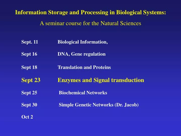
Information Storage and Processing in Biological Systems: A seminar course for the Natural Sciences. Sept. 11 Biological Information, Sept 16 DNA, Gene regulation Sept 18 Translation and Proteins Sept 23 Enzymes and Signal transduction Sept 25 Biochemical Networks

E N D
Information Storage and Processing in Biological Systems: A seminar course for the Natural Sciences Sept. 11 Biological Information, Sept 16 DNA, Gene regulation Sept 18 Translation and Proteins Sept 23 Enzymes and Signal transduction Sept 25 Biochemical Networks Sept 30 Simple Genetic Networks (Dr. Jacob) Oct 2
C - Introduction to Proteins / Protein Functions Proteins carry out a wide variety of functions in, on and outside the cell. For the purpose of this course, we will generalize these functions into three categories. These are not mutually exclusive and many proteins fit into more than one of these categories. 1 - Structural 2 - Enzymatic 3 - Signal Transduction (information processing)
C1 - Protein Functions: Structural Proteins can form large complexes that function primarily as structural elements: Protein coats of viruses. These are large, regular repeating structures composed of 100-1000’s of protein subunits. (Figs 6-74 and 6-72, Alberts). Electron micrographs of A) Phage T4, B) potato virus X, C) adenovirus, D) influenza virus. SV40 structure determined by X-ray crystallography.
Cytoskeleton in eukaryotic cells is responsible not only for determining shape but also in cell movement, mechanical sensing, intracellular trafficking and cell division. A human cell grown in tissue culture and stained for protein (such that only large regular structures are highlighted). Note the variety of structures (Fig 16-1, Alberts)
Microtubules form by the polymerization of tubulin subunits. Whether the polymer grows or shrinks is influenced conditions in the cell - Dynamic Instability (Fig 16-33, Alberts; for discussion of dynamic instability see Flyvbjerg H, Holy TE, Leibler S. Stochastic dynamics of microtubules: A model for caps and catastrophes. Phys Rev Lett. 1994 Oct 24;73(17):2372-2375.
X Y C2 - Protein Functions: Enzymatic Enzyme:a protein*that catalyzes a chemical reaction, where a catalyst is defined as a substance that accelerates a chemical reaction without itself undergoing change. * some RNA molecules can also be considered enzymes A A + B B C + D • Specificity • Accelerated reaction rates • Control (regulation) • Enzymes can only affect the rate (kinetics) of a reaction, they can not make a reaction more energetically favorable. • Enzymes can be saturated by substrate.
v = Vs (KM + s) Basics of Enzyme Kinetics Michaelis-Menton Kinetics - for a simple enzyme reaction, the interaction of enzyme and substrate is considered an equilibrium and the overall reaction as follows: E + S ES E + P k+1 k-1 k+2 v = velocity, reaction rate KM = Michaelis constant KM = k2 + k-1 k1
C3 - Protein Functions: Signal Transduction Signal Transduction - in general the relaying of a signal from one physical form to another - in biological terms, the process by which a cell responds to signals (can be intracellular, extracellular). Signal Transduction Input Output • Examples of ‘signals’ (inputs): • chemicals • light • temperature • electrical (ion gradients) • other cells (cell-cell contact) • mechanical sensing
Generalized Model of Response to Extracellular Signal Ligand Activated Receptor Receptor “Action” • Ligand can activate or inactivate receptor • Output (action) dependent on system and sometime cell type • In metazoans (multi-cellular eukaryotes), there are about 16 intercellular classes of signaling systems
Example 1: Transmembrane Tyrosine Kinase Receptors Ligand P~ ~P Activated Receptor Receptor “Action” • Ligand binding results in receptor dimerization • The cytoplasmic (intracellular) domains are tyrosinekinases which phosphorylate each other on Tyr residue side chains. • This sets off a series of intracellular events
Example 2 : Steroid Receptors Ligand Activated Receptor Receptor nucleus • The steroid binds to it’s receptor in the cytoplasm. • The steroid-receptor complex but not the free receptor can move into the nucleus . • The steroid-receptor complex binds to specific binding site(s) on the DNA to regulate gene expression.
Example 3. Heterotrimeric G-Proteins Ligand Activated Receptor GTP GDP GDP Receptor GTP GTP (a b g complex) • Ligand binding causes activation of the a subunit which promotes exchange of GDP for GTP • In the GTP form, the a subunit and the associated bg subunits dissociate from the complex. • Each subunit can go on to initiate a series of intracellular events.
D - Regulation of Protein Activity The concentration of a protein in the cell is a function of the rate of synthesis and the rate of degradation. Both these processes can be regulated. Synthesis Transcription Translation Degradation DNA RNA Protein Proteins are often regulated such that the ‘activity’ of a protein is not a constant function of its concentration. Protein Active Protein Inactive
Regulation of Enzyme Activity Negative Feedback (Product Inhibition) X A B X A B C D E F Mechanistically negative feedback can be by direct competition of the product with the substrate for the active site or it can be indirect through interaction wit the enzyme away from the active site (allosteric).
Regulation of Enzyme Activity Positive Feedback (Product Inhibition) X A B Positive Feedforward X A B
+ + Cooperativity / Allosteric Regulation Hypothetical examples of binding of a ligand to a dimeric protein. The binding curve is very sensitive to the effects on one site on the other. Two independent sites
+ + + + Cooperativity / Allosteric Regulation Hypothetical examples of binding of a ligand to a dimeric protein. The binding curve is very sensitive to the effects on one site on the other. Two independent sites Positive cooperativity
+ + + + + + Cooperativity / Allosteric Regulation Hypothetical examples of binding of a ligand to a dimeric protein. The binding curve is very sensitive to the effects on one site on the other. Two independent sites Positive cooperativity Negative cooperativity
+ + + + + + Cooperativity / Allosteric Regulation Hypothetical examples of binding of a ligand to a dimeric protein. The binding curve is very sensitive to the effects on one site on the other. Fraction bound vs ligand concentration Two independent sites Positive cooperativity Negative cooperativity
+ + + + + + Cooperativity / Allosteric Regulation Hypothetical examples of binding of a ligand to a dimeric protein. The binding curve is very sensitive to the effects on one site on the other. Two independent sites Positive cooperativity Positive Cooperativity (n=2, n=3) Negative cooperativity
+ + + + + + Cooperativity / Allosteric Regulation Hypothetical examples of binding of a ligand to a dimeric protein. The binding curve is very sensitive to the effects on one site on the other. Two independent sites Positive cooperativity Negative Cooperativity (n= 0.5) Negative cooperativity
Allosteric protein: a protein that changes from one conformation to another upon binding a ligand or when it is covalently (chemically) modified. The change in conformation alters the activity of the protein. Historically considered with multi-meric proteins (e.g. hemoglobin). Allosteric effector (positive) Ligand
Regulation of Protein Activity by Covalent Modification • The activity of a protein can modified by addition or removal of a chemical group to an amino acid side chain (i.e. - as a substrate for another enzyme). • The most common modifications are: • Methylation (-CH3) • Phosphorylation (-PO3) • Nucleotidyl • Fatty acid • Myristol • note that many proteins are modified in other ways such as addition of sugar groups (glycosylation) but these are not ‘regulatory’ modifications. Phosphorylation is the most common mechanism of regulation by covalent modification Kinase - an enzyme that phosphorylates Phosphatase - an enzyme that removes phosphate
Regulation by Localization Protein activity can be regulated by changing the localization of the protein. This turns out to be a common theme in eukaryotic signal transduction. Localization can be altered allosterically or by covalent modification. P~ ~P P~ ~P Addition of a fatty acid group can cause a cytoplasmic protein to associate with the cell membrane. Covalent modification of a protein can generate a binding site for another protein.
E - General Considerations Proteins have a diverse range of functions and a variety of mechanisms of regulation. The ability to form networks of proteins acting on proteins, the sharing of common reaction intermediates and forming multi-step chemical pathways allows for an endless number of possibilities. • Some general considerations about protein systems: • A reaction can behave as a step function (digital, boolean) if there is significant cooperativity in the system or if there modifying enzyme that works near saturation. • Since proteins can act in a catalytic manner, there can be signal amplification. • Many systems are adaptive, in that the response to signal is not necessarily constant over time (e.g. a signal transduction system may become desensitized and no loner respond to the presence of a ligand- c.f. heterotrimeric G protein).
Epidermal Growth Factor Signaling Pathway Not all pathways will operate in a single cell. • Protein interactions • Protein modification • (Activation/inhibition) • Protein re-localization • Transcriptional regulation http://www.grt.kyushu-u.ac.jp/spad/pathway/egf.html
Protein Stability: Robust to Site Mutations Protein function sensitive to changes in active site residues but relative robust with respect to ransom mutations. Tolerance for random mutations in proteins in general ~ 8 - 30% In some studies as much as 25% increased protein stability How does nature select for robustness? Is there a connection between robustness and evolvability?
Protein Dynamics in Living Cells- the use of GFP fusions Cell Division in Bacteria Time (30-40min) • Single cell elongates • At cell length = 2, divides at its midpoint
Protein Dynamics in Living Cells- the use of GFP fusions Cell Division in Bacteria • Division ring is formed by FtsZ • Positioning of FtsZ depends on Min proteins • MinD oscillates in the cell, MinE localizes to the division plane
Protein Dynamics in Living Cells- the use of GFP fusions Time-lapse fluorescence micrographs showing the dynamic behaviour of Gfp-MinC. MinC is a division inhibitor which associates, and co-oscillates, with MinD. Pole-to-pole oscillation of MinD, in turn, requires the activity of MinE Graphical view of dynamic Gfp-MinD distribution in cell
Protein Dynamics in Living Cells- the use of GFP fusions Time-lapse fluorescence micrographs showing the dynamic behavior of MinE-Gfp in normally dividing cells. Times are indicated in seconds. Note the accumulation of MinE-Gfp in the shape of a ring (the E ring)
Protein Dynamics in Living Cells- the use of GFP fusions Meinhardt and de Boer (2001) Proc. Natl. Acad. Sci. USA98 (25), 14202-14207.
Protein Dynamics in Living Cells- the use of GFP fusions The FtsZ ring
D. discoideum amoebae chemotaxing toward cAMP Cells are expressing a GFP-coronin fusion protein: coronin is a cytoskeletal protein that is localized in the extended pseudopods. G. Gerisch, Max Planck Institute, GDR