Download
1 / 63
670 likes | 956 Views
Nervous System: Nervous Tissue and Brain. Chapter 10. Lisa Ochs RN, BSN 2008. Structure of the Nervous System. Central Nervous System (CNS) Brain Spinal cord Peripheral Nervous System (PNS) Outside of the CNS Includes nerves that connect the CNS with the rest of the body.

E N D
Nervous System: Nervous Tissue and Brain Chapter 10 Lisa Ochs RN, BSN 2008
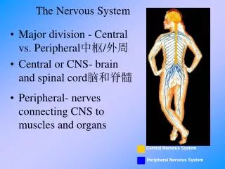
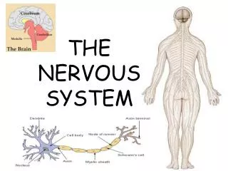
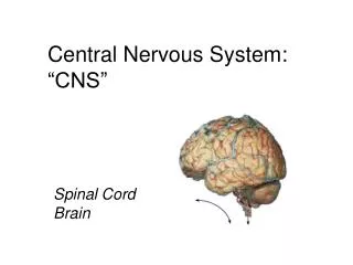
Structure of the Nervous System • Central Nervous System (CNS) • Brain • Spinal cord • Peripheral Nervous System (PNS) • Outside of the CNS • Includes nerves that connect the CNS with the rest of the body
Function of the Nervous System • Sensory • Gather information from inside and outside the body and transmit back to the CNS • Integrative • Processing and interpreting information received from the sensory nerves • Motor • Causing an action or movement in response to the information received
Types of Cells • Neuroglia • Also called glial cells; mostly found in the CNS • Support, protect, insulate and “care for” neurons • Do not conduct nerve impulses • Include astrocytes (most abundant), ependymal cells (secrete CSF), Schwann cells, oligodendrocytes
Types of Cells • Neurons • Transmits information in the form of nerve impulse • Various shapes, sizes and lengths • Do not replicate (nonmitotic); cannot replace themselves when injured
Types of Cells • Parts of a neuron • Dendrites • Tree like structures that receive information • Cell body • Contains the nucleus of the cell • Axon • Long extension that transmits information away from the cell body • Includes myelin sheath, nodes of Ranvier, & axon terminals
Types of Cells • Types of neurons • Sensory • Carries information from the periphery to the CNS • Also called afferent • Motor • Carries information from the CNS to the periphery • Also called efferent • Remember S.A.M.E.
Nerve Impulse • An electrical signal that conveys information along a neuron • Also called an action potential • Inside the cell, normal resting state is negative (polarized) • The cell changes to a positive state (depolarization) when stimulated • Returns to resting state of negative (repolarization)
Movement of Nerve Impulse • The nerve impulse travels along the length of the axon in a wave like manner • Each impulse depolarizes the next section of membrane • Each nerve impulse fires in an “all or nothing” manner; this ensures that the nerve impulse does not weaken as it travels along the axon
Speed of Nerve Impulse • Most axons are wrapped in myelin sheaths; the spaces between the myelin are the nodes of Ranvier; nerve impulses cannot travel through myelin • The nerve impulse jumps across the myelin to the bare nodes • This is called saltatory conduction • Myelinated fibers are considered fast-conducting fibers
Structure of Synapse • Synapse is the place where two neurons meet • Includes: • Synaptic cleft (space between the neuron & dendrite) • Neurotransmitters (most common is ACh and norepinepherine) • Inactivators (stop the activity of neurotransmitters) • Receptors (on the dendrite where the neurotransmitters attach)
Events at Synapse • The nerve impulse of the first (presynaptic) neuron causes the release of neurotransmitter into the synaptic cleft. • The neurotransmitter diffuses across the synaptic cleft and binds to the receptors on the second (postsynaptic) membrane. • The activation of the receptors stimulates a nerve impulse in the second neuron.
Brain Structure • Control center of the body- emotions, actions, memory, sleep/ wake, etc. • 2% of total body weight; requires 20% of body’s oxygen supply • Primary source of energy is glucose • 4 major areas: cerebrum, diencephalon, brain stem and cerebellum
Cerebrum • Largest portion of the brain • Right and left hemispheres joined by the corpus callosum (allows the separate sides to communicate) • Each hemisphere has four lobes • Frontal • Parietal • Temporal • Occipital (sound familiar?)
Lobes & Function • Frontal • Motor, personality, behavior, emotion, intellect • Parietal • Somatosensory (skin/ muscle, taste, speech, reading) • Temporal • Hearing, smell, memory, some speech • Occipital • Vision and vision functions (reading, distance, 3D)
Specific Functions • Decussation: crossing of fibers from one side of the brain to the other • Frontal lobe includes Broca’s area (left hemisphere); responsible for motor speech; patients with a stroke here suffer from expressive aphasia • Temporal & parietal lobes include Wernicke’sarea; broad area that involves translating thoughts into words; damage here can result in severe language deficits (Wernicke’s aphasia)
Markings on the Cerebrum • Gyri (sing. gyrus) • “bumps” or elevations on the surface • Sulci (sing. sulcus) • “grooves” • The extensive folding of the cerebral tissue increases its surface area; the greater the number of folds and grooves, the more intelligent the species
Hmmmm… • There are time-keeping neurons in our brains. Specifically in the prefrontal cortex and striatum of the cerebrum. Discovered recently in the brains of monkeys by researchers at MIT, these time-keeping neurons fire consistently at certain rhythms . . . • thus helping our brains to figure out when things are happening. This helps us with rhythmic activities, of course, but also with any number of tasks and memories that rely on knowing what came first, in what order, and so on.
Hmmmm… Researchers speculate that damage to these neurons, or damage to the mechanisms that read the timing pattern, may contribute to disorders (such as Parkinson Disease) that involve ill-timed movements and other functions. From http://theapprofessor.blogspot.com/, retrieved December 30, 2009
Hmmmm… In their paper, researchers failed to speculate whether this is why A&P students know exactly when to start slamming their books shut moments before a class is scheduled to end.
Diencephalon • 2nd main area of the brain, beneath the cerebrum and above the brain stem • Includes the thalamus and the hypothalamus
Diencephalon • Thalamus • Relay station for sensory fibers traveling from lower brain & spinal cord to sensory areas of cerebrum • Hypothalamus • Regulates many processes in the body- body temperature, water balance, metabolism • “thermostat” of the body
Brain Stem • Connects the spinal cord with higher brain structures • Includes midbrain, pons and medullaoblongata
Brain Stem • Midbrain • Relays sensory and motor information; reflex centers for vision and hearing • Pons • “bridge”; relays information; very important in regulating breathing rate and rhythm • Medulla Oblongata • Relays information; very important in regulating heart rate, blood pressure and respiration; called the “vital center”; also contains vomiting center
Cerebellum • Located under the occipital lobe and the base of the skull; “little brain” • Primarily concerned with coordination of voluntary muscle activity; balance and coordination • Persons with damage to the cerebellum may appear intoxicated
Other Structures • Not confined to specific lobes • Limbic system • “emotional brain”; responsible for various emotional states and behaviors • Reticular formation • Concerned with the sleep-wake cycle and consciousness • Sensitive to certain drugs and alcohol
Other Structures • Memory areas • Many lobes involved in recalling thoughts and images • Short term (seconds to hours) • Long term (years to decades)
Protection of the CNS • BONE • First layer of protection for the brain and spinal cord • Cranium and vertebral column
Protection of the CNS • MENINGES • Three layers of membranes that surround the brain and spinal cord • Dura mater (“tough mother”) • Tough connective tissue • Arachnoid (“spider-like”) • Weblike membrane • Pia mater (“soft mother”) • Delicate, thin; lies directly over the brain and spinal cord
Protection of the CNS • CEREBROSPINAL FLUID • Third layer of protection; formed from the blood within the brain; clear fluid that resembles plasma • Composed of water, glucose, protein and ions (esp. Na+ and Cl-) • Acts as a cushion and shock absorber, also delivers nutrients and removes waste.
Protection of the CNS • CEREBROSPINAL FLUID • Formed in the choroid plexus of the ventricles in the brain • Circulates around and through the brain and spinal cord; continuously produced and drained • Rate of production must equal rate of drainage, otherwise increased ICP will result
Protection of the CNS • BLOOD BRAIN BARRIER • An arrangement of cells (glial astrocytes) associated with blood vessels that supply the brain and spinal cord • Act as gate keepers- allows only certain substances to cross. Most harmful substances cannot cross this barrier • Pharmacology can be difficult- most antibiotics do not cross blood-brain barrier
NCLEX Question • After a brain stem infarction, a nurse would observe for which condition? • Aphasia • Bradypnea • Contralateralhemiplegia • Numbness and tingling in the face
Rationale • 2. Bradypnea, or slowed respiratory rate, would result from damage to the brain stem. The brain stem contains the control center for vital functions such as breathing, heart rate and blood pressure.
NCLEX Question • The nurse is caring for a client with a cerebral injury that has impaired his speech and hearing. The client most likely experienced damage to the: • Frontal lobe • Parietal lobe • Occipital lobe • Temporal lobe