Download
1 / 44

E N D
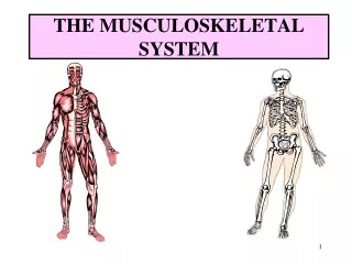
1. Chapter 52 Assessment of the Musculoskeletal System
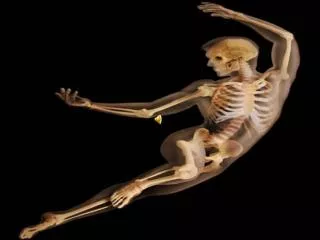
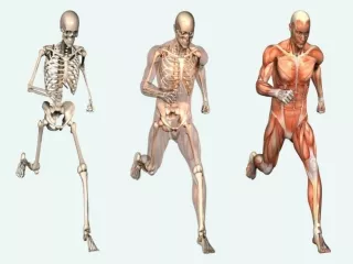
2. Skeletal System Bone types
Bone structure
Bone function
Bone growth and metabolism affected by calcium and phosphorus, calcitonin, vitamin D, parathyroid hormone, growth hormone, glucocorticoids, estrogens and androgens, thyroxine, and insulin
3. Bone Structure
4. Joints Types include synarthrodial, amphiarthrodial, diarthrodial.
Structure synovial joint.
Subtyped by anatomic structure:
Ball-and-socket
Hinge
Condylar
Biaxial
Pivot
5. Structure Diarthrodial Joint
6. Muscular System Assessment Patient history
Nutritional history
Family history and genetic risk
General inspection:
Posture and gait
7. Muscular System Assessment (Cont.)
8. Specific Assessments Face and neck
Spine
Hand
Hip
Ankles, feet
Neurovascular assessment
Psychosocial assessment
9. Diagnostic Assessment Laboratory tests�serum calcium and phosphorus, alkaline phosphatase, serum muscle enzymes
Radiographic examinations�standard radiography, bone density, tomography and xeroradiography, myelography, arthrography, and CT
Other diagnostic tests�bone and muscle biopsy
10. Electromyography EMG aids in the diagnosis of neuromuscular, lower motor neuron, and peripheral nerve disorders; usually with nerve conduction studies.
Low electrical currents are passed through flat electrodes placed along the nerve.
If needles are used, inspect needle sites for hematoma formation.
11. Arthroscopy Fiberoptic tube is inserted into a joint for direct visualization.
Patient must be able to flex the knee; exercises are prescribed for ROM.
Evaluate the neurovascular status of the affected limb frequently.
Analgesics are prescribed.
Monitor for complications.
12. Arthroscopy (Cont�d)
13. Other Tests Bone scan
Gallium or thallium scan
Magnetic resonance imaging
Ultrasonography
14. Chapter 53 Care of Patients with Musculoskeletal Problems
15. Osteoporosis Chronic metabolic disease, in which bone loss causes decreased density and possible fracture
Osteopenia (low bone mass), which occurs when osteoclastic activity is greater than osteoblastic activity
16. Osteoporosis (Cont�d)
17. Osteoporosis (Cont�d) Etiology and genetic risk
Genetic considerations
Incidence/prevalence
Cultural considerations
18. Classification of Osteoporosis Generalized osteoporosis occurs most commonly in postmenopausal women and men in their 60s and 70s.
Secondary osteoporosis results from an associated medical condition such as hyperparathyroidism, long-term drug therapy, long-term immobility.
Regional osteoporosis occurs when a limb is immobilized.
19. Health Promotion/Illness Prevention Teaching should begin with young women who begin to lose bone after 30 years of age.
The focus of osteoporosis prevention is to decrease modifiable risk factors.
Ensure adequate calcium intake.
Avoid sedentary lifestyle.
Continue program of weight-bearing exercises.
20. Assessment Physical assessment
Psychosocial assessment
Laboratory assessment
Imaging assessment:
DXA
QCT
QUS
21. Osteoporosis: Interventions Nutrition therapy
Exercise
Other lifestyle changes
22. Osteoporosis: Drug Therapy Calcium and vitamin D supplements
Estrogen or hormone therapy
Bisphosphonates
Selective estrogen receptor modulators
Calcitonin
Other agents used with varying results
23. Osteoporosis: Surgical Interventions Vertebroplasty
Kyphoplasty
24. Osteomalacia Loss of bone related to vitamin D deficiency
Bone softens because of inadequate deposits of calcium and phosphorus in the bone matrix
Rickets
25. Collaborative Care Assessment
The major treatment for osteomalacia is vitamin D
26. Paget�s Disease of the Bone Chronic metabolic disorder in which bone is excessively broken down and reformed
Genetic considerations
Collaborative care:
Physical assessment
Diagnostic assessment
27. Paget�s Disease: Nonsurgical Management Analgesics
Decrease bone resorption
Selected bisphosphonates
Calcitonin
Plicamycin
Diet therapy
Nonpharmacologic pain-relief measures
28. Paget�s Disease: Surgical Management Tibial osteotomy
Partial or total joint replacement
Surgical decompression and stabilization of the spine
29. Osteomyelitis Infection in bony tissue
30. Osteomyelitis: Collaborative Care Assessment
Antibiotic therapy
Hyperbaric oxygen therapy
Surgical management:
Sequestrectomy
Microvascular bone transfers
31. Benign Bone Tumors Often asymptomatic and may be discovered on routine x-ray or as a cause of pathologic fracture:
Chrondrogenic tumors�from cartilage
Osteogenic tumors�from bone
Fibrogenic tumors�from fibrous tissue; most commonly found in children
32. Interventions Non-drug pain-relief measures
Drug therapy�analgesics, NSAIDs
Surgical therapy�curettage (simple excision of the tumor tissue), joint replacement, or arthrodesis
33. Bone Cancer Primary tumors
Metastatic lesions
Pathophysiology
Assessment
Nonsurgical management:
Drug therapy
Radiation therapy
34. Bone Cancer: Surgical Management Preoperative care
Operative procedure
Postoperative care
35. Bone Cancer: Community-Based Care Home care management
Health teaching
Health care resources
36. Disorders of the Hand Dupuytren's contracture�slowly progressive contracture of the palmar fascia resulting in flexion of the fourth or fifth digit of the hand
37. Ganglion Round, benign cyst often found on a wrist or foot joint or tendon
39. Disorders of the Foot Hallux valgus
Hammertoe
Morton�s neuroma
Tarsal tunnel syndrome
Plantar fasciitis
Other problems of the foot
40. Foot
41. Scoliosis Changes in muscles and ligaments on the concave side of the spinal column
43. Scoliosis (Cont�d) Pathophysiology
History
Treatment of children
Treatment of adults
44. Progressive Muscular Dystrophies Pathophysiology
Genetic considerations
Diagnosis
Management
Nursing interventions