Download
1 / 1
10 likes | 107 Views
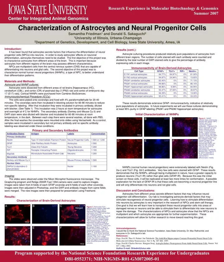
Research Experience in Molecular Biotechnology & Genomics Summer 2007. Center for Integrated Animal Genomics. Characterization of Astrocytes and Neural Progenitor Cells. Samantha Friedman 1 and Donald S. Sakaguchi 2 1 University of Illinois, Urbana-Champaign

E N D
Research Experience in Molecular Biotechnology & Genomics • Summer 2007 Center for Integrated Animal Genomics Characterization of Astrocytes and Neural Progenitor Cells Samantha Friedman1 and Donald S. Sakaguchi2 1University of Illinois, Urbana-Champaign 2Department of Genetics, Development, and Cell Biology, Iowa State University, Ames, IA Introduction: It has been found that astrocytes secrete factors that influence the differentiation of neural progenitor cells (NPCs) into neurons. In order to study astrocytes’ effects on neuronal differentiation, astrocytes themselves need to be studied. The first objective of this project was to characterize astrocytes from different areas of the brain. This is important because astrocytes from different regions of the brain may possess different characteristics. NPCs are multipotent cells from the central nervous system (CNS) that are capable of differentiating into neurons and glial cells. The second objective of this project was to characterize normal human neural progenitors (NHNPs), a type of NPC, to better understand their differentiation patterns. Materials and Methods: Astrocyte and NHNP cultures: Astrocytes were dissected from different areas of rat brains [hippocampus (HC), cerebellum (CBL), and cortex (CR) of postnatal day 2 (PN2) rats and cortex of embryonic day 17 (E17) rats]. Cortical NHNPs were purchased from Cambrex BioSciences. Immunocytochemical Procedure (ICCs): The cells were plated onto coverslips and fixed with 4% paraformaldehyde for 20-40 minutes. The coverslips were then incubated in blocking solution for 60-90 minutes to reduce non-specific labeling. After that incubation they were incubated in primary antibody, diluted with blocker. (See Table 1) This incubation was 2 hours at room temperature for astrocytes and overnight at 4C for NHNPs. The secondary antibody (Donkey anti-Mouse Cy-3) and DAPI stain were also diluted with blocker and incubated for 60-90 minutes at room temperature, in the dark. Between each step there were several washes, all done with PBS. After the final washes the coverslips were mounted onto slides using Vectashield.As a control, samples were incubated in secondary but not primary antibody and no specific antibody labeling was observed under these conditions. Imaging: The slides were observed under the Nikon Microphot fluorescence microscope. The Qcapturing program and Retiga 2000R Fast 1394 camera were used to capture images. Images were taken from 8 fields of each GFAP coverslip and 6 fields of each other coverslip. Images were then adjusted in Photoshop, and the DAPI and antibody images from same fields were merged. The new images were then prepared for presentation using Freehand. Results: Results (cont.): Astrocyte culturing procedures produced relatively pure populations of astrocytes from different brain regions. The number of cells stained with each antibody were counted and divided by the total number of DAPI stained cells to give the percentage of antibody expressing cells in each image. These results demonstrate extensive GFAP- immunoreactivity, indicative of relatively pure populations of astrocytes. In future experiments we will use those cultures demonstrating at least 95% purity in GFAP labeling (PN2#1 and PN2#2 hippocampal astrocytes). NHNPs (normal human neural progenitors) were extensively labeled with Nestin (Fig. 2a-c) and TUJ1 (Fig. 2d-f) antibodies. Very few cells were stained with GFAP. These results demonstrate that the NHNPs, although being multipotent in nature, have a greater capacity to produce neurons (TUJ1-IR) rather than glial cells (GFAP-IR). Because this was the initial screen on these cells, it will be replicated at least two more times for conformation. A possible explanation for the lack of GFAP-IR is that these cells are becoming a neuronal progenitor line and will only differentiate into neurons and not glial cells. Discussion and Conclusions: Astrocytes from different areas secrete different factors that may influence neural progenitor cell differentiation. Our lab is investigating astrocyte-derived factors that may stimulate neurogenesis of neural progenitor cells. Learning how to stimulate differentiation into neurons by astrocytes is very important in the research of NPCs and stem cell therapy. One goal is that we will learn how to transplant these neural progenitor cells into areas of damaged nerves or neurons and be able to stimulate them to differentiate into new neurons to repair the damage. The characterizations of NPCs and astrocytes show that the NPCs are multipotent and which astrocytes are appropriate for further experimentation. These characterizations will allow for further research to move toward reaching this goal. Acknowledgements: I would like to thank the National Science Foundation, Iowa State University, Dr. Max Rothschild, and everyone in Dr. Sakaguchi’s lab. References: Gage, Fred H. Theo O. Palmer. Jun Takahashi. The Adult Rat Hippocampus Contains Primordial Neural Stem Cells. Molecular & Cellular Neuroscience. Vol. 8. Pg. 387-404. 1997. Gage, Fred H. Charles Stevens. Hangjun Song. Astroglia Induce Neurogenesis From Adult Neural Stem Cells. Nature. Vol 417. 2 May 2002. Author E-mail: Sfriedm2@uiuc.edu Immunoreactivity of Brain-Derived Astrocytes Initial Characterization of NHNPs Figure 2: NHNP cultures were extensively labeled with Nestin (NPC marker) and TUJ1 (neuronal marker) antibodies.Figures A, B, and C represent an anti-Nestin stained culture, grown in maintenance media. Figure A shows the anti-Nestin staining, Figure B shows the DAPI staining, and Figure C is the merged image. The merged images show which cells (all are stained with DAPI) are stained with antibody. This can be used to determine the percentage of antibody-labeled cells. Figures D, E, and F represent a TUJ1 stained culture, maintained in differentiation media. Primary and Secondary Antibodies Characterization of Brain-Derived Astrocytes Figure 1:Astrocytes from different brain regions label with GFAP. Figures A, B, and C represent the hippocampal astrocytes from PN2 rat number one. Figure A shows the anti-GFAP staining, Figure B shows the DAPI staining, and Figure C is the merged image. The merged image is used to determine the percentage of cells in the field that are GFAP-labeled. Figures D, E, and F are all merged images. Figure D, GFAP-immunoreactivity (IR) in cortical astrocytes. Figure E, GFAP-IR cerebellar astrocytes. Figure F, TUJ1-IR for neuronal differentiation in E17 rat cortical cultures. This TUJ1 image shows that there is sometimes other cell-types contaminating what should be pure astrocyte cultures. Program supported by the National Science Foundation Research Experience for Undergraduates DBI-0552371; NIH-NIGMS-R01-GM072005-01