Download
1 / 1
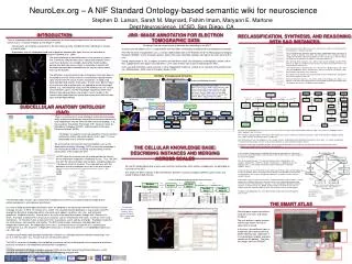
10 likes | 149 Views
Chemical Synapse. has aggregate part. has aggregate part. has aggregate part. has property. located in. Synaptic Site. Post-synaptic Density. Asymmetrical. Pre-synaptic Active Zone Component. Population (of Synaptic Vesicle). is population of. contains site. contains site.

E N D
Chemical Synapse has aggregate part has aggregate part has aggregate part has property located in Synaptic Site Post-synaptic Density Asymmetrical Pre-synaptic Active Zone Component Population (of Synaptic Vesicle) is population of contains site contains site contains site Synaptic Vesicle located in located in located in Pre-synaptic Site Synaptic Cleft Post-synaptic Site Pre-synaptic Active Zone contains site Workflow: Ontology-based Annotation Obtain data (CCDB) 1 Segment objects (Jinx) 2 3 Instance: relationships recorded Axon_0000 is_part_of Glomerulus_0000 Clathrin_Coated_Vesicle_0000 is_part_of Dendrite_0002 Synaptic_Bouton_0002 contacts_with Dendrite_0001 Synaptic_Bouton_0002 is_part_of Glomerulus_0000 Sub-surface_Cisternae_0000 is_part_of Axon_0000 Mitochondrion_0000 is_part_of Synaptic_Bouton_0000 Dendritte_0002 is_part_of Glomerulus_0000 Astrocyte_Process_0000 is_related_to Dendrite_0000 site adjacent to site adjacent to site adjacent to Internode Juxtaparanode Paranode Node of Ranvier is location of is location of is location of is location of Node of Ranvier Axon Internode Axon Paranode Axon Juxtaparanode Axon has molecular constituent has molecular constituent is location of Voltage Gated Na Channel K Channel has component 3d volume 2d image Oligodendrocyte Compact Myelin is location of Subplasma- lemmal Coating SAO-guided annotation 4 Instance is stored Oligodendrocyte Paranodal Termination Cellular Knowledge Base root related to is location of Peripheral Astrocyte Process Glomerulus_0000 is regional part of 5 Surface rendering generated part of contacts with Key: is regional part of restriction Oligodendrocyte non restriction Astrocyte Cell_Body 0001 Axon_0000 Synaptic_Bouton 0002 Dendrite_0001 Dendrite_0000 Dendrite_0002 is regional part of is regional part of is regional part of Axon Lysosome 0000 Mitochondrion 0001 Synaptic_Bouton 0000 Sub-surface_ Cisternae_0000 Synaptic_Bouton 0002 Vacuole_0000 Mitochondrion 0004 Clathrin_Coated_ Vesicle_0000 SER_0000 View relationships Mitochondrion 0000 Myelin_Sheath 0000 Astrocytic_ Process_0000 Cristae_0000 Compact_Myelin 0000 NeuroLex.org – A NIF Standard Ontology-based semantic wiki for neuroscience Stephen D. Larson, Sarah M. Maynard, Fahim Imam, Maryann E. MartoneDept Neuroscience, UCSD, San Diego, CA INTRODUCTION JINX: IMAGE ANNOTATION FOR ELECTRON TOMOGRAPHIC DATA RECLASSIFICATION, SYNTHESIS, ANDREASONING WITH SAO INSTANCES • There is a growing need to accumulate neuroscience data for sharing and merging within the neuroscience community, in a machine readable format (Bloom F, 2006). • Observations are made by neuroscientists but not always formally recorded for other individuals to analyze or expand upon. • Experiments are not standardized and tend to generate complex data types that are not amenable to efficient storage in a database or in computation (Martone et al., 2004). Challenge: How do we get people to describe data according to the SAO? Instances can be added via Jinx, a segmentation tool that allows microscopic image data to be ontologically annotated. The SAO has been incorporated in Jinx, a manual segmentation tool at the National Center for Microscopy and Imaging Research (NCMIR), so the annotation process follows the current workflow and does not require any extra work for the user. During segmentation in Jinx, as objects of interest are identified in each slice throughout a tomographic volume, rather than supplying their own object name identifiers, users select entities from a pick list defined by the SAO. Users can define whether a given instance is either independent or part of, related to, or contacts with another entity, e.g., Mitochondrion_0000 is part of Synaptic_Bouton_0000. We constructed some OWL classes that reclassify the neuron cell types based on their properties assigned by the SAO. We classified neurons based on neurotransmitter, morphological type or presence of spines simply by defining using OWL and Protégé that these categories ought to include any cell which had the main property of that category (e.g., that the neuron was known to use glutamate or GABA as a neurotransmitter, etc). After defining these categories, we used the open source ontology reasoner Pellet (Sirin et al, 2007) to transform the flat version of the SAO neuron type hierarchy (A) into the inferred hierarchy (B). The inferred hierarchy demonstrates that a cell like the a Medium Spiny cell is both spiny and GABAergic while a Dentate Gyrus granule cell can be classified as spiny, glutamatergic, and granule at the same time. Any arbitrary reclassification may be performed using the combinations of properties that suits the purpose of the user. Since the inferred hierarchy is not written back to the ontology, this allows us to maintain a hierarchy with single parents in the authored version of the ontology, while still allowing cells to exist in multiple inferred categories. The grand challenge of neuroinformatics is the creation of systems that seamlessly integrate data across spatial and temporal scales such that information, for example, about white matter bundles derived from diffusion tensor imagin is analyzable in context with electrophysiological data recorded from the neurons whose axons make up the bundles. The difficulties in performing this type of integration from data alone is illustrated on the left, which shows an intracellularly injected medium spiny neuron from the mouse nucleus accumbens, imaged using correlated light and electron microscopy. At each level, different types of visualization and analytical tools are applied to extract meaningful content, e.g., the branching structure of the dendritic tree, the surface area of dendritic spines. But the knowledge required to relate these different data representations and analysis results resides in the scientist who understands the relationship among these different data types and biological objects. SUBCELLULAR ANATOMY ONTOLOGY (SAO) • There is rising interest in using ontologies to formalize knowledge within a domain for exchange among the neuroscience community and incorporation into the semantic web and existing groups, e.g., Neuroscience Information Framework (NIF), National Center for Biomedical Ontologies (NCBO), and Biomedical Information Resource Network (BIRN). • “Ontology:” an explicit, formal representation of terms and their relationships within a particular domain that is both: 1) human interpretable and 2) machine readable • We are creating neuroscience-specific ontologies such as the Subcellular Anatomy Ontology (SAO) to formalize knowledge in the neuroscience field for classifying and describing scientific observations at the subcellular level. • The SAO takes the view that the cell should provide the rallying point for information integration in biological tissues. Thus, the SAO starts with the cell and models how cell parts, including molecules, fit into coarser levels of anatomy. This view contrasts with the approaches of many ontologies that start at the level of gross anatomy and traverse down to the level of the cell e.g., the Foundational Model of Anatomy (FMA); Rosse and Mejino, (2003) and BAMS; Bota et al., (2005). We have reassembled data instances in the cases of the chemical synapse (left) and the Node of Ranvier (right). The synapse is modeled using the object aggregate and site classes. We created an aggregate object consisting of a pre-synaptic part, a post-synaptic part and a junctional part, similar to the Synapse Ontology of Zhang et al., (2007) and then localize them to the synaptic site. Each of these parts have cell components, e.g., synaptic vesicles, located within them that define the extents of these parts, i.e., the pre-synaptic part is the part of the presynaptic structure (axon terminal, dendrite or soma) containing synaptic vesicles. Through the relationships encoded in the SAO, we can restrict the definition of the synapse to that part of the cellular structure where certain structures, e.g., synaptic vesicles, or molecules are localized. THE CELLULAR KNOWLEDGE BASE: DESCRIBING INSTANCES AND MERGING ACROSS SCALES In this example, we performed a straightforward rule-based reasoning task, inferring the presence of a connection between two brain areas from the presence of a single synapse in an electron micrograph. Prior to reasoning, Synapse_1 has two intercellular junction compartments, Pre-synaptic_compartment_1 and Post-synaptic_compartment_1. Applying the first two Algernon rules, it is discovered that the pre-synaptic compartment is related to Dendritic_Spine_1. Using rules that infer the presence of neurons from axon terminals, and a corresponding set of rules for dendritic spines, the synapse can be directly associated with the two neurons that participate in that synapse, through the properties Pre-synaptic_Neuron and Post-synaptic_Neuron. If two neurons share a synapse, then there is a connection between those neurons. If those neurons are in different brain areas, then those areas have a connection between them. Since the neurons that participated in Synapse_1 have already been identified, and their locations are known through has_BAMS_location relations, the third rule can make an explicit connection statement about the brain areas that the neurons are found within. Through a methodology like this, many different kinds of data can be brought together across scales 2D and 3D morphological data of nerve cells and their relationships within cellular microdomains are described as instances of the SAO. This allows the SAO instances to be intermediaries between structures imaged at different spatial scales and stored in different data formats: Instance of Medium Spiny Neuron Dendrite and Its Functional Compartments has Regional part has Regional part has Regional part has Regional part Medium Spiny Neuron Dendrite Dendritic Branch Algernon-J Rules used to produce inference diagrammed above. (http://algernon-j.sourceforge.net/) Dendritic Spine 1 … Dendritic Spine 32 properties ((:instance "sao:Chemical_Synapse" ?a) (has_Pre-synaptic_Neuron ?a ?b) (has_Post-synaptic_Neuron ?a ?c) (has_BAMS_Location ?b ?d) (has_BAMS_Location ?c ?e) (:add-instance (?f "bams:Connection_Statement") (:name ?f "") ("bams:reference" ?f "Inferred") ("bams:sending_Structure" ?f ?d) ("bams:receiving_Structure" ?f ?e) (example_Synapse ?f ?a))) properties properties ((:instance "sao:Chemical_Synapse" ?a) (:instance "sao:Pre-synaptic_Compartment" ?b) ("sao:intercellular_Junction_Compartment" ?a ?b) (:instance "sao:Axon_Terminal" ?c) ("sao:is_Intracellular_Junction_Compartment_Of" ?b ?c) (:instance "sao:Axon" ?d) ("sao:has_Compartment" ?d ?c) (:instance "sao:Neuron" ?e) ("sao:has_Compartment" ?e ?d) (has_Pre-synaptic_Neuron ?a ?e)) properties properties ((:instance "sao:Chemical_Synapse" ?a) (:instance "sao:Post-synaptic_Compartment" ?b) ("sao:intercellular_Junction_Compartment" ?a ?b) (:instance "sao:Neuron_Compartment" ?c) ("sao:is_Intracellular_Junction_Compartment_Of" ?b ?c) (:instance "sao:Neuron" ?d) ("sao:is_Compartment_Of" ?c ?d) (has_Post-synaptic_Neuron ?a ?d)) The SAO describes neurons, glia, multicellularmicrodomains and their associated functional compartments, cellular components, and molecular constituents. As a way to keep epistemological distinctions clear, we adopted as an organizing framework the Basic Formal Ontology version 1.0 (BFO 1.0; Grenon et al., 2003) The structure/function dichotomy is expressed in the BFO through the division of all possible entities into continuants (objects, qualities, sites, etc.) and occurrents (processes, temporal intervals). A continuant is an entity in the world that endures through time (Grenon et al., 2004). Examples of continuants are basic cell structures such as mitochondria and nuclei, as well as lumens and membranes. On the other hand, an occurrent refers to a process, event, activity, or change. Examples include the cell cycle phases, cell secretion, and motility. The BFO further divides continuants into dependent and independent continuants. An independent continuant is an entity that exists irrespective of its relationship to anything else, e.g., cell, organism. A dependent continuant is an entity that inheres in an independent continuant, e.g., color, age. Created using Protégé (http://protege.stanford.edu) and built as a concept hierarchy linked by relationships such as “is a” and “has part,” e.g., “neuron is a cell; cell has part nucleus.” The SAO is not meant to encode rules that define a canonical cell, but rather provides the framework to describe a particular instance of a neuroepithelial cell based on its properties. The SAO was built in the Web Ontology Language (OWL) format (http://www.w3.org/TR/owl-features)—a W3 standard, an extension of RDF, and compatible with the growing semantic web. THE SMART ATLAS Tomographic Reconstruction: Medium Spiny Neuron Dendrite, ACC1_2ma (from CCDB) Querying gene expression patterns using the smart atlas and spatial histogram. The user defines a spatial location and the type and/or intensity of signal to be returned. In this case, three different types of image data were returned for lhx5: protein labeling (top), radioactive in situ hybridization (middle) and gene specific cell fill (bottom). The latter two images are from GENSAT. REFERENCES Bloom F (2006). Prying Open the Black Box, Science, 314(5796):17. Bota, M., Dong, H.W., Swanson, L.W. (2005). Brain architecture management system. Neuroinformatics 3, 15-48. Grenon, P. (2003). BFO in a nutshell: a bi-categorialaxiomatization of BFO and comparison with DOLCE. IFOMIS, ISSN 1611-4019). Grenon, P., Smith, B., Goldberg, L. (2004). Biodynamic ontology: applying BFO in the biomedical domain. Stud Health Technol Inform 102, 20-38. Martone ME, Gupta A, and Ellisman MH (2004). E-Neuroscience: Challenges and Triumphs in Integrating Distributed Data from Molecules to Brains, Nat Neurosci, 7(5): 467-72. Rosse C, Mejino JL Jr. (2003) A reference ontology for biomedical informatics: the Foundational Model of Anatomy. J Biomed Inform. 36, 478-500. Sirin, E., Parsia, B., Grau, B.C, Kalyanpur, A. and Katz, Y. (2007). Pellet: a practical OWL-DL reasoner. Journal of Web Semantics 5. Zhang, W., Zhang, Y., Zheng, H., Zhang, C., Xiong, W., Olyarchuk, J.G., Walker, M., Xu, W., Zhao, M., Zhao, S., Zhou, Z., Wei, L. (2007). SynDB: a synapse protein database based on synapse ontology. Nucleic Acids Res 35, D737-41.


