Download
1 / 56
580 likes | 802 Views
Cell Reproduction and Differentiation. 0. 17. Cells Reproduce by Dividing in Two. Unicellular organisms: cell division is the mechanism of reproduction Multicellular organisms: cell division enables growth from a fertilized egg to a multicellular individual

E N D
Cells Reproduce by Dividing in Two Unicellular organisms: cell division is the mechanism of reproduction Multicellular organisms: cell division enables growth from a fertilized egg to a multicellular individual Mechanism of cell division is the same in all eukaryotes
The Cell Cycle Creates New Cells Cell cycle includes two major phases Interphase (between cell division) Mitotic phase (cell division) Interphase Long growth period between cell divisions G1 (first gap) primary growth phase, very active growth S (synthesis) synthesis of DNA for next cell division G2 (second gap) final growth phase before cell division
The Cell Cycle Creates New Cells Mitotic phase (cell division phase) Mitosis Nuclear division duplicated DNA is distributed between two daughter nuclei, nucleus divides Cytokinesis Cytoplasm divides Two new daughter cells are formed
Figure 17.1 CytokinesisCell divides MitosisNucleus divides MITOTICPHASE M G2 G1 Cell preparesfor division.Growth continuesslowly. Primary periodof cell growth. S DNA is duplicated.Growth continuesslowly. G0 INTERPHASE
The Cell Cycle Creates New Cells Complete cell cycle takes 18–24 hours Mitosis and cytokinesis takes less than one hour of the complete cell cycle Many cells enter a nondividing state, G0, either temporarily or permanently Neurons, osteocytes enter G0 after adolescence
Replication, Transcription, and Translation: An Overview Human DNA is organized into 46 separate chromosomes DNA replication Process of copying DNA prior to cell division This involves making exact copies of all 46 chromosomes Gene Short segment of DNA that contains the code, or recipe, for a protein Smallest functional unit of DNA
Figure 17.2 Double-strandedDNA Histones Chromatin material:not visible duringinterphase One chromatid Its sister chromatid Centromere Chromosome: visible during mitosis
Replication, Transcription, and Translation: An Overview Transcription Process of copying DNA of a gene into mRNA (messenger ribonucleic acid) Occurs within the cell nucleus Translation Process of converting the mRNA template into one or more proteins Occurs in the cytoplasm at ribosomes
Figure 17.3 Replication • Produces an exactcopy of DNA • Occurs prior toevery cell division DNA Transcription • Converts a portion ofdouble-stranded DNA toa single-stranded mRNA • Occurs within the nucleus mRNA Translation • Converts an mRNA codeinto one or more proteins • Takes place in thecytoplasm Protein
Replication: Copying DNA Before Cell Division Process DNA strands uncoil and “unzip” Each single strand serves as a template for the creation of a new complementary strand DNA nucleotides are positioned and linked by DNA polymerase Precise base-pairing (A-T, C-G) assures that an exact copy is made Centromere holds duplicate daughter chromosomes (sister chromatids) together
Figure 17.4 Replicationbubble Keys: Cytosine Adenine Guanine Thymine Parentstrands New complementarystrands 2 complete daughterDNA molecules Parent DNAmolecule The unwinding and the formation of new strandsoccur simultaneously at many sites on the DNAmolecule. The sites of replication expand outward untilthey join. For simplicity the two strands are shown asparallel in (b), but in actuality they form a helical shapeas shown in (a). Parent strand New (daughter)strands forming Parent strand The double-stranded DNA unwinds, and eachsingle strand serves as a template for a newcomplementary strand.
Figure 17.5 4 During mitosis the sister chromatidscondense and become visible. Then the sisterchromatids are pulled apart, with one going toeach daughter cell. When mitosis is complete thesingle chromosome again takes on the moredispersed form and is no longer visible. 3 After replication,the duplicatedchromosomesconsist of sisterchromatids heldtogether by acentromere. Singlechromosome 1 Sisterchromatids MITOTICPHASE During G1, DNAmolecules existas chromatinmaterial that cannot be seendistinctly underthe microscope. M Centromere G2 G1 S INTERPHASE 2 During the S phase, the two strandsof the DNA molecule unwind andreplicate. Replication occurs at multiplesites along the chromosome.
Mutations Are Alterations in DNA Mutations Alterations or “mistakes” in the DNA code Occur most frequently during DNA replication Causes Chemical and physical forces Effects of mutations Silent mutations: have no effect Many mutations are harmful, may result in cell death or cancer Some mutations are beneficial Some mutations are repaired by repair enzymes
Mechanism of DNA Repair Enzymes recognize errors Errors are cut out with enzymes Damaged section is replaced DNA backbone is reconnected Numerous different DNA repair enzymes Repair enzymes are most active between the time of DNA replication and the beginning of mitosis
Transcription: Converting Gene’s Code into mRNA Transcription DNA within the region of a gene unwinds RNA polymerase assists in copying base sequence in RNA nucleotides Primary transcript made, includes introns (intervening sequences) and exons Introns edited out Exons (carry genetic information) are spliced appropriately Messenger RNA strand produced
Figure 17.6 DNA Introns DNA strand notbeing transcribed Exons Assemblysite DNA template RNA primary transcriptis released from DNA Introns areedited out Codons mRNA The introns are editedout by enzymes to produce the final mRNA. RNA primary transcript The portion of the DNA moleculecorresponding to the gene unwindstemporarily, and a complementarystrand of RNA is produced from theDNA strand. The strand of the RNA released fromDNA is called a primary transcript.It contains secretions that carry geneticinformation, called exons, and sectionsthat are apparently useless, called introns.
Transcription: Converting a Gene’s Code into mRNA Genetic code The genetic code is a triplet code Codon Sequence of three mRNA bases Codes for amino acids Redundancy of the genetic code 64 different codons, but 20 different amino acids Several different codons encode each amino acid, with the exception of methionine (AUG, start codon)
Transcription: Converting a Gene’s Code into mRNA Genetic code Codon “grammar” Start codon AUG (methionine): begins all genes Stop codons UAA, UAG, UGA: one ends each gene
Figure 17.7 Second position U C A G UUUUUCUUAUUG UCAG UCUUCCUCAUCG UAUUACUAAUAG UGUUGCUGAUGG Phe Cys Tyr U Ser Stop Stop Leu Stop Trp CCUCCCCCACCG CAUCACCAACAG CGUCGCCGACGG UCAG CUUCUCCUACUG His C Arg Pro Leu Gln First position Third position AUUAUCAUAAUG ACUACCACAACG UCAG AAUAACAAAAAG AGUAGCAGAAGG Ser Asn Ile Thr A Arg Lys Met/start GUUGUCGUAGUG GAUGACGAAGAG GCUGCCGCAGCG GGUGGCGGAGGG UCAG Asp G Gly Val Ala Glu PhePhenylalanineLeuLeucine IleIsoleucine MetMethionine ValValine SerSerine GlnGlutamine AsnAsparagine LysLysine AspAspartic acid GluGlutamic acid ProProline ThrThreonine AlaAlanine TyrTyrosine HisHistidine CysCysteine TrpTryptophan ArgArginine GlyGlycine
Translation: Making a Protein from RNA Components Messenger RNA (mRNA): basically this is a copy of the “recipe” Transfer RNAs (tRNA): relatively small RNA molecules that escort amino acids to the ribosome (site of translation) Ribosomes made of ribosomal RNA (rRNA) and protein Contain sites for mRNA and incoming amino acid-tRNA Contains the enzymes/factors that catalyze the peptide bond formation
Translation: Making a Protein from RNA Process Initiation Initiator tRNA (carrying methionine), start codon on mRNA, and ribosomal subunits form an initiation complex Elongation tRNA brings specific amino acids to developing protein chain Chain elongates one amino acid at a time Termination Stop codon terminates developing chain, protein is released from ribosome
Figure 17.8 Amino acids tRNA capturesfree amino acid tRNA Completed protein Bond formsbetween amino acids Anticodon His tRNA anticodonbinds tomRNA codon Leu Val Val Met Peptide chaindetaches atstop codon tRNAis released “Stop” codon Ribosomalsubunits mRNA Ribosome movesalong mRNA “Start” codon 3 1 2 Initiation. A tRNAcarrying the “start”anticodon binds to thesmaller ribosomalsubunit and to the“start” codon of mRNA.The larger ribosomalsubunit joins them. Elongation. tRNA molecules capture freeamino acids and bring them to theappropriate codon on the mRNA. As mRNApasses between the two ribosomal subunits,the ribosome bonds to the tRNA andcatalyzes the formation of the bond betweensuccessive amino acids. The tRNA is thenreleased to find another amino acid. Termination.When a “stop”codon is reached,the ribosomalsubunits and thenewly formedprotein detachfrom the mRNA.
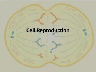
Cell Reproduction: One Cell Becomes Two Two types of cell reproduction processes Mitosis: generates new diploid cells Diploid—cell has two sets of chromosomes, one from the mother and one from the father Meiosis: generates haploid gametes Haploid—cell has only one set of chromosomes
Mitosis: Daughter Cells Are Identical to the Parent Cell Nuclear division (mitosis) followed by cytoplasmic division (cytokinesis) Daughter cells (offspring) are genetically identical to the parent cell Sequence of phases Prophase Metaphase Anaphase Telophase
Mitosis: Daughter Cells Are Identical to the Parent Cell Prophase Mitotic spindle formed Centrioles migrate to cell poles Chromatin condenses into visible chromosomes Nuclear membrane dissolves Metabolic activity decreases Metaphase Duplicate chromosomes form single line at the equator between centriole poles
Mitosis: Daughter Cells Are Identical to the Parent Cell Anaphase Duplicate chromosomes separate Daughter chromosomes are pulled toward poles by microtubules Telophase Reverse of prophase Mitotic spindle disintegrates Nuclear membrane reforms Chromosomes uncoil and revert to chromatin
Mitosis: Daughter Cells Are Identical to the Parent Cell Cytokinesis Contractile ring of filaments forms at midsection of cell and tightens, forming a cleavage furrow Two daughter cells formed as the contractile ring pinches them apart Result: two identical daughter cells (diploid)
Figure 17.9 MITOSIS CYTOKINESIS Nuclearmembraneforming Daughterchromosomes Centrioles Cleavagefurrow Mitotic spindle Mitotic spindle Sister chromatids Centromere Chromosomesuncoiling Anaphase Prophase Metaphase Telophase and cytokinesis MITOTIC PHASE G1 G2 S Interphase
Mitosis Produces Diploid Cells and Meiosis Produces Haploid Cells All cells in human body divide by mitosis, with the exception of the cells that form sperm and eggs All body cells other than sperm and eggs have 46 chromosomes (are diploid) These represent 23 pairs of chromosomes Gametes (sperm, eggs) have 23 chromosomes (are haploid) Reduction in chromosome number from diploid to haploid is accomplished by meiosis, a special cell division process that occurs in ovaries and testes
Meiosis: Preparing for Sexual Reproduction Meiosis includes two successive cell division processes Meiosis I Prophase I, metaphase I, anaphase I, telophase I, and cytokinesis Meiosis II Prophase II, metaphase II, anaphase II, telophase II, and cytokinesis Meiosis reduces chromosome number by half (reduction division) Daughter cells are haploid (n)
Meiosis: Preparing for Sexual Reproduction Meiosis I Prophase I Duplicated homologous chromosomes pair up and swap segments (crossing over) Metaphase Homologous pairs of chromosomes line up Double line of chromosome pairs Anaphase Pairs of chromosomes separated, but duplicated chromosomes stay intact Telophase I and cytokinesis End of Meiosis I: two haploid daughter cells, but chromosomes are still in duplicated state
Meiosis: Preparing for Sexual Reproduction Meiosis II Each of the two daughter cells from Meiosis I goes through Meiosis II Similar process to mitosis Prophase II Metaphase II Anaphase II Duplicated chromosomes (chromatids) separate Telophase II and cytokinesis Nuclei have the haploid chromosome number End of Meiosis II: four haploid daughter cells
Figure 17.11 MEIOSIS I MEIOSIS II Centrioles Prophase II Spindleapparatusforms. Crossing-overoccurs Spindleapparatusforms Prophase I Homologouschromosomespair. Metaphase II Chromosomesalign onequatorial plane. Metaphase I Homologouspairs align onequatorialplane. Anaphase I Homologouspairsseparate. Anaphase II Sister chromatidsseparate. Telophase I Telophase II Cytokinesis occurs.Each cell has ahaploid number ofchromosomes, eachconsisting of twosister chromatids. Nuclei form atopposite poles;cytokinesis occurs. Daughter cells Four haploid daughter cells
Sex Differences in Meiosis: Four Sperm Versus One Egg Males Four sperm produced from each cell entering meiosis All viable, functional Female Unequal cytokinesis during meiosis I and II One egg and three polar bodies produced from each cell entering meiosis Only the egg is viable
Figure 17.12 MALE FEMALE Granulosa cells Primaryoocyte 23 pairs ofduplicatedchromosomes Primaryspermatocyte Meiosis I Meiosis I Secondaryspermatocyte Polar body 23duplicatedchromosomes Secondaryoocyte Meiosis IIandmaturation Completionofmeiosis II Sperm Polarbodies Egg Sperm nucleus (23 chromosomes each) Fertilization Nucleus offertilized egg(23 pairs ofchromosomes)
Animation: Comparing Mitosis and Meiosis Right-click and select Play
How Cell Reproduction Is Regulated Internal surveillance and control mechanism Several key checkpoints where “go ahead” signals must be received in order for the cycle to progress to the next phase G1, G2, M checkpoints Outside influences Can modify cell cycle Hormones, growth factors, presence of other cells
Figure 17.13 M checkpoint G2 checkpoint M G2 G1 S G1 checkpoint
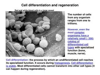
Environmental Factors Influence Cell Differentiation All body cells have the same DNA, yet there are great differences between the shape and function of different cell types Differentiation Process by which a cell becomes different from its parent or sister cell Differentiation is based on different gene expression
Differentiation in Early Development After fertilization of egg by sperm, zygote begins several cell divisions during which cells divide but don’t grow (up to 16 or 32 cells), and form a ball After eight-cell stage, cells are exposed to different environments inside versus outside the ball Cloning Can occur by embryo splitting at eight-cell stage because cells are not yet differentiated
Figure 17.14 Fertilized egg 2-cell stage 4-cell stage 8-cell stage Each cell is exposedto the same environment. A cell at the center of thisball will be exposed to adifferent environment thana cell on the surface. 16-cell stage Differentiation
Differentiation Later in Development Two factors Developmental history of earlier cells Local environment Genes are “turned on” or “turned off” at various stages of development
Differentiation Later in Development External substances harmful to fetuses: Cigarette smoke: retards growth Alcohol: fetal alcohol syndrome Medications: pass through placenta Illegal drugs: child born addicted Environmental chemicals: in air, water, soil Radiation: radon, X-rays Intrauterine infections: HIV, syphilis, rubella
Reproductive Cloning Requires an Undifferentiated Cell Reproductive cloning Producing a “copy” of an entire organism Requires a completely undifferentiated cell as the starting point Two methods Embryo splitting Somatic cell nuclear transfer