Download
1 / 69
750 likes | 944 Views
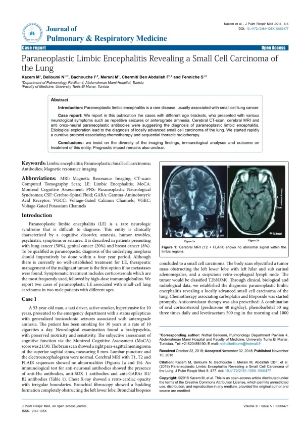
Limited-Stage Small Cell Lung Carcinoma: Overview with Focus on Management. John M. Watkins, M.D. Medical University of South Carolina Department of Radiation Oncology. Background. Small Cell Lung Cancer 15-20% of primary lung neoplasms Decreasing incidence Classically large, central mass

E N D
Limited-Stage Small Cell Lung Carcinoma: Overview with Focus on Management John M. Watkins, M.D. Medical University of South Carolina Department of Radiation Oncology
Background • Small Cell Lung Cancer • 15-20% of primary lung neoplasms • Decreasing incidence • Classically large, central mass • Rapid growth • High metastatic potential (liver, adrenals, bone, bone marrow, brain).
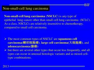
Classification • WHO (old classification) • Oat cell • Intermediate cell • Combined oat cell (with SqCC or adenoCA) • WHO/IASLC (1999) • Classical SCLC (most common) • Large cell neuroendocrine • Combined small-cell (predominant SCLC with areas of NSCLC)
Histopathology • Diagnosis by light microscopy often sufficient: • Small “blue” malignant cells • Sparse cytoplasm • Finely dispersed chromatin without nucleoli • High mitotic rate, necrosis common
Histopathology • Immunohistology • Epithelial • keratin • epithelial membrane Ag • Neuroendocrine* • NSE • chromogranin A *Pre-requisite for dx of large cell neuroendocrine but not small-cell.
Genetic Features • p53: mutated in 75-90% • 9p LOH: 90% • Rb: loss of transcripts (60%) or abnormal gene product (40%) • Telomerase: activated in 90% • c-kit: up-regulated in 80-90% • k-ras: mutation rare (unlike NSCLC)
Staging • VA Lung Study Group • Limited (60-70%): primary/nodal disease confined to ipsilateral hemithorax, within a single radiotherapy port • Extensive (30-40%): metastatic disease outside the ipsilateral hemithorax • IASLC: Limited (M0) vs extensive (M1) Lung Cancer 2002;37:271
Work-Up • H&P • Chest, liver, adrenal CT • Adrenal 17% FN rate by CT • Head MRI (or CT) • CNS mets in 20-30% overall; 15% detection in asymptomatic pts • BoneScan • +/- Marrow Bx • Involved in 15-30% at presentation, but solitary metastatic focus in only 2-6% Am J Roentgenol 1983;140:949 J Neurooncol 2000;48:243 Cancer 1989;63:763
Work-Up • Role of PET (and PET/CT) • Not presently reimbursable for staging SCLC • Small retrospective studies at present: • 4-11% upstaged • Management change in ~15% • RT nodal coverage changed in 25% Clin Lung Cancer 2008;9:30 Radiol Clin N Amer 2007;45:609
Work-Up • Tissue Diagnosis • Trans-Bronchial vs CT-Guided Approach • Core Biopsy vs FNA • Pleural Effusion
Prognostic Factors • Performance Status • Weight Loss • Stage • Early Limited > Limited > Extensive • Within extensive, number of involved sites • LDH (elevated = adverse) • Gender (women>men) • Paraneoplastic syndrome (adverse)
Treatment Paradigm • Interventions • Surgery (early) • Chemotherapy • Radiotherapy • Surgery (recent)
Background Classification Histopathology Genetic Features Staging Review Work-Up Prognostic Factors Management Early Studies Chemotherapy Radiotherapy Surgery Outline SHAMELESS PLUG
Limited Stage SCLC: Early Role of Surgery • 1960s: MRC (n=144, randomized) • Limited-Stage: Resection vs RT • Minimal active chemotherapy at the time • Similar poor outcomes (1-yr surv ~20%) • Resection out of favor with improvements in chemotherapy, advancements in radiotherapy Lancet 1973;2:63
Limited Stage SCLC:Chemotherapy • Due to systemic nature of SCLC, chemotherapy is considered an essential intervention • SCLC is very chemo-responsive, with response rates 80-90% for limited stage; however, very rarely sustained (median 6-8 months) • Upon recurrence, median survival 4 months • cCR ~50% in limited stg, ~15-25% extensive stg • cCR controversial prognostic indicator; pooled European trial data suggests cCR and KPS were independent predictive factors for survival >2yrs Cancer 2000;89:523
ChemotherapyRegimens • Cisplatin / Etoposide (EP) • Demonstrated equivalent survival to CAV (or CEV) chemo or EP/CAV alternation in extensive stage disease • EP/RT superior survival to VEC/RT in limited-stage • Most clinicians favor EP in order to avoid adriamycin-associated toxicities concurrent with XRT JCO 1992;10:282 JNCI 1991;83:855 JCO 2002;20:4665
Limited-Stage SCLC:Radiotherapy • Why XRT? • ~80-90% eventual local failure with chemo alone • cCR may improve long-term survival… so add RT to boost cCR rates • CALGB (Perry; n=399) • ChemoRT (cyc 1) vs ChemoRT (cyc 4) vs Chemo • Chemo = cyclophos/etoposide/vincristine q3wk x18mo (adria replaced etoposide every other cycle after cycle 6) • RT = 40 Gy to primary, mediast, bilat s-clavs + 10 Gy boost (also PCI to all pts, concurrent w/chemo) • Results: ChemoRT regimens improved cCR, FFF, OS NEJM 1987;316:912
Limited-Stage SCLC:Radiotherapy • META Analysis (Pignon, 13 randomized trials): • 2103 evaluable patients, mdn survivor f/u 43 months • Improved survival of 5.4% at 3yrs for chemoRT over chemo alone. • Benefit more apparent in younger patients (RR 0.72 for age <55y vs 1.07 for >70y) • Unable to discern benefit of “early” (w/in 60 days of chemo start) vs “late” RT initiation, or sequential vs concurrent. NEJM 1992;327:1618
Limited-Stage SCLC:Radiotherapy • Timing of XRT: JCOG9104 (Takada): • 231 pts w/LS-SCLC • Concurrent vs Sequential: • Cis/Etopo q3-4wk x4cyc, 45 Gy @ 1.5 Gy/fx BID w/cyc 1 vs after cyc 4 • Trend improved survival for concurrent chemoRT • Median Surv: 27.2 vs 19.7 months (p=0.097) • Increased leukopenia in concurrent, similar esophagitis JCO 2002;20:3054
Limited-Stage SCLC:Radiotherapy • Timing of XRT: META Analysis (Huncharek): • 1574 evaluable patients, 7 of 8 trials used platinum-based chemo regimen (3 cisplatin-etoposide alone) • Evaluation of 1-, 2-, and 3-year overall survivals of patients treated with early (cycle 1-2) vs late (>3 cycle, or sequential, or split) radiotherapy concurrent with chemotherapy. • Outcome: Early initiation results in 50-60% relative improvement in 3-year survival Oncologist 2004;9:665
Limited-Stage SCLC:Radiotherapy • Radiotherapy Dose / Fractionation • Present Acceptable Regimens • Daily: 50 – 70 Gy @ 2 Gy/fx once daily • BID: 45 Gy @ 1.5 Gy/fx twice daily • Concomitant Boost: 61 Gy @ 1.8 Gy/fx, BID over final 9 days
Limited-Stage SCLC:Radiotherapy • Rationale: • Linear cell kill even at low doses • No shoulder = minimal DNA repair capability • Pilot studies demonstrate efficacy & tolerability of BID chemoRT • 2y OS - 40% • Gr 3 esophagitis: ~35-40% • Smaller fraction size should spare late toxicity
Limited-Stage SCLC:Radiotherapy NEJM 1999;340:265
Limited-Stage SCLC:Radiotherapy • Design: • Limited Stage SCLC • Chemotherapy: Cisplatin (60 mg/m2) + Etoposide (120 mg/m2), q3wks x 4 cycles • Radiotherapy: 45 Gy delivered @ 1.8 Gy/fx daily (5 wks) versus 1.5 Gy/fx BID (3 wks) • RT begins with cycle 1 • Fields: gross tumor, ipsi hilar, bilat mediast • Ipsi S-clav only if clinically involved • Inf border ~5cm below carina (or ipsi hilum); otherwise, 1-1.5cm to block edge • Megavoltage linacs only (no Cobalt) • PCI (whole brain) given 25 Gy @ 2.5 Gy/fx if cCR
Limited Stage SCLC: BID vs Daily RT • Eligibility • SCLC limited to one hemithorax +/- ipsi s-clav • No pleural effusion (regardless of cytology) • Bone scan, bilat BM bx neg • Labs: plt (>100k), WBC (>4k), Cr (<1.5) • FEV1: >1L • Stratification • ECOG 0-1 vs 2 • Gender • Wt loss past 6 mo (<5% vs >5%)
Limited Stage SCLC: BID vs Daily RT • Enrollment • No Major differences • Majority ECOG PS 0-1 • Rare s-clav at pres’n • Majority whites • Majority <5% wt loss
Limited Stage SCLC: BID vs Daily RT • Toxicity • Higher Gr 3 esophagitis for BID, o/w no diff
Limited Stage SCLC: BID vs Daily RT • Clinical Response Rate • No difference between BID / QD
Limited Stage SCLC: BID vs Daily RT • Outcomes • At mdn f/u 8y (min 5y) • 5y OS 26% BID vs 16% QD • Patterns of Failure • Thoracic failure ~35% BID vs ~50% QD • Local + Distant ~5% BID vs ~25% QD • Subgroup Analysis • Worse PS & male gender assoc with worse px
Limited Stage SCLC: BID vs Daily RT • Issues: • Increased toxicity • Requirement of elective mediastinal nodal radiotherapy? • Identifiable factors associated with severe esophagitis? • Suboptimal local control • Was 45 Gy in daily fractionation a sufficient comparative arm? • Increased thoracic failures in QD arm • Reduction of thoracic failure is primary benefit of XRT; with chemo alone, ~90% local failure!! • Benefit of higher dose? • Benefit of increased dose intensification? • Other treatment-related factors impacting loco-regional control in BID group?
Limited Stage SCLC: RT Volume • Role of Elective Nodal RT (De Ruysscher) • Phase II Study (prospective) • RT Targets: Primary Tumor & +LNs (by CT) • 45 Gy @ 1.5 Gy BID (w/cyc 1-2) • Carbo/etopo chemo x5cyc • At median f/u 18 months, isolated nodal failure in 3/27 (11%) • All ipsilat supraclav! None mediastinum • Grade 3 esophagitis in 8/27 (30%) • “The safety of selective nodal RT… should not be extrapolated to patients with LD-SCLC until more data are available” Radiother Oncol 2006;80:307
Predictors of Severe Acute Esophagitis from Twice-Daily Thoracic Radiotherapy and Concurrent Chemotherapy for Small-Cell Lung Cancer John M. Watkins, M.D. Medical University of South Carolina Department of Radiation Oncology Charleston, South Carolina, U.S.A. Oral presentation at the 90th Annual Meeting of the American Radium Society, Laguna Niguel, California: 3 May 2008. Manuscript submitted to Int J Radiat Oncol Biol Phys, 13 Jun 2008.
Objectives • In SCLC patients undergoing twice-daily RT with concurrent platinum-based chemotherapy, describe: • Severe Acute Esophagitis (RTOG Grade >3) • Incidence • Treatment Delays (attributable to toxicity) • Associated Factors (Patient-, Tumor-, Treatment-, and Dosimetric-Related)
Design • Retrospective cohort descriptive series • Medical University of South Carolina • Ralph H. Johnson Veterans’ Affairs Medical Center (Charleston, SC)
Design • Inclusion Criteria: • Limited- or Extensive-Stage SCLC • Concurrent chemoRT with twice-daily RT at 1.5 Gy per fraction • Completion of >42 Gy • CT-based treatment planning (3D reconstruction) • Treatment conducted at MUSC
Design • Exclusion Criteria: • Treatment break >5 days (unless due to esophageal toxicity) • Machine maintenance, holidays considered treatment breaks • RT at another institution/facility • Insufficient post-chemoRT follow-up • Minimum 3 months post chemoRT completion
Methods • Retrospective analysis of QA Database • June 1999 through June 2007 • Definitions • Severe Acute Esophagitis: RTOG Grade >3 • Grade 3: Severe odynophagia requiring feeding tube, intravenous fluids, hyperalimentation, +/- >15% weight loss • Grade 4: Complete esophageal obstruction, ulceration, fistula, or perforation • Grade 5: Death due to esophagitis http://www.rtog.org/members/toxicity/acute.html
Definitions & Statistics Stepwise univariate logistic regression analyses of potential associated factors Secondary multivariate analysis for significant & marginally significant factors (multiple logistic regression model) Methods
Definitions & Statistics Variables analyzed: Elective mediastinal irradiation Days RT start to completion Esophageal volume Mean esophageal dose Esophageal Dosimetry (Relative Volume) Area Under Curve Dose Thresholds (V5V45) Methods • Age (</>65yrs, continuous) • Gender • Race • Tobacco Use (Active) • Tumor Site • Tumor Size (Max; </>3cm, continuous) • Number of beams
Study Population • Patient Characteristics • 48 patients included; median post-RT survivor follow-up 25.8 months (range 2.7-83.0)
Study Population • Tumor Characteristics *Of 43 patients, excluding 2 mediastinal primaries and 3 with indeterminate location by records. $By maximal dimension, of 41 patients with recorded data.
Study Population • Radiotherapy Characteristics *Initiated RT with AP/PA, changed at ~9 Gy to 6-field. $IL=ipsilateral, BL=bilateral, Mediast=mediastinum, SClav=supraclavicular.
Study Population • Chemotherapy Characteristics *Of 41 pts with chemo data; CE=cisplatin/etoposide; CbE= carboplatin/etoposide. Prior to concurrent therapy, one patient received paclitaxel with CbE for 3 cycles and another changed from CbE after 1 cycle of CE.
Study Population • Dosimetric Characteristics • 47 patients with contoured esophageal volumes and mean/maximal esophageal doses
Study Population • Dosimetric Characteristics • 38 patients with dose-volume histograms.
Results • Acute Toxicities • All 48 patients evaluable
Results • Univariate Analysis *Continuous variable. #RV-AUC=Relative Volume-Area Under Curve.
Results • Multivariate Analysis • Only RV-AUC remained significant:
Results • Association of Relative Volume by Absolute Dose Threshold 0% vs 48% for V15 </>50% *Odds ratio for a change of 10% (of patients experiencing grade 3 toxicity).