Download
1 / 11
110 likes | 572 Views
Equine Proximal Sesamoiditis. Accession # 70753 Christina Copple, DVM Radiology Resident NCSU CVM-VTH. Sesamoid Bones : embedded within a tendon and protect it Equine Metacarpo-/Metatarsophalangeal Joints = function to provide a frictionless surface along the flexor aspect

E N D
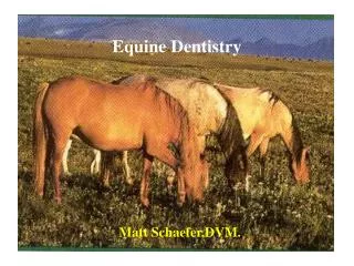
Equine Proximal Sesamoiditis Accession # 70753 Christina Copple, DVM Radiology Resident NCSU CVM-VTH
Sesamoid Bones: embedded within a tendon and protect it Equine Metacarpo-/Metatarsophalangeal Joints = function to provide a frictionless surface along the flexor aspect Sesamoiditis – ischemic disease of the proximal sesamoid bones resulting in lameness usually after hard exercise and pain on deep palpation of the abaxial surface of the proximal sesamoids
Anatomy • Proximal Sesamoid Bones are 3-sided pyramids with an apex, body, and base • Dorsal surface • Abaxial surface • Palmar surface
Sesamoidean Ligaments Collateral sesamoidean Intersesamoidean Superficial (straight) Middle (oblique) Deep (cruciate) Palmar annular ligament Anatomy
Anatomy Palmarolateral View
Anatomy Palmar View
Views Lateromedial (standing & flexed) Dorsopalmar/-plantar (standing & flexed) 45 degree DLPM/DMPL obliques Findings Marginal osteophytosis Abaxial enthesophytosis Enlarged vascular channels Focal osteolysis Radiography
Accession # 70753Blackis 15 yr old MC Walking HorseChronic left hindlimb 4/5 lameness, marked swelling palpable over metatarsophalangeal joint
References • Pasquini, Chris, D.V.M, V. Krishna Reddy, B.V.Sc., Ph.D., Marc H. Ratzlaff, D.V.M., Ph.D. Atlas of Equine Anatomy. 2nd ed. Sudz Publishing. Eureka, CA, 1983. • Dyce, K. M., D.V.M. &S., B.Sc., M.R.C.V.S., W.O. Sack, D.V.M., Ph.D., Dr. med. Vet., C. J. G. Wensing, D.V.M., Ph.D., Textbook of Veterinary Anatomy. 2nd ed. W.B. Saunders. Philadelphia, PA, 1996. • Ross, Mike W., DVM, Sue J. Dyson, MA, VetMB, PhD, DEO, FRCVS. Diagnosis and Management of Lameness In the Horse. Saunders. Philadelphia, PA, 2003. • Thrall, Donald E., DVM, PhD, DACVR. Textbook of Veterinary Diagnostic Radiology. 5th ed. Saunders. St. Louis, MO, 2007. • Butler et al. Clinical Radiology of the Horse. 2nd ed. Blackwell Science. Onsey Mead, Oxford, 2000. • Denoix, J.-M. The Equine Distal Limb Atlas of Clinical Anatomy and Comparative Imaging. Iowa State University Press. Ames, IA, 2000.