Download
1 / 36
380 likes | 556 Views
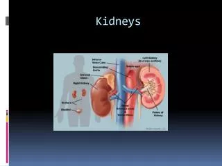
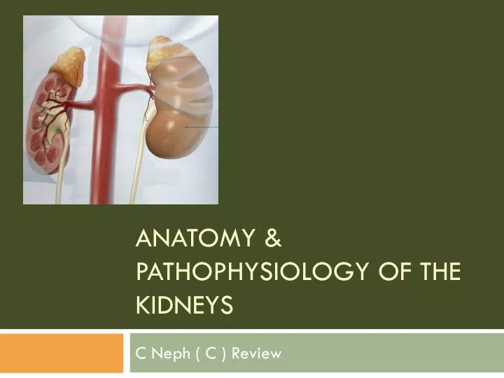
Anatomy & PathoPhysiology of the Kidneys. C Neph ( C ) Review. Function. Regulate the composition and concentration of the extracellular fluids surrounding the body cells. Function is accomplished by the production of urine.

E N D
Anatomy & PathoPhysiology of the Kidneys C Neph ( C ) Review
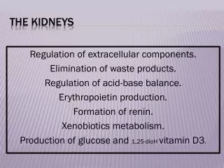
Function • Regulate the composition and concentration of the extracellular fluids surrounding the body cells. • Function is accomplished by the production of urine. • During the process of urine formation other functions are occurring: • Regulate volume of blood plasma • Control the concentration of waste products in the blood • Regulate concentration of the plasma’s electrolytes. • Contribute to the acid/base level (the pH) of the plasma
Find those kidneys! • To locate your kidneys, put your hands on your hips, and then slide your hands up until you can feel your ribs. Place your thumbs on your back and you have located the kidneys. You can’t feel them, but they are there.
The kidney: Paired bean-shaped organs. Size of your fist Each kidney weighs average 0.5% of your total body weight (113 – 170g), 10 - 12 cm long, 5 - 6cm wide, 5.5 cm thick. Gross Anatomy
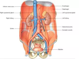
Behind the peritoneum at the back of the abdominal cavity. Referred to as retroperitoneal Symmetrical to the spinal column at the level of the 12th Thoracic vertebra (T12) to the 3rd lumbar vertebra (L3). Right kidney is lower than the left due to the location of the liver.
Each kidney covered by a fibrous capsule. This is covered by a perinephric fat & then by the perinephric (Perirenal Fascia, which also encloses the adrenal gland. Hilus Anatomy of the Kidney
Renal cortex is the outer zone of the kidney and the renal medulla is the inner zone made up of the renal pyramids. The cortex contains all the glomeruli and medulla contains the loops of Henle, the vasa recta, and final portions of the collecting ducts. Sagittal section Renal Medulla Renal Cortex Cortex Medulla
Draining System • The pelvis of the ureter divides into two or three major calyces • Subdivides into two or three minor calyces. • Each minor calyx contains a renal papilla, which is the apex of the medullary pyramid.
Ureter passes out of the kidney behind the peritoneum on the psoas muscle and then enters the pelvis in front of the sacroiliac joint. Moves down the the lateral pelivc wall towards the ischial spine and then turns forward and medially to enter the bladder. Draining System
Vessels & Nerves • Blood vessels and the ureter connect with the kidney at the renal hilus
Renal artery from the aorta and usually divides into three branches. • Two pass in front of the ureter • One goes behind • Renal Vein • Comprised of five or six small veins coming together entering the inferior vena cava.
Glomerulus Efferent Arterioles Renal Artery Interlobular Veins Arcuate Veins Segmental artery Interlobar artery Arcuate artery Interlobular Artery Afferent Arterioles Glomerulus Interlobar veins Segmental Vein Renal Vein
Nerves • Position of the lymphatic and renal sympathetic nerves is variable • Lymphatic drain to the aortic lymph nodes • Sympathetic nerves supply the renal vasculature, juxtaglomerular apparatus, and to a lesser extent rest of the nephron. • Afferent fibers enter the spinal cord at T10, T11, T12. • Afferent nerves from the ureter enter the spinal cord at T11, T12, L1, and L2. • The bladder connection is at S3, S4, and S5
Urine formed by filtration in the glomerulus: it is then modified in the tubules by the reabsorption and secretion of substances. Production of Urine
The Nephron Basic unit of the kidney Responsible for the actual purification and filtration of the blood About one million nephrons are in each kidney Microanatomy
Cortical nephrons: located throughout the renal cortex and have short loops of Henle Juxtamedullarynephronsbegin near the corticomedullary junction and have long loops of Henle, descend deep into the medulla and enable to concentrate urine effectively. Cortical nephronsvsjuxtamedullary 7:1 ratio Types of nephrons
Each nephron is made up of a very small filter called a glomerulus Each glomerulus is attached to a tubule Renal Corpuscles
Osmotic Gradient Proximal convoluted tubule Peritubular capillary Passive Transport Cl- Na+ Accumulation of Na+ Active Transport stimulates
Proximal Convoluted tubules Peritubular H2O Na Cl Na Cl H2O Na Cl Na Na Cl Cl H2O Na Na Cl Cl Na Cl
Juxtaglomerular Apparatus • Consist of Macular densa (specialised cells) and renin releasing cells. They are responsible for maintaining a constant blood flow though the glomerulus and thus a constant GFR despite fluctuations in arterial pressure. • Localized feedback system
Hormone Activity • Anitdiuretic hormone (ADH) • Opens membrane pores in the collecting duct and allows water to pass • Produced by neurons in the hypothalamus • Secreted by the posterior lobe of the pituitary gland • Stimulated when chemical receptors in the hypothalmus respond to an increase or decrease of sodium and other ions in the blood.
Aldosterone • In the distal convoluted tubules • Reabsorption of sodium ions • Reabsorption of water • Stimulates secretion of potassium from blood into the fluid of the distal convoluted tubule. • Secreted by the cortex of the adrenal gland.
Urine • 95% water • 5% solids (organic wastes, ions, salts) • Organic wastes • Urea: product of liver metabolism • Produced during the conversion of amino acids to energy-supplying compounds • Ammonia: • Uric acid: (from nucleic acid breakdown) • Creatinine: (product of creatine phosphate utilization in the muscle cells) • Ketones: resulting breakdown of fat molecules
Ions • Cations (sodium, potassium, magnesium, and calcium) • Anions (chloride, sulfate, and phosphate)
Characteristics of Human Urine • Clarity: Transparent or clear: becomes cloudy on standing • Specific Gravity: 1.015 to 1.020 • Color: Amber or straw-colored • Amount in 24 hours: About 3 pints (1500ml); varies according to fluid intake, amount of perspiration, and other factors • pH: Acidic, may be alkaline ;normal pH range is 4.6 to 8.0; average, about 6. • Odor: Characteristics urine odor; develops ammonia on standing from formation of ammonium compounds
Small Intestine: Approximately 20 feet (6m) long Coiled tube contains roughly 2700 square feet (300m2) of surface area for absorbing nutrients into the bloodstream Excretory Organs
Large Intestine: Functions as a receptacle where water and electrolytes can be reabsorbed into the bloodstream and undigested material is compacted and stored as feces. Excretory Organs
Excretory Organs • Liver • Metabolizes the products of hemoglobin or red blood cells and excretes the products of hemoglobin breakdown as bile pigments. • Lungs • Excrete carbon dioxide and give off some water • Skin • Excretory organ it excretes perspiration during sweating