Download
1 / 31
310 likes | 335 Views
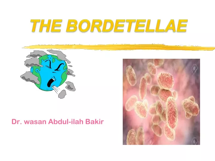
THE BORDETELLAE. Dr. wasan Abdul-ilah Bakir. THE BORDETELLAE. There are several species of Bordetella : 1- Bordetella pertussis , a highly communicable and important pathogen of humans, causes whooping cough (pertussis).

E N D
THE BORDETELLAE Dr. wasan Abdul-ilah Bakir
THE BORDETELLAE There are several species of Bordetella : 1- Bordetella pertussis, a highly communicable and important pathogen of humans, causes whooping cough (pertussis). 2- BordetellaparapertussisIt can cause a milder pertussis-like disease in humans, but Bordetella pertussis is the most serious human pathogen in this genus
Bordetella pertussis • The organisms are minute, gram-negative coccobacilli • A capsule is present. • aerobic, non-spore forming, • Specific to Humans • Colonizes the respiratory tract • B. pertussis invades its human host through entry into the respiratory tract where it colonizes to cause whooping cough, also known as pertussis, which was at one time a very common and potentially life threatening infection for children
Spread • Pertussis is highly contagious • Pertussis is generally transmitted from person to person via respiratory droplets, but direct contact with respiratory secretions from infected individuals may also lead to the disease. • 90% of nonimmune household contacts acquire the disease • Adolescents and adults are the major source of infection in unvaccinated children • Infants and young children are infected by older siblings who have mild to asymptomatic disease.
Spread Freshly contaminated articles (such as clothing) from the infected person can also contain infectious respiratory secretions, allowing pertussis to be passed indirectly from the infected person to a susceptible host who comes in direct contact with these items.
Pathogenesis, Pathology and Virulence factors • B. pertussis invades the human host through the inhalation of respiratory droplets adheres to the ciliated epithelium of the respiratory tract but do not invade the underlying tissue. • It was believed that B. pertussis was an entirely extracellular pathogen, but it has recently been shown that B. pertussis can invade aveolar macrophages. • This pathogen can multiply rapidly on the mucosal membrane of the upper respiratory tract, producing adhesions that allow it to colonize by adhering to the ciliated epithelium.
Pathogenesis, Pathology and Virulence factors • B. pertussis must survive within the hostile environment of its human host by producing a variety of virulence factors in an attempt to evade or counter the immune system of the host as it tries to clear the infection. • B pertussis survives for only brief periods outside the human host. There are no vectors. • Transmission is largely by the respiratory route from early cases and possibly via carriers.
Pathogenesis, Pathology and Virulence factors • B pertussis produces a number of virulence factors that are involved in the pathogenesis of disease. Adhesions such as: • filamentous hemagglutinin • agglutinogens • peractin • fimbriae • a number of toxins including: • pertussis toxin • adenylatecyclase toxin (ACT) • trachaelcytotoxin • dermonecrotic toxin (DNT) • heat-labile toxin • lipopolysaccharide coatthat acts as an endotoxin and can aid colonization by agglutinating human cells.
Pathogenesis, Pathology and Virulence factors • Filamentous hemagglutinin, a large surface protein, and fimbriae (surface appendages) mediate adhesion to ciliated epithelial cells and are essential for tracheal colonization. • Pertussis toxin (a classic A/B structure toxin) promotes lymphocytosis, sensitization to histamine, and enhanced insulin secretion that disrupts function of signal transduction in many cell types. • The filamentous hemagglutinin and pertussis toxin are secreted proteins and are found outside of the B pertussis cells.
Pathogenesis, Pathology and Virulence factors • Adenylatecyclasetoxin (ACT), dermonecrotic toxin (DNT), and hemolysin. ACT is an important virulence factor that inhibits phagocyte function but the role of DNT in pertussis is unknown. • The tracheal cytotoxinkills respiratory epithelial cells in vitro. • The lipooligosaccharidein the cell wall may also be important in causing damage to the epithelial cells of the upper respiratory tract.
Pathogenesis, Pathology and Virulence factors • The organism adheresto and multipliesrapidly on the epithelial surface of the trachea and bronchi and interfereswith ciliary action. The bacteria liberatethe toxins and substances that irritate surface cells, causing coughing and marked lymphocytosis. Later, there may be necrosis of parts of the epithelium and polymorphonuclear infiltration, with peribronchial inflammation and interstitial pneumonia.
Pathogenesis, Pathology and Virulence factors
Pathogenesis, Pathology and Virulence factors
Pathogenesis, Pathology and Virulence factors • Secondary invaders such as staphylococci or H influenzaemay give rise to bacterial pneumonia. • Obstruction of the smaller bronchioles by mucous plugs results in diminished oxygenation of the blood. This probably contributes to the frequency of convulsions in infants with whooping cough.
Clinical Symptoms • After an incubation period of about 2 weeks, the “catarrhal stage” develops, with mild coughing and sneezing. • During this stage, large numbers of organisms are sprayed in droplets, and the patient is highly infectious but not very ill.
Clinical Symptoms • During the “paroxysmal” stage, the cough develops its explosive character and the characteristic “whoop” upon inhalation.This leads to rapid exhaustion and may be associated with vomiting, cyanosis, and convulsions. The “whoop” and major complications occur predominantly in infants. Paroxysmal coughing predominates in older children and adults.
Clinical Symptoms • The white blood count is high (16,000–30,000/μL), with an absolute lymphocytosis. • Convalescence is slow. B pertussis is a common cause of prolonged cough in adults. ends with the convalescence stage is characterized by fewer paroxysmal coughing episodes and usually disappears in 2-3 weeks, but may continue for months. Fever minimal to absent. Symptoms subside gradually over months (convalescent stage 1-2 wks)
Lab. Diagnosis • A. Specimens • Nasopharyngeal (NP) swabs or NP aspirates. • Nasal swabs are not acceptable • For adults, cough droplets expelled directly onto a “cough plate” held in front of the patient’s mouth, during a paroxysm is a less desirable method of specimen collection.
Lab. Diagnosis • B. Direct Fluorescent Antibody Test • The fluorescent antibody (FA) reagent can be used to examine nasopharyngeal swab specimens. However, false-positive and false-negative results may occur; the sensitivity is about 50%. • The FA test is most useful in identifying B pertussis after culture on solid media.
Lab. Diagnosis • C. Culture • NP aspirates or swabs are cultured on solid media (Bordet-Gengou medium) with high percentage of blood (20%-30%) to inactivate inhibitors in the agar, and also contain potato and glycerol. • The antibiotics in the media tend to inhibit other respiratory microbiota but permit growth of B pertussis. Selective media for B. pertussis includes Regan-Lowe, Bordet-Gengou, or charcoal agar. Successful isolation declines with pervious exposure to antibiotic therapy effective against pertussis or if specimens are collected beyond the first two weeks of illness. Isolation is also difficult for vaccinated patients.
Lab. Diagnosis • F. Serology • Production of IgA, IgG, and IgM antibodies occurs after exposure to B pertussis and these antibodies can be detected by enzyme immunoassays. • Serologic tests on patients are of little diagnostic help acutely because a rise in agglutinating or precipitating antibodies does not occur until the third week of illness. • D. immunofluorescence staining or by slide agglutination with specific antiserum. • E. Polymerase Chain Reaction
Optimal Timing in Weeks for Diagnostic Testing Week Cough Onset 0 2 4 6 8 10 12 Culture PCR Serology
Immunity • Recovery from whooping cough or immunization is followed by immunity that is not lifelong. • Second infections may occur but are usually milder; reinfections occurring years later in adults may be severe. It is probable that the first defense against B pertussis infection is the antibody that prevents attachment of the bacteria to the cilia of the respiratory epithelium. • Antibodies to PT are highly immunogenic
Treatment • Administration of erythromycin during the catarrhal stage of disease promotes elimination of the organisms and may have prophylactic value. • Treatment after onset of the paroxysmal phase rarely alters the clinical course. • Oxygen inhalation and sedation may prevent anoxic damage to the brain.
Treatment • Azithromycin is the drug of choice. Note that azithromycin reduces the number of organisms in the throat and decreases the risk of secondary complications but has little effect on the course of the disease at the “prolonged cough” stage because the toxins have already damaged the respiratory mucosa. • Supportive care (e.g., oxygen therapy and suction of mucus during the paroxysmal stage is important, especially in infants).
Prevention • There are two types of vaccines: 1 . an acellular vaccine containing purified proteins from the organism. 2. a killed vaccine containing inactivated B. pertussis organisms. • The acellular vaccine has fewer • side effects than the killed vaccine but has a shorter duration of immunity.
Prevention • The pertussis vaccine is usually given combined with diphtheria and tetanus toxoids (DTaP) in three doses beginning at 2 months of age. A booster at 12 to 15 months of age and another at the time of entering school are recommended. • Because outbreaks of pertussis have occurred among teenagers, a booster for those between 10 and 18 years old is recommended. This vaccine, called Boostrix, contains diphtheria and tetanus toxoids also.
To protect newborns, pregnant women should receive pertussis vaccine. AntipertussisIgG will pass the placenta and protect the newborn. • The killed vaccine is no longer used in the United States because it is suspected of causing various side effects, including postvaccine encephalopathy at a rate of about one case per million doses administered. • Azithromycin is useful in prevention of disease in exposed, unimmunized individuals. It should also be given to immunized children younger than 4 years who have been exposed because vaccine-induced immunity is not completely protective.
BORDETELLA PARAPERTUSSIS • This organism may produce a disease similar to whooping cough, but it is generally less severe. • The infection is often subclinical. • B parapertussis grows more rapidly than typical B pertussis and produces larger colonies. • It also grows on blood agar. • B parapertussis has a silent copy of the pertussis toxin gene.