Download

1 / 64
710 likes | 1.06k Views
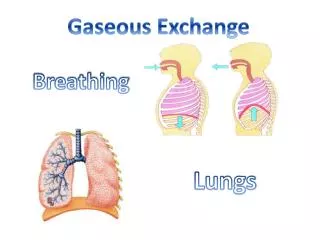
The Lungs. The Lungs Left and right lungs Are in left and right pleural cavities The base Inferior portion of each lung rests on superior surface of diaphragm Lobes of the lungs Lungs have lobes separated by deep fissures . The Lungs. The right lung has three lobes

E N D
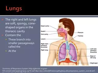
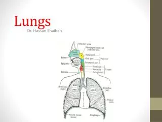
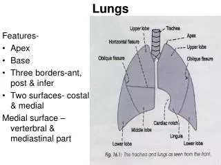
The Lungs • The Lungs • Left and right lungs • Are in left and right pleural cavities • The base • Inferior portion of each lung rests on superior surface of diaphragm • Lobes of the lungs • Lungs have lobes separated by deep fissures
The Lungs • The right lung has three lobes • Superior, middle, and inferior • Separated by horizontal and oblique fissures • The left lung has two lobes • Superior and inferior • Separated by an oblique fissure
The Lungs • Lung Shape • Right lung • Is wider • Is displaced upward by liver • Left lung • Is longer • Is displaced leftward by the heart forming the cardiac notch
The Lungs Figure 21–7a The Gross Anatomy of the Lungs.
The Lungs Figure 21–7b The Gross Anatomy of the Lungs.
The Lungs Figure 21–7b The Gross Anatomy of the Lungs.
The Lungs Figure 21–8 The Relationship between the Lungs and Heart.
The Lungs • The Bronchial Tree • Is formed by the primary bronchi and their branches • Extrapulmonary Bronchi • The left and right bronchi branches outside the lungs • Intrapulmonary Bronchi • Branches within the lungs
The Lungs • A Primary Bronchus • Branches to form secondary bronchi (lobar bronchi) • One secondary bronchus goes to each lobe • Secondary Bronchi • Branch to form tertiary bronchi, also called the segmental bronchi • Each segmental bronchus • Supplies air to a single bronchopulmonary segment
The Lungs • Bronchopulmonary Segments • The right lung has 10 • The left lung has 8 or 9 • Bronchial Structure • The walls of primary, secondary, and tertiary bronchi • Contain progressively less cartilage and more smooth muscle • Increased smooth muscle tension affects airway constriction and resistance
The Lungs • Bronchitis • Inflammation of bronchial walls • Causes constriction and breathing difficulty
The Lungs • The Bronchioles • Each tertiary bronchus branches into multiple bronchioles • Bronchioles branch into terminal bronchioles • One tertiary bronchus forms about 6500 terminal bronchioles • Bronchiole Structure • Bronchioles • Have no cartilage • Are dominated by smooth muscle
The Lungs • Autonomic Control • Regulates smooth muscle • Controls diameter of bronchioles • Controls airflow and resistance in lungs • Bronchodilation • Dilation of bronchial airways • Caused by sympathetic ANS activation • Reduces resistance
The Lungs • Bronchoconstriction • Constricts bronchi • Caused by: • parasympathetic ANS activation • histamine release (allergic reactions)
The Lungs • Asthma • Excessive stimulation and bronchoconstriction • Stimulation severely restricts airflow • Trabeculae • Fibrous connective tissue partitions from root of lung • Contain supportive tissues and lymphatic vessels • Branch repeatedly • Divide lobes into increasingly smallercompartments
The Lungs • Pulmonary Lobules • Are the smallest compartments of the lung • Are divided by the smallest trabecular partitions (interlobular septa)
The Lungs Figure 21–9 The Bronchi and Lobules of the Lung.
The Lungs Figure 21–9 The Bronchi and Lobules of the Lung.
The Lungs • Surfaces of the Lungs • Each terminal bronchiole delivers air to a single pulmonary lobule • Each pulmonary lobule is supplied by pulmonary arteries and veins • Exchange surfaces within the lobule • Each terminal bronchiole branches to form several respiratory bronchioles, where gas exchange takes place
The Lungs • An Alveolus • Respiratory bronchioles are connected to alveoli along alveolar ducts • Alveolar ducts end at alveolar sacs • Common chambers connected to many individual alveoli • Has an extensive network of capillaries • Is surrounded by elastic fibers
The Lungs Figure 21–10 Respiratory Tissue.
The Lungs Figure 21–11a Alveolar Organization: Basic Structure of a Portion of Single Lobule.
The Lungs Figure 21–11b Alveolar Organization: A Diagrammatic View of Structure.
The Lungs • Alveolar Epithelium • Consists of simple squamous epithelium • Consists of thin, delicate pneumocytes type I • Patrolled by alveolar macrophages, also called dust cells • Contains pneumocytes type II (septal cells) that produce surfactant
The Lungs • Surfactant • Is an oily secretion • Contains phospholipids and proteins • Coats alveolar surfaces and reduces surface tension
The Lungs • Respiratory Distress • Difficult respiration • Due to alveolar collapse • Caused when pneumocytes type II do not produce enough surfactant • Respiratory Membrane • The thin membrane of alveoli where gas exchange takes place
The Lungs • Three Layers of the Respiratory Membrane • Squamous epithelial lining of alveolus • Endothelial cells lining an adjacent capillary • Fused basal laminae between alveolar and endothelial cells
The Lungs Figure 21–11c Alveolar Organization: The Respiratory Membrane.
The Lungs • Diffusion • Across respiratory membrane is very rapid • Because distance is short • Gases (O2 and CO2) are lipid soluble • Inflammation of Lobules • Also called pneumonia • Causes fluid to leak into alveoli • Compromises function of respiratory membrane
The Lungs • Blood Supply to Respiratory Surfaces • Each lobule receives an arteriole and a venule • Respiratory exchange surfaces receive blood: • From arteries of pulmonary circuit • A capillary network surrounds each alveolus: • As part of the respiratory membrane • Blood from alveolar capillaries: • Passes through pulmonary venules and veins • Returns to left atrium
The Lungs • Blood Supply to the Lungs • Capillaries supplied by bronchial arteries • Provide oxygen and nutrients to tissues of conducting passageways of lung • Venous blood bypasses the systemic circuit and flows into pulmonary veins
The Lungs • Blood Pressure • In pulmonary circuit is low (30 mm Hg) • Pulmonary vessels are easily blocked by blood clots, fat, or air bubbles, causing pulmonary embolism
The Lungs • The Pleural Cavities and Pleural Membranes • Two pleural cavities • Are separated by the mediastinum • Each pleural cavity • Holds a lung • Is lined with a serous membrane (the pleura)
The Lungs • The Pleura • Consists of two layers • Parietal pleura • Visceral pleura • Pleural fluid • Lubricates space between two layers
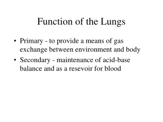
Introduction to Gas Exchange • Respiration refers to two integrated processes • External respiration • Includes all processes involved in exchanging O2 and CO2 with the environment • Internal respiration • Also called cellular respiration • Involves the uptake of O2 and production of CO2 within individual cells
Introduction to Gas Exchange Figure 21–12 An Overview of the Key Steps in External Respiration.
Introduction to Gas Exchange • Three Processes of External Respiration • Pulmonary ventilation (breathing) • Gas diffusion: • Across membranes and capillaries • Transport of O2 and CO2: • Between alveolar capillaries • Between capillary beds in other tissues
Pulmonary Ventilation • Pulmonary Ventilation • Is the physical movement of air in and out of respiratory tract • Provides alveolar ventilation • Atmospheric Pressure • The weight of air • Has several important physiological effects
Pulmonary Ventilation • Boyle’s Law • Defines the relationship between gas pressure and volume: P = 1/V • In a contained gas • External pressure forces molecules closer together • Movement of gas molecules exerts pressure on container
Pulmonary Ventilation Figure 21–13 Gas Pressure and Volume Relationships.
Pulmonary Ventilation • Pressure and Airflow to the Lungs • Air flows from area of higher pressure to area of lower pressure • A Respiratory Cycle • Consists of • An inspiration (inhalation) • An expiration (exhalation)
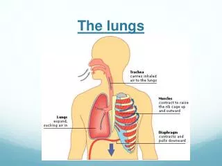
Pulmonary Ventilation • Pulmonary Ventilation • Causes volume changes that create changes in pressure • Volume of thoracic cavity changes • With expansion or contraction of diaphragm or rib cage
Pulmonary Ventilation Figure 21–14 Mechanisms of Pulmonary Ventilation.
Pulmonary Ventilation • Compliance • An indicator of expandability • Low compliance requires greater force • High compliance requires less force • Factors That Affect Compliance • Connective tissue structure of the lungs • Level of surfactant production • Mobility of the thoracic cage
Pulmonary Ventilation • Pressure Changes during Inhalation and Exhalation • Can be measured inside or outside the lungs • Normal atmospheric pressure: • 1 atm or Patm at sea level: 760 mm Hg
Pulmonary Ventilation • The Intrapulmonary Pressure • Also called intra-alveolar pressure • Is relative to Patm • In relaxed breathing, the difference between Patm and intrapulmonary pressure is small • About -1 mm Hg on inhalation or +1 mm Hg on exhalation
Pulmonary Ventilation • Maximum Intrapulmonary Pressure • Maximum straining, a dangerous activity, can increase range • From -30 mm Hg to +100 mm Hg
Pulmonary Ventilation • The Intrapleural Pressure • Pressure in space between parietal and visceral pleura • Averages -4 mm Hg • Maximum of -18 mm Hg • Remains below Patm throughout respiratory cycle