Download
1 / 55
550 likes | 699 Views
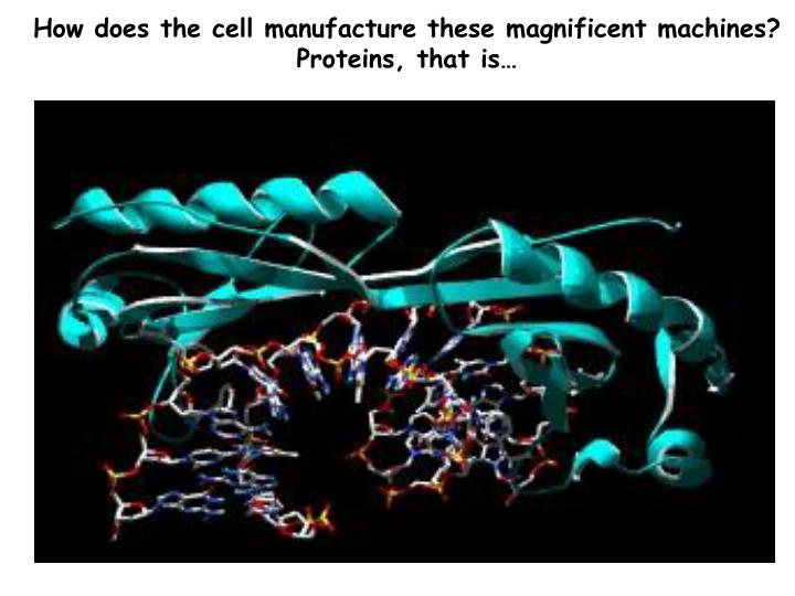
How does the cell manufacture these magnificent machines? Proteins, that is…. Proteins. Long polymers of amino acids, joined by peptide (amide) bonds are called polypeptides Polypeptides fold into stable three- dimensional shapes and are called proteins

E N D
How does the cell manufacture these magnificent machines? Proteins, that is…
Proteins • Long polymers of amino acids, joined by peptide (amide) bonds are called polypeptides • Polypeptides fold into stable three- dimensional shapes and are called proteins • Shape determines the function of proteins (active sites are on the surface)
Proteins - classified by functions Enzymes-catalytic activity and function Transport Proteins- bind & carry ligands Storage Proteins-ovalbumin, gluten, casein, ferretin Contractile(Motor): can contract, change shape, elements of cytoskeleton (actin, myosin, tubulin) Structural(Support):collagen of tendons & cartilage, elastin of ligaments (tropoelastin), keratin of hair, feathers, & nails, fibroin of silk & webs Defensive (Protect):antibodies (IgG), fibrinogen & thrombin, snake venoms, bacterial toxins Regulatory(Signal): regulate metabolic processes, hormones, transcription factors & enhancers, growth factor proteins Receptors(detect stimuli): light & rhodopsin, membrane receptor proteins and acetylcholine or insulin.
Structure of Proteins the Variety of Protein Structures may be INFINITE... average protein has 300-400 amino acid's & has a MW of 30kD to 45kD a PROTEIN of 300 amino acids made with 20 different kinds of amino acids can have 20300different linear arrays of aa's [10390 different proteins] 1st protein sequenced was BeefInsulin by Fred Sanger - 1958 Nobel Prize winner to date about 100,000 proteins have been sequenced only about 10,000structures known [2K/yr] E. coli make about 3,000 proteins, humansmake about 100,000 proteins from about 30,000 genes
4 levels of protein structure are recognized primary - linear sequence of aa's secondary - regular, recurring orientation of aa in a peptide chain due to H-bond tertiary - complete 3-D shape of a peptide quaternary - spatial relationships between different polypeptides or subunits Start with the building blocks: amino acids (aa’s)
There are three types of side chains…. • Nonpolar (hydrophobic) • Polar uncharged (hydrophilic) • Polar charged (hydrophilic)
Primary sequence… Linear sequence of amino acids in a polypeptide repeated peptide bonds form the back bone of the polypeptide chain R side groups project outward on alternate side Chain... one end of polypeptide chain has a free (unlinked) amine group: N-terminus other end has a free (unlinked) carboxyl group: C-terminus N-C-C-N-C-C-N-C-C-N-C-C-N-C-C-N-C-C Size… protein size is specified by mass (MW in daltons = 1 amu) average MW of a single amino acid ≈ 113 Da thus if a protein is determined to have a mass of 5,763 Da ≈ 51 amino acids average yeast protein = 52,728 Da [52.7 kDa] with about 466 amino acids Protein Primary Sequence today is determined by reading the GENOME Sequence Function is derived from the 3D structure (conformation) specified by the primary amino acid sequence and the local environs interactions.
-helix = Pitch 3.6 aa per turn -helices obey the n + 4 rule: H-bonding from C=O….H-N 1 4
-pleated sheet In a Beta sheet, R-groups of alternating amino acids protrude above and below the sheet
Proteins are 3-dimensional molecules Primary structure = Amino acid sequence -sheet Secondary structure = Alpha helix Beta sheet Tertiary structure = 3-D shape Quaternary structure = ?? -helix
Supersecondary structures: - - motif…. …and -meander
-barrel Supersecondary structures:
Supersecondary structures: Greek Key motif formed folding -sheets And here is a unique family of -helical Greek Key proteins
The Helix-turn-helix (H-T-H) is a common DNA-binding motif A movie showing the engrailed homeodomain H-T-H motif
Engrailed controls a key process in animal development The engrailed gene, a "segment-polarity" gene, divides each of unit, or segment, into anterior and posterior compartments. The fourteen narrow compartments shown here correspond to specific segments of the embryo. There are three head segments (H, top right), three thoracic segments (T, lower right), and eight abdominal segments (a, from bottom right to upper right).
Hydrophobic interactions bring together two subunits via the Leucine Zipper motif Leucine zipper (dimerization) --every 7th aa is a LEU residue DNA binding domain Leucine zippers are an example of amphipathic -helices
The Estrogen receptor binds DNA via 4 Zinc finger domains -- the zinc finger -helix binds DNA in the major groove
Tertiary level level most responsible for3-D orientationof proteins in space is the thermodynamically most stable conformation of a protein... and is due to – weak non-covalent interactions - hydrophobic interior & hydrophilic exterior -via H-bonds - & S-S bridges results in Protein Foldingintospecific 3D shapes &unique binding sites
Disulfide bridge formation stabilizes protein structure Cys - S - S - Cys Cys - S - H + H - S - Cys
a-Hemolysin from Staphylococcus aureus a-toxin (a-hemolysin) The best characterized and most potent membrane-damaging toxin of S. aureus is a-toxin. It is expressed as a monomer that binds to the membrane of susceptible cells. Subunits then oligomerize to form heptameric rings with a central pore through which cellular contents leak. In humans, platelets and monocytes are particularly sensitive to a-toxin. Susceptible cells have a specific receptor for a-toxin which allows the toxin to bind causing small pores through which monovalent cations can pass. The mode of action of alpha hemolysin is likely by osmotic lysis.