Download
1 / 41
570 likes | 1.64k Views
The Head and Neck, Pharynx, and Oral Cavity. Neck Regions. When dissecting the neck from the anterior surface, you will come across the HYOID BONE. The hyoid bone is the only bone in the body that does not articulate with other bones. The base of the tongue is attached to the hyoid bone.

E N D
Neck Regions • When dissecting the neck from the anterior surface, you will come across the HYOID BONE. • The hyoid bone is the only bone in the body that does not articulate with other bones.
The base of the tongue is attached to the hyoid bone. • The hyoid bone is attached by muscles to the skull, providing for flexibility for speech. • It also attaches to the styloid process of the TEMPORAL BONE. • The muscles in the neck are named for their points of attachment.
From the STERNUM to the THYROID CARTILAGE lies the STERNOTHYROID MUSCLE. This muscle lies lateral and deep to the STERNOHYOID MUSCLE. • From the STERNUM to the MASTOID PROCESS lies the STERNOMASTOID MUSCLE.
From the CLAVICLE to the MASTOID PROCESS lies the CLEIDOMASTOID MUSCLE. • In humans, the STERNOMASTOID and the CLEIDOMASTOID are one muscle called the STERNOCLEIDOMASTOID MUSCLE.
The DIGASTRIC MUSCLE opens the jaw and attaches to the mastoid process and the inner border of the mandible at the central portion (midline fusion area).
stylohyoid digastric sternohyoid omohyoid sternocleidomastoid thyrohyoid trapezius clavicle
Regions of the Neck • Anterior Triangle -bounded by sternohyoid muscle, the digastric muscle, and the sternomastoid muscle. • Posterior Triangle - bounded by the cleidomastoid muscle, the clavotrapezius muscle, and the clavicle.
Anterior Triangle • Thyroid Gland -bilobate -endocrine gland • Common Carotid Arteries and Branches - cranial thyroid artery - muscular branch (lateral to the CTA) - cranial laryngeal artery - lingual artery (sublingual artery) - internal (brain) and external carotid arteries (passes deep to the digastric muscle
INTERNAL JUGULAR VEIN runs with the common carotid artery in the anterior triangle. • The HYPOGLOSSAL NERVE (cranial nerve XII) runs with the sublingual artery. • The SPINAL ACCESSORY NERVE (cranial nerve XI) innervates the cleidomastoid and trapezius muscles. • The nerve that runs with the common carotid artery is the VAGUS NERVE (cranial nerve X) which is joined by the SYMPATHETIC TRUNK.
Posterior Triangle • The EXTERNAL JUGULAR VEIN runs obliquely across this triangle. • The SPINAL ACCESSORY NERVE runs from the cleidomastoid and clavotrapezius. This nerve is the only structure found in both triangles. • The SUBCLAVIAN ARTERY and VEIN are found deep to the clavicle.
Deep to the clavicle you will find the BRACHIAL PLEXUS. This complex of nerves are VENTRAL RAMI from C5-T1. It innervates the muscles and provides sensation to the upper extremity.
Muscles of the Posterior Neck Identify the Following: Splenius capitis Rhomboid major Rhomboid minor Supraspinal ligament Trapezius Semispinalis capitis Splenius cervicis Sternocleidomastoid
The Larynx and Thyroid Gland • The LARYNX (voice box) is a modified portion of the trachea. • It is superior to the trachea. • There are cartilaginous rings that are connected by dense connective tissue forming a tube.
The TYROID CARTILAGE is shaped like a shield when viewed from the anterior surface. This is the ADAM’S APPLE. This is not a complete ring. • On the posterior side of the thyroid cartilage, the CRICOID CARTILAGE extends superiorly to where the thyroid cartilage would be. This cartilage is sometimes called the SIGNET RING CARTILAGE.
It has a narrow band across the anterior side. • It is superficial to the thyroid cartilage posteriorly where it is connected by the CRICOTHYROID LIGAMENT (dense connective tissue). • Inferiorly, the cricoid is attached to the first ring to the trachea by dense CT.
The EPIGLOTTIS is a spade shade cartilage that is important during swallowing. • It tips inferiorly to seal off the glottis and prevents food from entering the trachea. • The ARYTENOID CARTILAGE is below the epiglottis at the entrance to the GLOTTIS. • The GLOTTIS is a passageway into the trachea. The thyroid cartilage forms the walls of the glottis. The arytenoid cartilage extends inferiorly into the glottis.
The arytenoid cartilages anchor the vocal cords. • The true vocal cords are located inferiorly inside the glottis. • As air passes over the vocal cords they flutter, producing sound from the vibration. • Pitch can be changed by tightening or loosening the cords. • In humans, the tongue is used to make sense of the sounds (make words). You cannot talk if your tongue is not functioning. • There are folds covering part of the epiglottis called FALSE VOCAL CORDS.
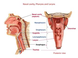
Pharynx and Oral Cavity • Superior to the ORAL CAVITY is the HARD PALATE composed of the MAXILLARY and PALATINE bones. • Superior and posterior to the oral cavity are the INTERNAL NARES. • From the internal nares, if we go anteriorly we will find the EXTERNAL NARES or NOSTRILS.
Posterior to the hard palate is the SOFT PALATE. This is muscular tissue that is moved during swallowing. • Hanging from the soft palate is a conical structure called the UVULA. • The two nasal cavities are separated by the NASAL SEPTUM which is formed by the union of the VOMER and PERPENDICULAR PLATE of the ETHMOID bones.
3 Areas of the Oral Cavity • OROPHARYNX -soft palate to epiglottis -two sets of TONSILS a. Palatine b. Lingual -the tonsils remove pathogens that enter the pharynx. They contain lymphocytes
2. NASOPHARYNX -located superior and posterior to the soft palate. -contains the PHARYNGEAL TONSILS and TUBAL TONSILS
3. LARYNGOPHARYNX -inferior to the epiglottis and posterior to the larynx. - this division opens into the esophagus and larynx.
Sagital section of cadaver head Notice the nasal conchae. They serve to expand the surface area to warm and moisten breathed air. Also, notice the position of the spinal cord within the vertebral canal.
Epiglottis Hyoid Bone Thyrohyoid Ligament Thyroid Cartilage Aryetnoid cartilage Cricoid cartilage ANTERIOR trachea Tracheal rings POSTERIOR
5 Openings into the Pharynx • Mouth • Left and right nasal passages • Eustachian tubes (connect middle ear to the throat) • Larynx • Esophagus
Swallowing • Is a reflex. • When the mouth closes, the soft palate is pushed superiorly and closes the nasal passages • A sphincter valve closes off the eustachian tubes • The glottis closes and respiration stops. The glottis also bends and closes the entrance into the larynx. • The esophagus is opened by pressure of the food. This allows the epiglottis to open. • Food then enters the esophagus.
Teeth • Very similar to bone. • Three major components: 1. hydroxyapatite Ca5(PO4)3(OH) 2. bone collagen 3. cells
The pH of the mouth is usually 7.2 • There are acids in the mouth that come from three sources: 1. stomach acid during vomiting 2. foods 3. waste products of mouth bacteria
Types of Teeth • INCISORS – chisel shaped for nipping food. • CANINES – cone shaped for tearing • PREMOLARS – • MOLARS - grinding food • 32 teeth in the Permanent Dentition • 20 teeth in the Deciduous Dentition
Tooth Anatomy • Enamel: hardest substance in the body • Pulp Cavity: contains arteries, veins, and nerves. • Alveolus: made of alveolar bone • Root: made of dentin • Gingiva: gum • Periodontal membrane: periosteum found around the tooth • Cementum: material that holds the tooth in the alveolus.
Identify the Following: Incisors Molar Premolars Canines
Salivary Glands • When you dissect your cat, you will notice two muscles on the inside of the cheek. • The DIGASTRIC MUSCLE opens the jaw. • The MASSETER MUSCLE closes the jaw.
The masseter inserts on the mandible. • Superficial to part of the masseter and anterior to the ear is the large PAROTID GLAND. This gland produces SALIVARY AMYLASE (ptyalin), a digestive enzyme. • The parotid gland is GRANULAR, it is attached by fascia. It is also the largest of the salivary glands.
The parotid empties into the PAROTID DUCT which empties between the last two molars at the angle of the jaw. • The parotid gland is an EXOCRINE GLAND. Exocrine glands empty via a duct to a specific location. The other type of gland is an ENDOCRINE GLAND that empties directly into the bloodstream.
Caudal and ventral to the parotid gland is the SUBMANDIBULAR GLAND (SUBMAXILLARY). • The SUBMAXILLARY DUCT empties this gland. It runs on the lateral aspect of the digastric muscle. • This gland carries saliva into the angle of the lower jaw.
The SUBLINGUAL GLAND is on the submaxillary duct. It is wedge shaped and it is lateral to the digastric muscle. • The DORSAL and VENTRAL FACIAL NERVES run around the outline of the masseter muscle. These nerves come out in front of the ear from the STYLOMASTOID FORAMEN and branch across the face.
Parotid Duct Masseter muscle Parotid gland Submandibular gland Sublingual gland Submandibular Duct