Download
1 / 39
400 likes | 656 Views
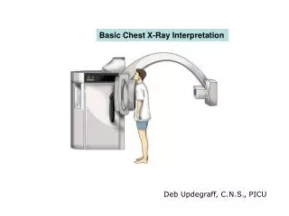
Basic Chest X-Ray Interpretation. Deb Updegraff, C.N.S., PICU. X-rays- describe radiation which is part of the spectrum which includes visible light, gamma rays and cosmic radiation. Unlike visible light, radiation passes through stuff. When you shine a beam of X-Ray at a person

E N D
Basic Chest X-Ray Interpretation Deb Updegraff, C.N.S., PICU
X-rays- describe radiation which is part of the spectrum which includes visible light, gamma rays and cosmic radiation. Unlike visible light, radiation passes through stuff. When you shine a beam of X-Ray at a person and put a film on the other side of them a shadow is produced of the inside of their body.
Different tissues in our body absorb X-rays at different extents: • Bone- high absorption (white) • Tissue- somewhere in the middle absorption (grey) • Air- low absorption (black)
Be systematic : 1) Check the quality of the film
Film Quality • First determine is the film a PA or AP view. PA- the x-rays penetrate through the back of the patient on to the film AP-the x-rays penetrate through the front of the patient on to the film. All x-rays in the PICU are portable and are AP view
Film Quality (cont) • Was film taken under full inspiration? -10 posterior ribs should be visible. Why do I say posterior here? When X-ray beams pass through the anterior chest on to the film Under the patient, the ribs closer to the film (posterior) are most apparent. A really good film will show anterior ribs too, there should Be 6 to qualify as a good inspiratory film.
Quality (cont.) • Is the film over or under penetrated if under penetrated you will not be able to see the thoracic vertebrae.
Quality (cont) • Check for rotation • Does the thoracic spine align in the center of the sternum and between the clavicles? • Are the clavicles level?
Verify Right and Left sides • Gastric bubble should be on the left
Now you are ready • Look at the diaphram: for tenting free air abnormal elevation • Margins should be sharp (the right hemidiaphram is usually slightly higher than the left)
Check the Heart • Size • Shape • Silhouette-margins should be sharp • Diameter (>1/2 thoracic diameter is enlarged heart) Remember: AP views make heart appear larger than it actually is.
Cardiac Silhouette • R Atrium • R Ventricle • 3. Apex of L Ventricle • Superior Vena Cava • Inferior Vena Cava • 6. Tricuspid Valve • Pulmonary Valve • Pulmonary Trunk • 9. R PA 10. L PA
Check the costophrenic angles Margins should be sharp
Check the hilar region • The hilar – the large blood vessels going to and from the lung at the root of each lung where it meets the heart. • Check for size and shape of aorta, nodes,enlarged vessels
Finally, Check the Lung Fields • Infiltrates • Increased interstitial markings • Masses • Absence of normal margins • Air bronchograms • Increased vascularity