Download

1 / 59
650 likes | 2.4k Views
Infusion Therapy. Metropolitan Community College Fall 2013 Jane Miller. Infusion Therapy. PARENTAL – the gastrointestinal system is by-passed Medications and solutions are delivered by Intravenous (IV) routes

E N D
Infusion Therapy Metropolitan Community College Fall 2013 Jane Miller
Infusion Therapy • PARENTAL – the gastrointestinal system is by-passed • Medications and solutions are delivered by Intravenous (IV) routes • Non-vascular routes - (i.e. - Subcutaneous (SQ), Intramuscular (IM), Intraosseous (IO), Intraspinal, or Intrathecal routes.
Vascular Access Device (VAD) • Device or catheter introduced through the skin and into the vascular network. • Peripheral Vascular Access Devices (PVAD) • Inserted in upper extremities and/or lower extremities • Central Vascular Access Devices (CVAD) • Inserted into a Centrally or Peripherally Located Vein with the tip residing in the vena cava
Vascular Access Devices (VAD) • Bevel – the slant or angle at the end of the needle • Insertion is Bevel up – facilitates puncture and cannulation of the vascular lumen. • Flashback chamber – small space in the hub of the stylet that allows confirmation of the presence of blood and indicated access to the vascular lumen • Self-sheathing stylet - the needle to becomes encased in a protective chamber upon removal
Types of Peripheral Access Devices • Winged Steel • Over-the-needle Peripheral Short Catheters • Through-the-needle Peripheral Short Catheters • Midline Catheters
Peripheral Vascular Access Devices (PVAD) –Peripheral-Short Catheters • Most - < 7.5 cm (3.5 inches) in length and Gauge (needle size) 25 (smallest) to 10 gauge (largest). • Locations of insertion and length of insertion time: • Inserted in upper extremities mostly (lower extremities - per policy) of adults patients. • Pediatric patients – insertions are upper extremities, occipital, superficial temporal, posterior auricular(ear) and saphenous veins. • Dwell times are 72 to 96 hours, in most cases.
Winged Steel Infusion Set • Winged Steel – flexible plastic attachments “wings” that extend from either side of the steel needle to facilitate insertion. • Length – 3/8 to 1.5 inches • Gauges – 19 to 27 gauge • Attached to the needle is plastic tubing extending from several inches to 12 inches with an adaptor attachment on the end for infusion administration equipment. • Manufactured with safety sheaths. • Complications – infiltration - due to needle rigidity • Temporary / short term infusions < 4hours – supportive devices – arm boards/splints
Over-the-needle and Through-the-needle Peripheral Short Catheters • OVER-THE-NEEDLE – MOST COMMON • Hollow bore needle (stylet) – inserted through the lumen of a flexible catheter – needle (stylet) is then withdrawn as the flexible catheter is advanced forward into the vein • THROUGH-THE-NEEDLE • Allows passage of a catheter through a steel introducer – introducer needle is then withdrawn from the vessel lumen-and then protected by a cover or sleeve to prevent sheering or patient injury
Through-the-needle short catheterComplication-catheter shearing
Peripheral VAD - Midline • Peripherally inserted but is approximately 7.5 cm to 20 cm (3.1 inches to 8.0 inches) • Longer dwell periods but not to exceed 4 weeks • Insertion sites – • Adults - antecubital fossa • Pediatrics – antecubital fossa, saphenous, posterior auricular (ear) superficial temporal • Distal tip – dwells in basilic, cephalic, or brachial veins – level with the axilla and distal to the shoulder • Inserted using an introducer-once vein is canulated the needle (stylet) is withdrawn and the midline catheter is threaded though the introducer –then introducer is peeled apart or separated and withdrawn. • Therapies for midlines-restricted to pH 5-9, osmolarities of <500mOsm/L
Peripheral IV Catheter Insertion/Removal • RN’s, LPN-C – trained in IV insertion using standard precautions and aseptic technique, RN’s only can be specially trained to insert midlines. • RN’s, LPN-C and LPN – trained in IV removal using standard precautions and aseptic technique • Assess/observe for phlebitis, drainage-apply sterile dressing till hemostasis is observed. • Inspect cannula, for midline – length of catheter should be measured and be same length as insertion.
CENTRAL VASCULAR ACCESS DEVICE (CVAD) • Catheter inserted into a centrally located vein with the tip in residing in the vena cava. • Tip in vena cave must be confirmed with radiologic examination (X-Ray) • Three major CVAD Categories • Nontunneled and noncuffed • Tunneled and cuffed • Implanted Ports
Nontunneled and Noncuffed CVAD • Inserted percutaneuosly via direct skin puncture with passage of catheter into the vasculature. • Single or multiple lumens • Indentified by insertion site (i.e. – Subclavian, Jugular, Femoral) • Inserted by physicians or advanced practice clinicians • Insertion requires – sterile technique and maximum barrier precautions • Complications – pneumothorax, air embolism, arrhythmias • Once inserted secured – sutures, stat locks • Catheters are not tunneled under the skin – less dwell times than tunneled • Examples – TRIPLE LUMEN NONCUFFED (ARROW)
Removal of Nontunneled and Noncuffed CVAD’s (including PICC) • May be done by a nurse skilled and educated in the procedure • Standard precaution and aseptic technique • Recumbent position (preferred) • Removal of occlusive dressing and securing devices • Instruct Patient to perform the valsalva maneuver - to prevent air from entering – causing air embolism • Pressure is applied 30 minutes to achieve hemostasis and sterile occlusive dressing applied • Post removal catheter length measured and compared to insertion length (same)
Peripherally Inserted Central Catheter (PICC) • Nontunneled and non cuffed • Long-term catheter – several weeks to one year. • Therapies with extreme variations in solution pH (>5 and < 9) and osmolarities >600 mOsm/L with irritant and Vesicant properties • Therapies – Total Parental Nutrition (TPN), Antineoplastic, Anti-infective and Inotropic • Radiologic confirmation (X-Ray) required for tip location – vena cava • Complications – device fracture, tip malposition, device occlusion, thrombus formation • Removal – same as central insertion central venous access devices
Tunneled and Cuffed CVAD • Cuff – piece of the catheter for stabilization and reduces migration of organisms • Anti-thrombogenic catheters • Tunneled under the skin – reduces infection risk • Inserted in surgical or radiologic suite • Inserted by physicians, surgeons, vascular surgeons and advanced practice clinicians trained in procedure • Long term therapies • Single or multi-lumens • Examples: Broviac, Hickman, Raaf, Groshong
Implanted Ports • Long term – chronic therapies • Chambered device, double or single reservoirs with an attached single or double lumen catheter • Catheter tip dwells in a designated structure depending on therapy – could be vascular or non-vascular (CVAD PORTS – Vena Cava) • Inserted by physicians, surgeons, vascular surgeons and advanced practice clinicians trained in procedure in a surgical or radiological suite • The septum is the access point-damage to the septum could cause air embolism, infiltration, extravasion, infection • Access of septum of port requires a noncoring (Huber needle) needle with opening on the side and preventing coring of the septum • Therapies – antineoplastics, inotropics, TPN, Antibiotics
Additional Info on VAD’s • Tunneled and cuffed and Implanted port removal only by physicians, advanced clinicians trained in procedure and not included in the scope of practice for nurses • CVAD – Lines flushed and patency checked q shift with 10ml of Normal Saline (Heparin per institution) whereas PVAD’s are flushed and checked for patency q shift with 3-5 ml Normal Saline.
SPECIALIZED INFUSION CATHETERS • Hemodynamic Monitoring – Swan Ganz • Dialysis and Pheresis Catheters • Arterial – Venous Shunts • Arterial Catheters • Hepatic Artery Catheters
Swan-Ganz Catheter • Exclusively for cardiac monitoring and hemodynamic analysis • Used to measure • Central Venous Pressure (CVP) • Pulmonary Artery Pressure (PA) • Cardiac Output (CO) • Pulmonary Artery Wedge Pressure (PAWP) • Temporary Pacing • Parental Therapy Administrations
Hemodialsys and Pheresis Catheters • RARELY considered for infusions • Used for procedures using large amounts of blood – Dialysis, Plasmapheresis, Aquapheresis • Dialysis Catheters – temporary till a shunt can be placed • Catheters are large (12 to 16 G) for blood to flow in both directions on dialysis or pheresis machine, dwell in the vena cava • Surgical Placement – OR, Radiological suite or at bedside by vascular or general surgeon or advanced practice clinician trained in procedure • Examples – Quinton, Hohn, Tesio, Perma-Cath, and Trialysis* *(Trialysis-can be used for infusions)
Arterial-Venous Shunts • Anastomose venous and arterial structures • Arm – radial and cephalic and brachial veins • Dialysis or large blood volume exchanges • Accessed with large bore needle • Can be used for other infusion- rare • Patency assessed by auscultation bruit and palpating vibration • No IV sticks, phlebotomy or Blood Pressure on Arm with AV Shunt • If AV shunt is damaged – only surgical repair or replacement is the option
Arterial Catheters • Used for ONLY two reasons: • Invasive Blood Pressure Monitoring • Blood Draws • Catheters are made of stiff polyurethane material to withstand the high pressure of the artery and blood pressure monitoring attached to pressure transducers and monitor, and to limiting kinking of line. • Inserted in Radial or Femoral Arteries • Inserted by physician or advanced practice clinician in OR, or Radiological suite or at bedside.
Hepatic Artery Catheters • Specialized Catheter Inserted in Hepatic Artery • Only for regional anti-neoplastic therapy for Liver Cancer Patients
NONVASCULAR ACCESS DEVICES • Subcutaneous Infusion Therapy • Intraosseous Therapy • Intraspinal Catheters • Intrathecal Therapy
Subcutaneous Infusion Therapy • Hypodermoclysis – infusing large amounts of isotonic solutions in the subcutaneous to be absorbed • Continuous Subcutaneous Infusion – infusing small amounts of medications like pain medications and insulin • Use very small gages of 25-27G and only ½ inch in length • Needle attached to a adhesive disk of may appear as a winged steel infusion set • May be inserted by a nurse in locations of adequate subcutaneous tissue • Site is prepared with antiseptic agent, allowed to dry and then aspirated to confirm absence of blood • Device is then secured with sterile dressing • Complications – localized skin irritation, erythemia, itching, infection and dislodgement of device
Intraosseous therapy • Insertion of hollow bore needle into the bone accessing the marrow space • Screws into long bones of the leg, iliac crest or sternum • Allows for rapid infusions of large volumes of fluids • Complications – bone fracture, infiltration, osteomyelitis, cellulitis, occlusions, or needle breakage • Ideal for patients with difficult venous access • Being used more by EMT, Paramedics
Intraspinal Catheters • Catheters inserted within the spinal spaces – epidural and intrathecal • Delivery of anesthesia and infusions • Diagnostic testing • 22 to 26 Gauge, 10 to 30 inches in length • Catheter inserted percutaneously or tunneled • Infusates, drugs and diluents must be preservative free due to neurotoxic effects • Epidural space usually cannot accommodate infusion rates greater 15ml/min • Cross the dura mater and have direct effect on the CNS • Complications: malposition and infection • Sterile technique required • Usually inserted by anesthesiology or neurology
Intrathecal Therapy • Administration of medications directly into the Cerebral Spinal Fluid via an implanted reservoir • Catheter dwells (terminates) in the ventricular space of the cerebrum • Ommaya Reservoir • Delivers preservative free opioids • Antineoplastics • Measures CSF pressures • Drains excess CSF • Obtaining CSF samples for Lab • Surgically places via burr hole in the skull and skin secured over the device • Standard precautions and sterile technique
INFUSION DELIVERYFluid Containers • Glass • Can be heavy • Requires venting • Accurate to measure • Plastic • Portable, easy to store, inexpensive • Flexibility may cause volume and dosage administration to vary
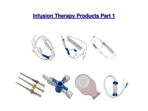
Administration Sets • The tubing that delivers fluid from the container to the catheter • Primary set = main tubing • Secondary set = tubing that attaches to the primary set (piggy backed on the primary set) • Metered-Volume Chambered Set – neonates and pediatric patients and critical care units for close observation and monitoring with fluid control issues
Add On Devices • Flow-control device – controls flow when used with gravity IV set • Stopcocks – manually directs flow • Extension sets – adds length to tubing, provides additional stopcocks, access sites • Multi-flow adaptors and Y sets – multiple access on one catheter • Injection ports, caps or hub – sites for IV catheter access • Filters – remove particulate, precipitate
ADD ON DEVICES Flow Control Device Stopcock
Add On Devices Extension Set Multiflow Adaptor