Download
1 / 37
400 likes | 1.24k Views
Anemia. Ramzi Shawahna , PhD An- Najah National University. Course outlines. Investigation of anemia Thalassemia Iron deficiency anemia Anemia of chronic diseases. Anaemia.

E N D
Anemia RamziShawahna, PhD An-Najah National University
Course outlines • Investigation of anemia • Thalassemia • Iron deficiency anemia • Anemia of chronicdiseases
Anaemia • Anaemia is one of the most common clinical conditions encountered. It can vary from a mild, clinically quiescent condition to a serious, incapacitating disability. • Its causes vary from a modest mismatch between the iron requirements and intakes of a pregnant woman to the only overt sign of advanced malignant or other systemic disease.
Definition Anemia (a decrease in the number of RBCs, Hb content, or Hematocrit) below the lower limit of the normal range for the age and sex of the individual. • In adults, the lower extreme of the normal haemoglobin is taken as 13.0 g/ dl for males and 11.5 g/dl for females. • Newborn infants have higher haemoglobin level and, therefore, 15 g/dl is taken as the lower limit at birth,
Treatment may be simply an oral course of haematinics - the substances required for maintenance of a normal blood count such as iron, vitamin B12 and folic acid. It may require life-long parenteral administration of vitamin B12 or it may consist of effective treatment of the underlying aetiology. Strictly speaking, anaemia is not a disease per se. It is a clinical feature of other conditions such as haemorrhage, chronic renal disease or infection, liver disease or dietary deficiency.
A pseudo-anaemia can occur during pregnancy when expansion of plasma volume occurs without a corresponding increase in red cell numbers. This is not a true anaemia as the tables of normal ranges used to define anaemia stipulate distinct values for pregnant women.
Haemoglobin • Haemoglobin (Hb) is an iron-containing pigment, which binds to and transports oxygen. Haemoglobin levels are determined by measuring the colour intensity of a blood sample in which the red cells have been lysed (broken open). • This is compared with an internal machine standard and the result is reported as either g/L or g/dl. The official SI unit for Hb is g/L but the g/dl notation is probably still more commonly used - values in g/dl are exactly 10 times smaller than g/L; an Hb of 150g/L is equal to 15g/dl.
Although other definitions are encountered, a reduced Hb level is the defining element of anaemia. The WHO defines anaemia as: ‘a haemoglobin (Hb) concentration in blood that is below the expected value, when age, gender, pregnancy and certain environmental factors, such as altitude, are taken into account’.
Red cell count (RBC) • The red cell count (RBC) is an estimation of the number of red cells present in 1ml of whole blood. The exact method used to count the red cells varies between different designs of automated cell counter. The normal red cell count varies according to age and sex. If performed on an electronic counter this is a very accurate estimation. The principle source of error is the quality of the blood sample submitted.
Mean cell volume (MCV) • Mean cell volume (MCV) is a measurement of the average size of the red cells. The normal range of the MCV is between 80 and 99fl. • The initial classification of anaemia is typically based in part on the MCV. A reduced MCV is termed microcytosis. An increased MCV is called macrocytosis. Where the MCV is within normal limits the anaemia is termed normocytic.
Packed cell volume (PCV) • The packed cell volume (PCV) is often referred to as the haematocrit. It measures what percentage of a given volume of blood is made up of red blood cells. On automated counters the PCV is usually a calculated value derived from the RBC and the MCV. The PCV is affected by many conditions other than anaemia. Certainly the PCV alone can never be used to diagnose anaemia. The PCV may be reported as a percentage or as litres/litre.
Red cell indices • The red cell indices are not directly measured properties of the red cell. They are values that are calculated from the results of the automated cell count. The most widely used are the mean cell haemoglobin (MCH) and the mean cell haemoglobin concentration (MCHC). The MCH is a calculated value based on the RBC and the Hb level. It is the amount of haemoglobin in an average red cell and is measured in picograms. The MCHC is again a calculated value based on the Hb and the PCV. It reflects the extent to which the red cell is ‘packed’ with haemoglobin - the units are g/dl.
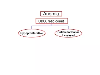
Reticulocyte count • Normal red cells undergo a maturation process within the bone marrow. During this process the cells shed their nuclei and all cytoplasmic structures concerned with protein synthesis (a mature RBC cannot synthesise proteins). • A small percentage of relatively immature RBCs are normally present in circulating blood. These have shed their nuclei but they still retain ribonucleic acid (RNA). They are known as reticulocytes and they offer a useful indirect index of marrow erythropoiesis (red cell formation).
Reticulocytes may be visible in a routine blood film as large, slightly blue-staining, RBCs. To count reticulocytes requires a special stain - many modern haematologyanalysers can offer an automated reticulocyte count. • Manual counts (by microscopy) are usually reported as percentages (of all RBCs). A correction must be applied where anaemia is severe. Automated counts are typically reported as absolute numbers of reticulocytes per unit volume, reflecting the greater accuracy of such counts.
Signs and Symptoms of Anemia • Dependent on the degree of anemia, the rate at it evolved, and the oxygen demand • Normally, RBCs carry oxygen linked to Hgb from the lung to tissue capillaries, where oxygen is released • Symptoms result from decreased oxygen delivery or acute blood loss (hypovolemia) • Compensatory mechanisms allow one to tolerated lower levels of Hgb/Hct • Increase in stroke volume, HR ( increased CO) • Enhanced oxygen extraction by the tissues
In most laboratories the initial investigation and tentative diagnosis is made with a relatively small number of tests. The precise diagnosis is made with further special tests. Screening is usually done with the CBC or "complete blood count". The exact procedures in a CBC depends upon the instrumentation in the laboratory. Most laboratories now use automated, multiparameter instruments which will provide results for the following parameters: hemoglobin hematocrit red cell count MCV , MCH ,MCHC RDW white cell and platelet count
Normal hemoglobin values: • Men 14-17 gm% • Women 13-15 gm% • Infants 14-19gm% • Children (1year) 11-13gm% • Children (10-12 years0 12-14gm%
Clinical significance of Hb measurement: A decrease or increase in hemoglobin concentration must be reported ,as it is a sign of disease requiring investigations • A decrease in Hb concentration is a sign of anemia • While an increase can occur due to; • Haemochromatosis (loss of body fluid as in severe diarrhea) • Reduced oxygen supply (congenital heart disease , emphysema) • Polycythemia
Haematocrit or Packed Cell Volume It is the amount of packed red blood cell, following centrifugation, expressed as a total blood volume • Normal value • Male: 42-52 % • Female: 36-49% • Roughly, the haematocrit value is 3 times the Hb concentration
Clinical significance A decrease in the haematocrit value is a suitable measurement for detection of anaemia, also in case of hydremia (excessive fluid in blood as in pregnancy) • An increase is an indication decrease oxygen supply (as in congenital heart disease, emphysema) or as in polycythemia and dehydration • The value of haematocrit is used with haemoglobin and red cell count for the calculation of MCV, MCH and MCHC
RED CELL INDICES The type of anemia may be indicated by the RBC indices: • mean corpuscular volume (MCV), • mean corpuscular Hb (MCH), and • mean corpuscular Hb concentration (MCHC). • RBC populations are termed microcytic (MCV < 80 fl) or macrocytic (MCV > 95 fl). • The term hypochromia refers to RBC populations with MCH < 27 pg/RBC or MCHC < 30%. • These quantitative relationships can usually be recognized on a peripheral blood smear and, together with the indices, permit a classification of anemias that correlates with etiologic classification and greatly aids diagnosis.
Mean Cell Volume(MCV) • It is calculated from PCV and red cell count as follows: • MCV = PCV/RBC fl A femtoliter (fl) is 10 15 of a liter • Normal value: 80-95 fl • It decrease in iron deficiency anaemia and haemoglopinopathies • It is increase in megaloblastic anaemia and chronic haemolytic anaemia
Mean Cell Haemoglobin Concentration (MCHC) • It is calculated from the haemoglobin and PCV as follows: • MCHC = Hb/PCV g/dl • Normal value: 32-35.5 g/dl • It is usually decrease in iron deficiency anaemia (microcytic hypochromic anaemia)
Mean Cell Haemoglobin (MCH) • It is calculated from the haemoglobin and erythrocyte count as follows: • MCH = Hbx10/RBC pg A pictogram (pg) is 10-12 of a gram • Normal value: 27-32 pg • It is decrease in iron deficiency anaemia and thalassaemia (microcytic hypochromic anaemia) • It is recognized by the pale colour of the red cell in the peripheral blood film • It is increase in microcytic anaemia (vitamin B 12 and folic acid)
Red Cell Distribution width (RDW) • RDW reflects the variation of RBCs volume it is usually performed by modern analysers • Normal RDW varies between 12 to 17 • Severe iron deficiency anemia is associated with increased RDW • Thalassemia and anemia of chronic disease are associated with normal RDW
IRON METABOLISM Iron is present in the diet in many forms. Haem is the most important source. Vegans may need to supplement their dietary intake with non-organic iron. A normal adult requires 15-20 mg of iron per day to remain in balance.
Iron is normally absorbed by active transport across the wall of the duodenum and upper part of thejejunum. If large amounts of iron are ingested the active transport mechanism is overtaken by passive diffusion.. Disease of the upper small gut can lead to malabsorption of iron, e.g. coeliac disease or tropical sprue.
Iron is best absorbed in the ferrous (reduced) form (Fe++). Absorption is improved by reducing substances, e.g. ascorbic acid (vitamin C). Absorption is also increased by certain iron chelators and by alcohol.
Iron absorption is normally relative to the needs of the body. About 10% of dietary iron is usually taken up by the body but this can increase several-fold in iron deficiency, or reduce if the body has a surplus.
Most iron in the body is in the form of haem; present in large amounts in red cells, muscle and liver where it is essential for oxygen supply. Iron is also present in many enzyme systems, e.g. electron transport systems. The transport and storage of iron mainly involves three proteins: transferrin transferrin receptor (TfR) ferritin
Transferrin actively binds and transports iron in the body and can be estimated by measuring the serum total iron binding capacity (TIBC). Transferrin increases in iron deficiency and decreases with iron overload, liver disease, infection, malignancy and protein deficiency.
Excess iron is stored mainly in macrophages as haemosiderin; an insoluble protein-iron complex formed by lysosomal degeneration of ferritin. Ferritin is the water soluble protein-iron complex formed when iron combines with apoferritin. Iron in ferritin is in the ferric form (Fe+++) and must be reduced before it can be utilised.
LABORATORY INVESTIGATION OF IRON DEFICIENCY ANAEMIA • Full Blood Count • Serum Ferritin • Serum Iron & Total Iron Binding Capacity • Serum Transferrin • Bone Marrow
FULL BLOOD COUNT can be suggestive but not diagnostic of iron deficiency. Negative iron balance produces microcytosis (low MCV) and hypochromasia (low MCH). Red cell morphology varies from mild anisocytosis to marked anisopoikilocytosis. Thrombocytosis is common. Leucocytes are usually normal.
SERUM FERRITIN is now a standard diagnostic test for IDA; only iron deficiency will give a low result. Normally the level of serum ferritin reflects the body iron stores (100 μg/L = 800 mg of iron). A value <15 μg/L is diagnostic of IDA.
Circumstances in which the serum ferritin is normal or high in the presence of IDA: • Liver dysfunction; ferritin is released when hepatocytes are damaged • Increased haem turnover; haemolysis and trauma (including surgery) • Inflammatory lesions; malignancy, infection and inflammation
SERUM IRON (SI) and TOTAL IRON BINDING CAPACITY (TIBC) In iron deficiency the SI is low (<10 μmol/L) and the TIBC is usually raised (>70 μmol/L).