Download
1 / 81
840 likes | 1.38k Views
Placenta and its functions. Remarkable organ Undergoes differentiation and maturation during the short life span. DEVELOPMENT OF THE PLACENTA. Fertilization Zygote ( diploid cell with 46 chromosomes) undergoes cleavage Morula ( 16 cell stage)

E N D
Remarkable organ • Undergoes differentiation and maturation during the short life span
Fertilization Zygote ( diploid cell with 46 chromosomes) undergoes cleavage Morula ( 16 cell stage) Enters the uterine cavity 3 days after fertilization gradual accumulation of fluid between the cells of morula ( early blastocyst) 58 cell blastocyst outer cells – trophoblast inner cells - form the embryo
Trophoblast • Helps in implantation • Important role in the nutrition of the conceptus • Endocrine organ
Blastocyst implantation 3 steps • Apposition – initial adhesion of the blastocyst to the uterine wall • Adhesion – increased contact between the blastocyst and uterine epithelium • Invasion – penetration and invasion of the syncitiotrophoblast and cytotrophoblast into the endometrium , inner third of the myometrium and uterine vasculature
Trophoblastic differentiation 8th day postfertilization , after initial implantation … differentiates into • Outer multinucleated syncytium – primitive syncytiotrophoblast • Inner layer of primitive mononuclear cells – cytotrophoblast
After the implantation is complete… Trophoblast further differentiates into • Villous trophoblast give rise to chorionic villi - transports nutrients and oxygen between the mother and the fetus • Extravilloustrophoblast migrates into decidua and myometrium and penetrates into the maternal vasculature endovascular trophoblast interstitial trophoblast
Interstitial trophoblast • invades the decidua and penetrates the myometrium to form the placental bed giant cells • Surrounded by spiral arteries Endovascular trophoblast • Penetrates the lumen of the spiral arteries
10 th day • Gentle erosion between the epithelial cells of the surface of endometrium , the invading trophoblasts burrow deeper and the blastocysts become totally encased within the endometrium
Blastocyst wall • facing the uterine lumen – single layer of flattened cells • Opposite side – thicker wall trophoblast embryo forming inner cell mass primitive ectoderm underlying endoderm (Hypoblast) (Epiblast)
A small cavity develops within the epiblast forming the amniotic cavity • Epiblast + hypoblast embryonic disc • Cells rising from the hypoblast form an exocoelomic membrane ( Heuser’s membrane) that lines the inner part of the cytotrophoblast and together with the cytotrophoblast forms the exocoelomic activity or the primitive yolk sac
The syncytium between the cytotrophoblastic shells and the decidua degenerates and is replaced by a fibrinoid material called the NITABUCH’S MEMBRANE • This limits the penetration of the trophoblast in to the myometrium • Absence can lead to various degrees of adherent placenta
Day 12 after conception • The syncytiotrophoblast of the trophoblastic shell is permeated by system of intercommunicating channels called the trophoblastic lacunae • The lacunae are separated for each other by trabaculae • The lacunae communicate with each other and eventually a large space is formed . Now each trabeculae is surrounded by this lacunar space
The syncytiotrophoblast grows into the endometrium • The endometrium is eroded and blood fills up in the lacunar spaces • Cytotrophoblast begin to multiply and grow into each trabeculus • PRIMARY VILLI
Extra – embryonic mesoderm invades the centre of each primary villus • SECONDARY VILLI • Angiogenesis • TERTIARY VILLI
Development of chorion and decidua • As the blastocyst with its surrounding trophoblast expands into the decidua, • one pole extends outward toward the endometrial cavity • Opposite side will form placenta from villous trophoblasts and anchoring cytotrohoblast
Chorionic villi in contact with deciduabasalis proliferate to form chorionfrondosum (fetal component of placenta) • As the growth of embryonic and extra-embryonic tissue continues , the blood supply of the chorion facing the endometrial cavity is restricted • Deciduacapsularis cease to grow and degenerate
This portion of chorion becomes avascular fetal membrane that abuts the deciduaparietalis End of third month , chorionic laeve is separated from the amnion by exocoelomic cavity Intimate contact to form avascularamniochorion
Invasion of spiral arteries • Modification of spiral arteries are carried out by 2 populations endovascular trophoblast • Interstitial trophoblast(penetrate spiral artery lumen) (surrounds the spiral arteries)
Interstitial trophoblast • Penetrates decidua and myometrium • Aggregates around the spiral arteries • Functions: vessel preparation for endovascular trophoblastic invasion
Endovascular trophoblast • enters the lumen of the spiral arteries and forms cellular plugs Destroys the vascular endothelium by apoptosis Invades and modifies the vascular media with fibrinoid material Spiral arteries later regenerate the endometrium • Invading endovascualrtrophoblast can extend several cms along the lumen and migrate against arterial flow • Vascular changes are not observed in deciduaparietalis
Placental growth • In the first trimester the placental growth is more rapid than the fetus • By term , placental weight = 1/6 th of fetal wt
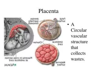
Maternal surface: • Slightly elevated convex areas ( lobes) – varies from 10-38 • Lobes are incompletely separated by grooves • Grossly visible lobes are referred to as cotyledons • Total number of lobes remain the same throughout gestation and individual lobes continue to grow
Fetal surface • Covered by smooth amnion with umbilical cord attached at or near the centre • Branches of the umbilical vessels are visible beneath the amnion as they radiate from the insertion of the cord
Maternal circulation • Arterial circulation : • About 120-200 spiral arteries open into the intervillous spaces by piercing the basal plate • In 12 weeks of pregnancy , there is a cytotrophoblastic invasion into the spiral arteries upto the intra-decidual portion • Endothelial and the muscular layer are replaced by fibrinoid material • Between 12-16 weeks, there is a secondary invasion of the trophoblast extending upto the radial arteries within the myometrium • Spiral arteries are converted to uteroplacental arteries
Circulation of the intervillous spaces • The arterial blood enters the space under pressure • Lateral dispersion occurs after it reaches the chorionic villi • Villi pulsation with uterine contraction – help in the migration of the blood towards the basal plate • During uterine contraction, the veins are occluded and the arterial blood is forced into the intervillous space, while uterine relaxation facilitates venous drainage
Fetal circulation • 2 umbilical arteries carry impure blood from the fetus enter the chorionic plate underneath the amnion , supplying each half of the placenta Artery breaks into smaller branches which enter the stems of chorionic villi Divides into primary , secondary and tertiary vessels of the corresponding villi • Maternal and fetal blood flow side by side but never in opposite direction • Fetal blood flow through the placenta – 400 mL/min
Placental barrier • Partition between the fetal and maternal circulation • In later part of pregnancy the membrane thickness reduces from 0.025mm to0.002mm • In early pregnancy • Syncytiotrophoblast • Cytotrophoblast • Basement membrane • Stromal tissue • Endothelium of the fetal capillary wall with its basement membrane
At term , there is attenuation of the syncytial layer • Sparse cytotrophoblast and distended fetal capillaries fill the villus
Teratogenicity • Diethylstilbestrol – vaginal adenosis , cervical hoods , uterine hypoplasia of the female offspring • Lithium – Ebstein’sanomaly,fetal diabetes insipedus , polyhydraminos, neonataalgoitre • Sodium valprovate – increased neural tube defects • Aminoglycosides – nephrotoxicity , ototoxicity in preterm infants • Phenetoin- cleft lip/palate, hypertelorism , finger nail hyperplasia
PLACENTAL FUNCTIONS • Transfer of nutrients • Respiratory , excretory , nutritive • Endocrine function – produces steroid and peptide hormones • Immunological function • Barrier function
Respiratory function: • No gaseous exchange • Intake of O2 and output of CO2 takes place by simple diffusion • Pressure gradient – driving force • O2 supply to the fetus – 8mL/kg/min and this is achieved with cord blood flow of 165-330mL/min • Excretory function: • Waste products such as urea , uric acid and creatinine are excreted by simple diffusion
Nutritive function • Glucose – glucose transporters GLUT1 by facilitated diffusion, glucose level : fetus > maternal • Lipid –transferred or synthesized , triglycerides and fatty acids are transported in early pregnancy , later synthesised , cholesterol – direct transfer • Amino acids- active transport, amino acid concentration : fetus > mother
Water and electrolytes • Sodium , potassium and chloride – by simple diffusion • Calcium, phosphorus and iron – by active transport ( fetal > maternal) • Water soluble vitamins – by active transport • Fat soluble vitamins – transferred slowly • Hormones • Insulin , steroids from the adrenals, thyroid , chorionic gonadotropin , or placental lactogen cross at a very slow rate
PLACENTAL GRADING • Grade 0 -Smooth chorionic plate, homogenous echotexture of substance of placenta. Most common in 1st trimester and early 2nd trimester (8-20weeks).
Grade I -Small intraplacental calcification randomly dispersed within the substance of a placenta. May appear as early as 14 weeks, and is most common until 34 weeks.
Grade II -Calcification of basal plate. Does not usually appear until after 30 weeks.
Grade III - Calcified indentation of placenta extending from the basal plate to the chorionic plate (placental cotyledons ). Usually not seen until 35 weeks. Found in 30% of term placentas