Download
1 / 41
410 likes | 609 Views
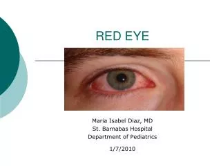
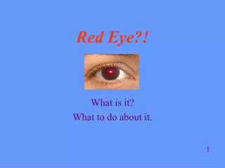
The Red Eye. EM Rounds Colleen Carey, BA, MD, CCFP (EM) July 31, 2008 Thanks to Dr. Jean Chuo, UBC Ophthalmology Resident. Goals. Hx Exam Most common etiologies Traumatic versus atraumatic Diagnosis Treatment When to get help. History. Trauma

E N D
The Red Eye EM Rounds Colleen Carey, BA, MD, CCFP (EM) July 31, 2008 Thanks to Dr. Jean Chuo, UBC Ophthalmology Resident
Goals • Hx • Exam • Most common etiologies • Traumatic versus atraumatic • Diagnosis • Treatment • When to get help
History • Trauma • Consider unrecognized trauma- awoke with symptoms • Pain? Itch? FB sensation? • Visual acuity changes, halos • Contact lenses- ? Overwear • Sick contacts/Viral symptoms • Prior surgery or eye disorders • Systemic disease
Red eye exam • Visual acuity • Visual fields • Pupil shape and reactivity • Lid closure • Foreign bodies • Ciliary flare • Foggy cornea (edema) • Corneal infiltrate • Fluorescein- corneal defects, Sidel’s sign • Anterior chamber cells • Intraocular pressure
Foreign body • Projectile metallic FB • Get orbital Xray • Rust ring • Visual axis involved?- refer if unable to completely remove • Burr • Tetanus status • Antibiotic prophylaxis?
Corneal abrasion • Get help if • not healing • corneal ulcer • large surface area • infringing on visual axis
Hyphema • Usually due to blunt trauma and immediate • Gross: layers out • Microscopic: cells in anterior chamber • Always refer • Tx: cycloplegics, steroids, serial IOP monitoring, sleep sitting upright, avoid valsalva, avoid anticoagulants, hard shield, avoid exertion • Complications: • Iritis • Synechiae, glaucoma • Rebleeding
Globe rupture • Penetrating FB • Blunt trauma by an object smaller than a fist • Blunt trauma with an orbital fracture • Prior open globe surgery • All must be repaired to prevent sympathetic ophthalmia • Need a hard shield. • Emergency referral, poor prognosis
Endophthalmitis • Red, painful, decreased vision • Anterior chamber cells+/- hypopion • Almost exclusively post-surgical complication • Rare: 1:100,000 cataract surgeries • Urgent referral
Superficial punctate keratitis • Very common problem • Mild itch, dry, gritty sandpaper sensation • Many causes: • Contact lens overwear • Dry Calgary air • Preservatives, antibiotic eye drops • Incomplete lid closure • Rule out other problems • Discontinue cause, moisturize, follow up in ER
Conjunctivitis/keratoconjunctivitis • Allergic • Viral • Bacterial • Irritative • Treat bacterial conjunctivitis with flouroquinolone or erythromycin drops. • Treat allergic with antihistamines, nasal steroid spray, allergen avoidance, cromolyn drops • Refer any keratitis
What is this? • Short fat branches with bulbs
Herpes Simplex Virus • HSV keratitis • Dendriticfluoroscein enhancing lesion • Hypoesthetic cornea • +/- periocular HSV vesicles • Tx is acyclovir +/- viroptic drops • HSV can affect any part of the eye • Next day referral as long as Tx started
What is this? • Long thin tapered branches
Herpes Zoster Ophthalmicus • HHV 3 (VZV) • V1 (opthalmic branch of CN V) • Macular rash =>vesicular lesions • Conjunctivitis • Keratitis • Uveitis/iritis +/- retinal necrosis • Cranial nerve palsies 3,4,6 • Cxns: Chronic ocular inflammation, vision loss, neuralgia, late corneal sequelae
Acute angle closure glaucoma • Risk Fx:Family Hx, contralateral eye, hyperopia, Asian race, age • Hx: Sudden eye pain, photophobia, halos • PE: Shallow anterior chamber, iris bombe, middilated pupil, hazy cornea, elevated IOP • Tx: one drop each of: 0.5% timolol 1%, apraclonidine, and 2% pilocarpine. Oral acetazolamide, IV mannitol • Ensure pressure drops within an hour
Acute angle closure glaucoma • Complete occlusion of the anterior chamber angle by iris tissue
Iritis • Causes: • Infections, eye disorders, systemic disorders • Trauma, autoimmune disorders, VZV, lyme disease, leukemia/lymphoma, idiopathic • Photophobia and dull ache • Urgent referral to ophtho • Get baseline IOP and start Predforte drops and cycloplegics
Ciliary flare of iritis • Intense injection at limbus
Subconjunctival hemorrhage • Causes • Valsalva • Coagulopathy • Presentation • Visual acuity • Absence of pain • Absence of photophobia • Absence of discharge • Should resorb in 1-2 weeks
This eye is not red • And that is the problem. • Alkali chemical burn- large corneal epithelial defect and scleral ischemia.
Of all the conditions you have seen today, this is the fastest to destroy an eye, and can have the worst prognosis • You have only minutes to diagnose and irrigate • Morgan lens, many litres • Afterward:confirm pH, slit lamp exam for corneal defect, r/o deposits in conjunctival recesses.
Chemosis • Insidious onset • Consider retro-orbital causes: mass, aneurysm.
Blepharitis • Chronic recurrent eyelid inflammation • Staph aureus or seborrhea (pityrosporum) • Warm lid compresses • Topical antibiotic eyedrops+/- ointment • Dandruff shampoos to scalp to eradicate pityrosporum • Slow response
Stye • Hordeolum- acute, painful • Chalzion- chronic, non painful • Hot compresses, milking • Refer if not resolving for I+C • Chronic lesions- ? Biopsy to r/o CA
Corneal edema • Note irregular corneal light reflex