Download
1 / 1
10 likes | 117 Views
(a). siRNA-D5. ns-siRNA. untreated. PSA/TBP. p38MAPK. C13orf19/TBP. β-Actin. phospho-p38MAPK. (b). 3.6 %. 3.9 %. 3.5 %. time (h). time (h). β-Actin. b). 72 h. a).

E N D
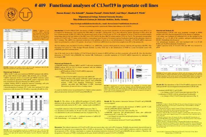
(a) siRNA-D5 ns-siRNA untreated PSA/TBP p38MAPK C13orf19/TBP β-Actin phospho-p38MAPK (b) 3.6 % 3.9 % 3.5 % time (h) time (h) β-Actin b) 72 h a) TABLEI Inhibition of the C13orf19 mRNA expression after transfection with 250 nM siRNA‑D5 in the cell line PC‑3. The results are indicated relative to the reference gene PBGD and normalized to ns‑siRNA control. 1.3 % 1.6 % 0.8 % untreated ns-siRNA siRNA-D5 FIGURE 4 C13orf19 mRNA expression in LNCaP cells after treatment with the synthetic androgen R1881 or combined with R1881 and the antiandrogen bicalutamide. In contrast the androgen dependence of the prostate specific antigen (PSA) mRNA expression measured in the same experiment. Both transcript numbers were normalized to the reference gene TBP. • Material and Methods II: • p38MAPK inhibitor treatment: BPH-1 and PC-3 cells were incubated in DMEM without FCS for 24‑72 h before treatment with the inhibitor SB203580 (0.5‑8.0 µM) for 1 h • → qPCR analyses of the C13orf19 mRNA expression • (TBP as reference gene) • inhibition of the C13orf19 mRNA expression with siRNA-D5 • → Western blotting with primary antibodies against phospho‑p38 MAP kinase and p38 MAP kinase (detects total p38MAPK protein; 1:1000; Cell Signaling Technology), secondary antibody: anti-rabbit IgG HRP-linked (1:1000, Amersham Biosciences); -actin served as a loading control • a yeast two-hybrid screen was performed by U. Stelzlto detect putative physical interactions between C13orf19 and p38MAPK [5] FIGURE 1 Relative cell viability of PC-3 cells 48 h after transfection with 125 nM siRNA‑D5 normalized to cells treated with ns‑siRNA (100%). FIGURE 3 Examination of C13orf19 interaction with p38MAPK. (a) C13orf19 mRNA expression in BPH‑1 and PC‑3 cells after treatment with the p38MAPK inhibitor SB203580 relative to the reference gene TBP normalized to DMSO-treated cells. (b) Western blot analysis of PC-3 cells 72 h after transfection with siRNA‑D5. The antibodies recognized total (upper) or phosphorylated (lower) p38MAPK protein. • Results I:The effects of the siRNA‑D5-mediated C13orf19 mRNA inhibition in the prostate cell line BPH‑1 and the PCa cell line PC‑3 were examined (all results shown for PC-3 cells – similar results with BPH-1): • remarkable reduction of the C13orf19 mRNA expression (Tab. 1) • no alterations in cell viability 24, 48 and 72 h after transfection (Fig. 1) • no adjustments in cell cycle distribution and rate of apoptosis (Fig. 2) • low apoptosis rate in PC‑3 cells → combined treatment of siRNA-D5 with chemotherapy → no synergistic effects • Results II: The putative interaction between C13orf19 and p38MAPK was investigated: • C13orf19 mRNA expression after treatment of BPH‑1 and PC‑3 cells with the p38MAPK inhibitor SB203580: • → no alterations in the gene expression (Fig. 3a) • p38MAPK expression and phosphorylation status after siRNA‑D5 transfection in PC‑3 cells (Fig. 3b): • → not changes in expression and phosphorylation of p38MAPK • yeast two-hybrid screen: • → no interactions between C13orf19 and p38MAPK • Functional analyses of C13orf19 in prostate cell lines • Doreen Kunze1, Uta Schmidt1*, Susanne Fuessel1, Ulrich Stelzl2, Axel Meye1, Manfred P. Wirth1 • 1Department of Urology, Technical University Dresden; • 2Max-Delbrueck-Centrum for Molecular Medicine, Berlin, Germany # 409 http://urologie.uniklinikum-dresden.de, e-mail: doreen.kunze@uniklinikum-dresden.de This study was partially supported by a grant from the Wilhelm-Sander-Stiftung (to A.M. & M.P.W.). • Material and Methods III: • hormonsensitive LNCaP cells were incubated overnight in RPMI medium with 10% charcoal-stripped FCS in the absence of androgen • cell culture was continued for additional 48 h in this medium in the presence of 1 or 10 nM of the synthetic androgen methyltrienolone (R1881, PerkinElmer Life Sciences) • for antiandrogen treatment, cells were incubated with 1 or 10 nM R1881 and 10µM bicalutamide (AstraZeneca) • mRNA expression levels of C13orf19, PSA and TBP were measured by qPCR Introduction: C13orf19 (NM 017569), also known as P38IP or FAM48A, was previously identified to be down-regulated in prostate cancer (PCa) [1]. It is localized on chromosome 13q13 between the TSGs BRCA-2 and RB-1. Chromosome 13q is often affected by genetic aberrations in PCa, above all loss of heterozygosity (LOH) [2]. LOH in the chromosomal region of the C13orf19 gene in 47% of the patients for seven C13orf19-associated markers being higher than for the four markers of the RB1 locus (39%) and for the three BRCA2-related markers (25%) on 13q [3]. The presumed corresponding protein of C13orf19 with 733 amino acids resembles a transcription factor because of its putative nuclear localization signal and the glutamine clusters at the C terminus. The mRNA was expressed exclusively in the epithelia as shown by in situ hybridization [1]. Recently, a C13orf19 mRNA expression study was performed in a cohort of 61 patients. An at least 1.5-fold lower mRNA level was observed in samples from 46% of the patients [4]. The first database entry described C13orf19 as P38IP (acc. no. AF093250), a protein which putatively interacts with the p38 map kinase (MAPK). This description was given by J. Han (Scripps Research Institute, La Jolla, USA) due to the identification of P38IP in a yeast two-hybrid screen with p38MAPK (personal communication). Aim of the study was to show whether a C13orf19 knock-down by means of siRNA will have an effect on prostate cell growth (I). Also, the described interaction of C13orf19 with p38MAPK was checked (II). Furthermore, a possible regulation of C13orf19 mRNA expression by androgens was investigated (III). • Material and Methods I: • BPH-1 & PC-3 cells were transfected DOTAP-mediated with siRNA-D5 (nt 1288‑1306 [C13orf19 mRNA: NM_017569]; 125 or 250 nM) • mRNA expression levels of C13orf19 and PBGD (reference gene) were measured by quantitative PCR (qPCR; relative expression values were normalized to the non-silencing (ns) siRNA-control) • cellular viability (WST‑1), apoptosis (annexin V-staining), cell cycle distribution and clonogenic survival were examined • chemotherapeutical treatment: PC-3 cells were incubated 24 h after transfection with docetaxel (0,5-40 nM) or etoposide (20 µM) for 24 h • Results III: The dependence of the C13orf19 mRNA expression on the • androgen R1881 was investigated in hormonsensitive LNCaP cells: • treatment with R1881 (1 and 10 nM): • → no changes of the C13orf19 transcript expression(Fig. 4) • combination of R1881 with the antiandrogen bicalutamide: • → no changes of the C13orf19 transcript expression (Fig. 4) References: 1 Schmidt U, Fiedler U, Pilarsky CP, Ehlers W, Fuessel S, Haase M, Faller G, Sauter G and Wirth MP: Identification of a novel gene on chromosome 13 between BRCA‑2 and RB‑1. Prostate 47: 91-101, 2001. 2 von Knobloch R, Konrad L, Barth PJ, Brandt H, Wille S, Heidenreich A, Moll R and Hofmann R: Genetic pathways and new progression markers for prostate cancer suggested by microsatellite allelotyping. Clin Cancer Res 10: 1064-1073, 2004. 3 Fiedler U, Ehlers W, Meye A, Fuessel S, Schmidt U and Wirth MP: LOH analyses in the region of the putative tumor suppressor gene C13 on chromosome 13q13. Anticancer Res 27: 2341‑2350, 2001. 4 Schmidt U, Fuessel S, Haase M, Kraemer K, Meye A and Wirth MP: Quantification of C13orf19/P38IP mRNA expression by quantitative real-time PCR in patients with urological malignancies. Cancer Lett 225: 253‑260, 2005. 5 Stelzl U, Worm U, Lalowski M, Haenig C, Brembeck FH, Goehler H, Stroedicke M, Zenkner M, Schoenherr A, Koeppen S, Timm J, Mintzlaff S, Abraham C, Bock N, Kietzmann S, Goedde A, Toksoz E, Droege A, Krobitsch S, Korn B, Birchmeier W, Lehrach H, Wanker EE: A human protein-protein interaction network: a resource for annotating the proteome. Cell 122: 957-968, 2005. * present address: Department of Physiological Chemistry, University of Ulm, Albert- Einstein-Allee 11, D-89081 Ulm Discussion: The reduced expression of C13orf19 in PCa does not seem to play a key role in PCa formation. Probably the previously detected decrease in expression [4] represents an accompanying effect of LOH on chromosome 13q. Furthermore, the C13orf19 mRNA reduction could be associated with the progression of PCa. All three experiments to examine the postulated interaction between C13orf19 and p38MAPK did not confirm it. Therefore, the designation of the gene as P38IP is misleading and the gene should forthwith be named C13orf19 or FAM48A. FIGURE 2 Cell cycle distribution (a) and rate of apoptosis (b) of PC-3 cells 48 h after transfection with 125 nM siRNA‑D5. The proportions (%) of early (annexin V-positive, PI-negative; lower right) and late apoptotic cells (annexin V-positive, PI-positive; upper right) are shown.